Promotional price valid on web orders only. Your contract pricing may differ. Interested in signing up for a dedicated account number?
Learn More
Learn More
Melan-A/MART-1 Antibody (M2-9E3), Novus Biologicals™

Mouse Monoclonal Antibody has been used in 2 publications
$524.00
Specifications
| Antigen | Melan-A/MART-1 |
|---|---|
| Clone | M2-9E3 |
| Concentration | 0.2mg/mL |
| Dilution | Western Blot 0.5-1ug/ml, Flow Cytometry 0.5-1ug/million cells, Immunocytochemistry/Immunofluorescence 1-2ug/ml, Immunoprecipitation 0.5-1ug/500ug protein lysate, Immunohistochemistry-Paraffin 0.5-1ug/ml, Immunohistochemistry-Frozen 0.5-1ug/ml, SDS-Page |
| Classification | Monoclonal |
| Catalog Number | Mfr. No. | Quantity | Price | Quantity & Availability | |||||
|---|---|---|---|---|---|---|---|---|---|
| Catalog Number | Mfr. No. | Quantity | Price | Quantity & Availability | |||||
|
NBP21519801
|
Novus Biologicals
NBP2151980.1MG |
0.1 mg |
Each of 1 for $524.00
|
|
|||||
Description
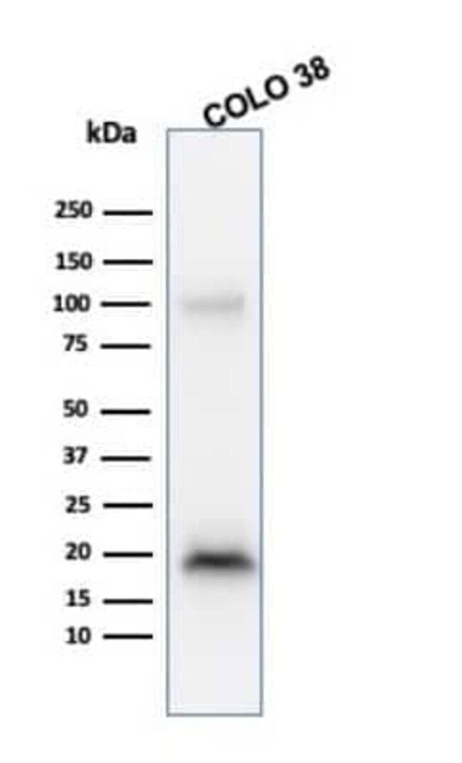
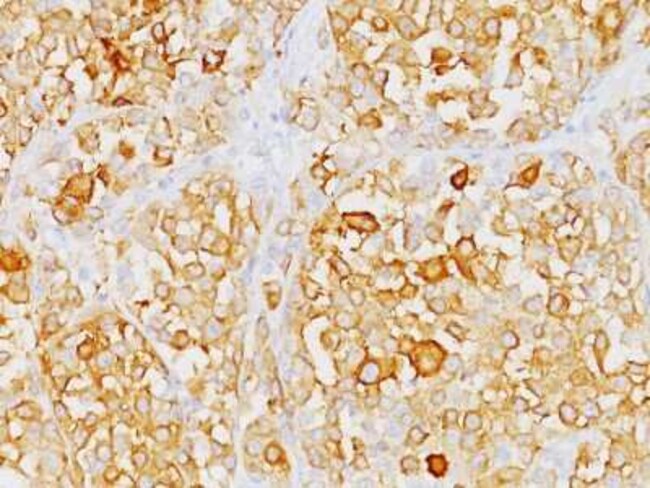
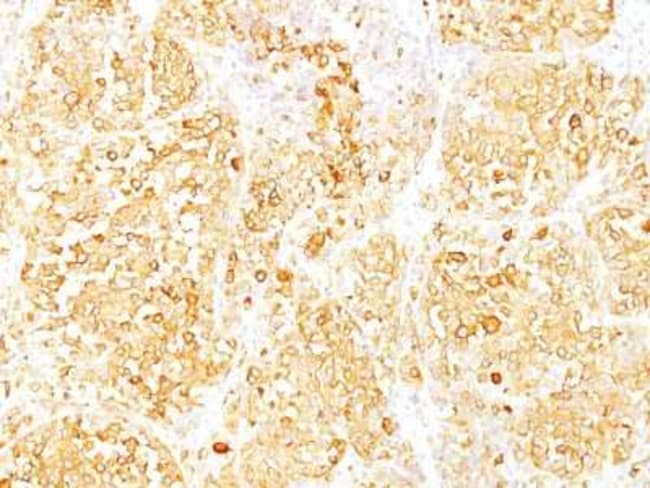
Melan-A/MART-1 Monoclonal specifically detects Melan-A/MART-1 in Human, Mouse, Rat samples. It is validated for Western Blot, Flow Cytometry, Immunohistochemistry, Immunocytochemistry/Immunofluorescence, Immunohistochemistry-Paraffin.Specifications
| Melan-A/MART-1 | |
| 0.2mg/mL | |
| Monoclonal | |
| Purified | |
| RUO | |
| Q16655 | |
| 2315 | |
| Recombinant human Melan-A protein was used as immunogen to generate the Melan-A antibody. | |
| Primary | |
| Store at 4C. |
| M2-9E3 | |
| Western Blot 0.5-1ug/ml, Flow Cytometry 0.5-1ug/million cells, Immunocytochemistry/Immunofluorescence 1-2ug/ml, Immunoprecipitation 0.5-1ug/500ug protein lysate, Immunohistochemistry-Paraffin 0.5-1ug/ml, Immunohistochemistry-Frozen 0.5-1ug/ml, SDS-Page | |
| Unconjugated | |
| Mouse | |
| PBS with 0.05% BSA. with 0.05% Sodium Azide | |
| Antigen LB39-AA, Antigen SK29-AA, Mart 1 Melan A, MART1MART-1, melan-A, melanoma antigen recognized by T-cells 1, Protein Melan-A | |
| MLANA | |
| IgG2b κ | |
| Protein G purified | |
| This antibody recognizes a protein doublet of 20-22kDa, identified as MART-1 (Melanoma Antigen Recognized by T cells 1) or Melan-A. MART-1 is a newly identified melanocyte differentiation antigen recognized by autologous cytotoxic T lymphocytes. Seven other melanoma associated antigens recognized by autologous cytotoxic T cells include MAGE-1, MAGE-3, tyrosinase, gp100, gp75, BAGE-1, and GAGE-1. Subcellular fractionation shows that MART-1 is present in melanosomes and endoplasmic reticulum. This MAb labels melanomas and other tumors showing melanocytic differentiation. It is also a useful positive-marker for angiomyolipomas. It does not stain tumor cells of epithelial, lymphoid, glial, or mesenchymal origin. |
For Research Use Only
Spot an opportunity for improvement?Share a Content Correction
Product Content Correction
Your input is important to us. Please complete this form to provide feedback related to the content on this product.
Product Title