
Donkey Secondary Antibodies


Biotium ALDH1A1 (Aldehyde Dehydrogenase 1A1) (ALDH1A1/1381), CF647 conjugate, 0.1mg/mL
Aldehyde dehydrogenase 1 family member A1 (ALDH1A1), also known as retinal dehydrogenase 1, belongs to the aldehyde dehydrogenase enzyme family, which is involved in the metabolism of alcohol. ALDH1A1 is predominantly expressed in the epithelium of testis, brain, eye, liver, kidney, as well as neural and hematopoietic stem cells. Reportedly, high ALDH1A1 expression is found in solitary fibrous tumor (SFT) and hemangiopericytoma (HPC), compared to meningiomas and synovial sarcomas. In combination with CD34, ALDH1A1 may be useful for the differentiation among SFT, HPC, meningioma, and synovial sarcoma.Primary antibodies are available purified, or with a selection of fluorescent CF Dyes and other labels. CF Dyes offer exceptional brightness and photostability. Note: Conjugates of blue fluorescent dyes like CF405S and CF405M are not recommended for detecting low abundance targets, because blue dyes have lower fluorescence and can give higher non-specific background than othe

Biotium EGFR (Epidermal Growth Factor Receptor) (GFR/2968R), CF647 conjugate, 0.1mg/mL
This MAb recognizes a protein of 170 kDa, identified as EGFR. EGFR is type I receptor tyrosine kinase with sequence homology to erbB-1, -2, -3 -4 or HER-1, -2, -3 -4. It binds to Epidermal Growth Factor (EGF), Transforming Growth Factor-a (TGF-a), Heparin-binding EGF (HB-EGF), amphiregulin, betacellulin and epiregulin. EGFR is overexpressed in tumors of breast, brain, bladder, lung, gastric, head & neck, esophagus, cervix, vulva, ovary, and endometrium. It is predominantly present in squamous cell carcinomas.Primary antibodies are available purified, or with a selection of fluorescent CF Dyes and other labels. CF Dyes offer exceptional brightness and photostability. Note: Conjugates of blue fluorescent dyes like CF405S and CF405M are not recommended for detecting low abundance targets, because blue dyes have lower fluorescence and can give higher non-specific background than other dye colors.
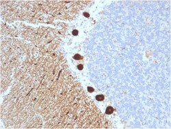
Biotium Carbonic Anhydrase VIII(CPTC-CA8-2), CF647 conjugate, 0.1mg/mL
Carbonic anhydrase VIII (carbonic anhydrase 8) was named because of sequence similarity to other known carbonic anhydrase genes. However, the gene product lacks carbonic anhydrase activity (i. e. , the reversible hydration of carbon dioxide). The gene product continues to carry a carbonic anhydrase designation based on clear sequence identity to other members of the carbonic anhydrase gene family. Mutations in the gene cause neurological disorders in mice. Primary antibodies are available purified, or with a selection of fluorescent CF Dyes and other labels. CF Dyes offer exceptional brightness and photostability. Note: Conjugates of blue fluorescent dyes like CF405S and CF405M are not recommended for detecting low abundance targets, because blue dyes have lower fluorescence and can give higher non-specific background than other dye colors.
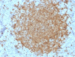
Biotium BOB.1 (B-Cell Marker)(BOB1/2422), CF647 conjugate, 0.1mg/mL
BOB-1 is a transcriptional coactivator that specifically associates with either POU2F1/OCT1 or POU2F2/OCT2, boosting the promoter activity of those complexes. It is essential for the response of B-cells to antigens, required for the formation of germinal centers, and regilates IL-6 expression. BOB-1 expression in a variety of established B-cell lines, representing different stages of B-cell development, has suggested a constitutive, B-cell-specific expression pattern. LP cells in nodular lymphocyte predominant Hodgkin lymphoma, because they are germinal center-derived, are consistently immuno-positive for BOB-1. Conversely, only some cases of classical Hodgkin lymphoma show BOB-1 immuno-reactivity within the Hodgkin and Reed-Sternberg cells. Expression of BOB-1 has been reported in follicular center cell lymphoma, diffuse large B-cell lymphoma and some cases of acute myeloid leukemia. B-CLL, marginal zone lymphoma, and mantle cell lymphoma may show weak to moderate immunoreactivit
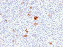
Biotium Bcl-X (Apoptosis Marker)(BCL2L1/2406), CF647 conjugate, 0.1mg/mL
Bcl-x belongs to the Bcl-2 family of proteins and plays a crucial role in apoptosis. Bcl-X has two isoforms: Bcl-XL (long), a 26 kDa protein which suppresses cell death, and Bcl-XS (short), a 178 amino acid protein lacking a 63 amino acid domain which functions as a dominant inhibitor of Bcl-2 and promotes apoptosis. This MAb reacts with both Bcl-XS and Bcl-XL proteins, and shows no cross-reaction with Bcl-2 or Bax proteins.Primary antibodies are available purified, or with a selection of fluorescent CF Dyes and other labels. CF Dyes offer exceptional brightness and photostability. Note: Conjugates of blue fluorescent dyes like CF405S and CF405M are not recommended for detecting low abundance targets, because blue dyes have lower fluorescence and can give higher non-specific background than other dye colors.
Biotium Alpha-1-Antichymotrypsin (SERPINA3) (Histiocytoma Marker) (AACT/1451), CF647 conjugate, 0.1mg/mL
Alpha-1 Antichymotrypsin (AACT) is a plasma protease inhibitor synthesized in the liver as a single glycopeptide chain. In human, the normal serum level of AACT is about one-tenth that of alpha-1-antitrypsin (AAT), with which it shares nucleic acid and protein sequence homology. Both are major acute phase reactants; their concentrations in plasma increase in response to trauma, surgery and infection. Elevated levels of AACT are widely, but not universally, reported in the cerebrospinal fluid and plasma of AD patients. Prostate-specific antigen (PSA) and its SDS-stable complex with AACT are in widespread use as markers for the diagnosis of prostate cancer. AACT deficiency may also be a possible cause of chronic liver disease. AACT antibody reacts with histiocytes and histiocytic neoplasms. It is widely used to identify histiocytes and tumors derived from them. Acinar tumors of the pancreas and salivary gland may also exhibit AACT positivity. Primary antibodies are available
Biotium CD81 / TAPA-1 (rC81/3442), CF647 conjugate, 0.1mg/mL
This antibody recognizes a protein of 26 kDa, identified as CD81 (Workshop VI; Code CD81.1)). CD81 has a very broad cellular distribution, being expressed on T- and B-lymphocytes, NK cells, thymocytes, eosinophils, fibroblasts, epithelial and endothelial cells. Neutrophils, erythrocytes and platelets are negative, while monocytes are variably positive. CD81 is a member of a family of tetraspanin transmembrane proteins, including CD9, CD37, CD53, CD63, and CD82. It associates with CD19, CD21, Leu 13, and integrins on cell membrane and is involved in signal transduction in B lymphocyte development and cell adhesion. CD81 also acts as a receptor for the envelope protein E2 of chronic hepatitis C virus. Antibodies to CD81 have anti-proliferative effects on different lymphoid cell lines, particularly those derived from large cell lymphomas.Primary antibodies are available purified, or with a selection of fluorescent CF Dyes and other labels. CF Dyes offer exceptional brightness a
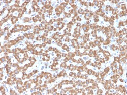
Biotium Prohibitin (Mitochondrial Marker)(PHB/3229), CF647 conjugate, 0.1mg/mL
Recognizes a protein of 30 kDa which is identified as Prohibitin, an evolutionarily conserved protein with homologues found in yeast to man. It is located in the inner membrane of mitochondria. Although prohibitin mRNA and protein expression occurs throughout the cell cycle, maximum levels are detected during the G1/S phase transition and minimum levels are seen in S phase and the G2/mitosis boundary. Prohibitin is located exclusively in the mitochondria with the highest concentration on the inner membrane. Prohibitin is an ideal mitochondrial marker. It shows antiproliferative activity and has been proposed to play a role in normal cell cycle regulation, replicative senescence, cellular immortalization, and tumor suppression. Primary antibodies are available purified, or with a selection of fluorescent CF Dyes and other labels. CF Dyes offer exceptional brightness and photostability. Note: Conjugates of blue fluorescent dyes like CF405S and CF405M are not recommended fo
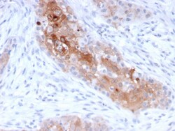
Biotium Desmoglein-1 (DSG1)(32-2B), CF647 conjugate, 0.1mg/mL
Recognizes a protein of ~150 kDa, identified as Desmoglein-1 (DSG1). Desmoglein-1 is a member of the desmosomal cadherin family. Desmosomes are intercellular adhering junctions that represent cell surface attachment sites for intermediate filament. Desmocollins and desmogleins are the main desmosomal transmembrane proteins. Desmogleins consist of Dsg1, Dsg2, Dsg3, and Dsg4 isoforms. Within the desmosome, the extracellular domain of desmoglein is essential for calcium dependent heterophilic binding to the desmocollins, whereas the intracellular domain is essential for binding to the desmosomal plaque protein, plakoglobin. Desmoglein 1 is synthesized exclusively in the suprabasal layers. Intact and functionally active desmoglein 1 is essential to epidermal integrity. Primary antibodies are available purified, or with a selection of fluorescent CF Dyes and other labels. CF Dyes offer exceptional brightness and photostability. Note: Conjugates of blue fluorescent dyes like CF
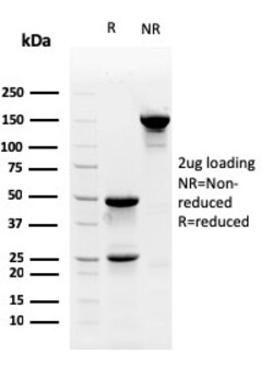
Biotium Myofibroblast Marker(PR 2D3), CF647 conjugate, 0.1mg/mL
Myofibroblasts are a unique group of smooth muscle-like fibroblasts that play an important role in oncogenesis, inflammation, repair, wound contraction and fibrosis. Like smooth muscle (SM) cells, myofibroblasts contain microfilament bundles and express -SM Actin, the Actin isoform that is present in myoepithelial cells and SM cells and especially abundant in vascular SM cells. Myofibroblasts secrete inflammatory and anti-inflammatory cytokines, chemokines, growth factors and lipid and gaseous inflammatory mediators, as well as extracellular matrix proteins and proteases in most organs and tissues. Besides being temporarily present following tissue injuries and fibrocontractive diseases, myofibroblasts are also present under normal conditions in regions such as the skin, pulmonary septa and periodontal ligaments. Stem cell factor and platelet-derived growth factor (PDGF) are two secreted proteins responsible for differentiating myofibroblasts from embryological stem cells. PR
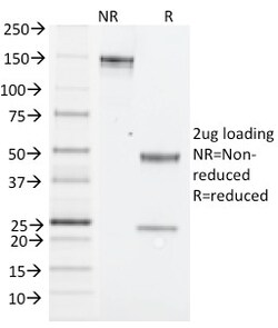
Biotium NK1.1 / CD161c / Klrb1c, Mouse(PK136), CF647 conjugate, 0.1mg/mL
NK1. 1 is a type II integral membrane glycoprotein with a C-type lectin domain and is encoded by theKlrb1c/NKR-P1Cgene. It is predominantly expressed as a disulfide-linked homodimer on NK cells however; it is also expressed on NK-T cells, a rare population of T lymphocytes. NK1. 1 is expressed in some mouse strains, including C57BL/6, FVB/N, and NZB, but not AKR, BALB/c, CBA/J, C3H, DBA/1, DBA/2, NOD, SJL, and 129. Expression of NKR-P1C antigen has been correlated with lysis of tumor cells in vitro and rejection of bone marrow allograftsin vivo. NK-1. 1 has also been shown to play a role in NK cell activation, IFN- production, and cytotoxic granule release. NK-1. 1 is commonly used as a mouse NK cell marker. Primary antibodies are available purified, or with a selection of fluorescent CF Dyes and other labels. CF Dyes offer exceptional brightness and photostability. Note: Conjugates of blue fluorescent dyes like CF405S and CF405M are not recommended for detecting low abu
Biotium CDX2 (GI Epithelial Marker) (CDX2/2951R), CF647 conjugate, 0.1mg/mL
The intestine-specific transcription factors CDX1 and CDX2 are important for directing intestinal development, differentiation, proliferation and maintenance of the intestinal phenotype. CDX2 protein expression has been seen in GI carcinomas. Anti-CDX2 has been useful to establish GI origin of metastatic adenocarcinomas and carcinoids and is especially useful to distinguish metastatic colorectal adenocarcinoma from lung adenocarcinoma. However, mucinous carcinomas of the ovary also express CDX2 protein. It limits the usefulness of this marker in the distinction of metastatic colorectal adenocarcinoma from mucinous carcinoma of the ovary.Primary antibodies are available purified, or with a selection of fluorescent CF Dyes and other labels. CF Dyes offer exceptional brightness and photostability. Note: Conjugates of blue fluorescent dyes like CF405S and CF405M are not recommended for detecting low abundance targets, because blue dyes have lower fluorescence and can give higher
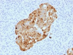
Biotium Chromogranin A / CHGA (Neuroendocrine Marker) (CHGA/1731R), CF647 conjugate, 0.1mg/mL
Chromogranin A is present in neuroendocrine cells throughout the body, including the neuroendocrine cells of the large and small intestine, adrenal medulla and pancreatic islets. It is an excellent marker for carcinoid tumors, pheochromocytomas, paragangliomas, and other neuroendocrine tumors. Co-expression of chromogranin A and neuron specific enolase (NSE) is common in neuroendocrine neoplasms. Reportedly, co-expression of certain keratins and chromogranin indicates neuroendocrine lineage. The presence of strong anti-chromogranin staining and absence of anti-keratin staining should raise the possibility of paraganglioma. The co-expression of chromogranin and NSE is typical of neuroendocrine neoplasms. Most pituitary adenomas and prolactinomas readily express chromogranin.Primary antibodies are available purified, or with a selection of fluorescent CF Dyes and other labels. CF Dyes offer exceptional brightness and photostability. Note: Conjugates of blue fluorescent dyes
Biotium CELA3B / ELA3B (Pancreatic Function Marker) (CELA3B/1811), CF647 conjugate, 0.1mg/mL
This MAb recognizes a protein of ~30 kDa, identified as CELA3B (Chymotrypsin like elastase family member 3B). Elastases form a subfamily of serine proteases that hydrolyze many proteins in addition to elastin. Humans have six elastase genes which encode the structurally similar proteins elastase 1, 2, 2A, 2B, 3A, and 3B. Unlike other elastases, elastase 3B has little elastolytic activity. Like most of the human elastases, elastase 3B is secreted from the pancreas as a zymogen and, like other serine proteases such as trypsin, chymotrypsin and kallikrein; it has a digestive function in the intestine. Elastase 3B preferentially cleaves proteins after alanine residues. Elastase 3B may also function in the intestinal transport and metabolism of cholesterol. Both elastase 3A and elastase 3B have been referred to as protease E and as elastase 1, and excretion of this protein in fecal material is frequently used as a measure of pancreatic function.Primary antibodies are available pur
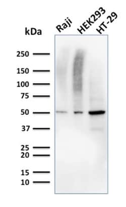
Biotium MMP3 (Marker of Metastasis and Rheumatoid Arthritis)(MMP3/2655), CF647 conjugate, 0.1mg/mL
The matrix metalloproteinases (MMP) are a family of peptidase enzymes responsible for the degradation of extracellular matrix components, including collagen, gelatin, fibronectin, laminin and proteoglycan. Transcription of MMP genes is differentially activated by phorbol ester, lipopolysaccharide (LPS) or staphylococcal enterotoxin B (SEB). MMP catalysis requires both calcium and zinc. MMP-3, MMP-10 and MMP-11 (also designated stromelysin-1, 2 and 3, respectively) activate procollagenase. MMP-3 activation of procollagenase can occur via two pathways. Direct activation by MMP-3 is slow and activation by MMP-3 in conjunction with tissue or plasma proteinases is rapid. MMP-10 is expressed in small intestine, and at lower levels in lung and heart. MMP-11 is specifically expressed in stromal cells of breast carcinomas and contributes to epithelial cell malignancies. Primary antibodies are available purified, or with a selection of fluorescent CF Dyes and other labels. CF Dyes of