
Donkey Secondary Antibodies


Jackson Immuno Research Labs AffiniPure Donkey Anti-Mouse IgM, µ chain specific, 1mg
AffiniPure Donkey Anti-Mouse IgM, u Chain Specific

Jackson Immuno Research Labs Alexa Fluor 488 AffiniPure F(ab')₂ Fragment Donkey Anti-Mouse IgG (H+L), .3mg
Alexa Fluor 488-AffiniPure F(ab')2 Fragment Donkey Anti-Mouse IgG (H+L) (min X Bov,Ck,Gt,GP,Sy Hms,Hrs,Hu,Rb,Rat,Shp Sr Prot)
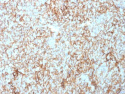
Biotium CD73 (Immuno-Oncology Target)(NT5E/2503), CF647 conjugate, 0.1mg/mL
CD73 (also designated ecto-5'-nucleotidase, E5NT, NT, NT5, NTE, eN and eNT) is a glycosyl-phosphatidylinositol (GPI)-anchored adhesion protein that catalyzes the dephosphorylation of extracellular purine and pyrimidine nucleotides to their corresponding bioactive nucleosides. CD73 is a dimer of two identical subunits that depends on GPI to link with the external face of the plasma membrane. Similar to other GPI-anchored proteins, CD73 mediates co-stimulatory signals in T cell activation. CD73 has few structural variants, yet elicits diverse biological function through differential regulation in endothelial cells (EC), subpopulations of B and T cells, germinal center follicular dendritic cells and on thymic medullary reticular fibroblasts. For example, IgG mediated neutralization of CD73 interferes with lymphocyte adhesion to EC, and blocks aggregation of germinal center B cells and follicular dendritic cells. Furthermore, IgG-mediated targeting of lymphocyte CD73, but not of
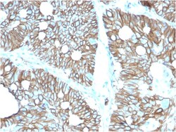
Biotium Cadherin 17 / LI Cadherin (Liver-Intestine Marker)(CDH17/2616), CF647 conjugate, 0.1mg/mL
Cadherin 17, also known as LI cadherin (for liver-intestine cadherin), is a member of the cadherin family of calcium-dependent adhesion molecules. Cadherins mediate cell-cell interactions via hemophilic binding, and are critical to the maintenance of tissue structure and morphogenesis. Cadherins each contain a large extracellular domain at the amino terminus, which is characterized by a series of five homologous repeats, the most distal of which is thought to be responsible for binding specificity. The relatively short carboxy terminal, intracellular domain interacts with a variety of cytoplasmic proteins, including beta-catenin, to regulate cadherin function. Cadherin 17 expression is restricted to liver and intestine tissues and is specifically localized to the basolateral domain of hepatocytes and enterocytes. Primary antibodies are available purified, or with a selection of fluorescent CF Dyes and other labels. CF Dyes offer exceptional brightness and photostability. N
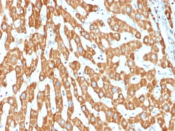
Biotium Prohibitin (Mitochondrial Marker)(PHB/1882), CF647 conjugate, 0.1mg/mL
Recognizes a protein of 30 kDa which is identified as Prohibitin, an evolutionarily conserved protein with homologues found in yeast to man. It is located in the inner membrane of mitochondria. Although prohibitin mRNA and protein expression occurs throughout the cell cycle, maximum levels are detected during the G1/S phase transition and minimum levels are seen in S phase and the G2/mitosis boundary. Prohibitin is located exclusively in the mitochondria with the highest concentration on the inner membrane. Prohibitin is an ideal mitochondrial marker. It shows antiproliferative activity and has been proposed to play a role in normal cell cycle regulation, replicative senescence, cellular immortalization, and tumor suppression. Primary antibodies are available purified, or with a selection of fluorescent CF Dyes and other labels. CF Dyes offer exceptional brightness and photostability. Note: Conjugates of blue fluorescent dyes like CF405S and CF405M are not recommended fo

Biotium Cytokeratin 19 (Pancreatic Stem Cell Marker) (rKRT19/800), CF488A conjugate, 0.1mg/mL
This antibody recognizes a protein of 40 kDa, identified as cytokeratin-19 (CK19), which is expressed in sweat gland, mammary gland ductal and secretory cells, bile ducts, gastrointestinal tract, bladder urothelium, oral epithelia, esophagus, and ectocervical epithelium. Anti-CK19 reacts with a wide variety of epithelial malignancies including adenocarcinomas of the colon, stomach, pancreas, biliary tract, liver, and breast. Perhaps the most useful application is the identification of thyroid carcinoma of the papillary type, although 50%-60% of follicular carcinomas are also labeled. Anti-CK19 is a useful marker for detection of tumor cells in lymph nodes, peripheral blood, bone marrow and breast cancer.Primary antibodies are available purified, or with a selection of fluorescent CF Dyes and other labels. CF Dyes offer exceptional brightness and photostability. Note: Conjugates of blue fluorescent dyes like CF405S and CF405M are not recommended for detecting low abundanc
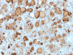
Biotium Thyroglobulin (Thyroidal Cell Marker) (rTGB24), CF647 conjugate, 0.1mg/mL
Thyroglobulin is a 660 kDa dimeric pre-protein with multiple glycosylation sites. It is produced by and processed within the thyroid gland to produce the hormone thyroxine and triiodothyronine. Prior to forming dimers, thyroglobulin monomers undergo conformational maturation in the endoplasmic reticulation. The vast majority of follicular carcinomas of the thyroid will give positive immunoreactivity for anti-thyroglobulin even though sometimes only focally. Poorly differentiated carcinomas of the thyroid are frequently anti-thyroglobulin negative. Adenocarcinomas of other-than-thyroid origin do not react with this antibody. This antibody is useful in identification of thyroid carcinoma of the papillary and follicular types. Presence of thyroglobulin in metastatic lesions establishes the thyroid origin of tumor. Anti-thyroglobulin, combined with anti-calcitonin, can identify medullary carcinomas of the thyroid. Furthermore, anti-thyroglobulin, combined with anti-TTF1, can be a
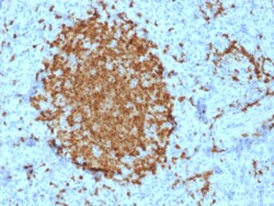
Biotium BOB.1 (B-Cell Marker)(BOB1/2421), CF647 conjugate, 0.1mg/mL
BOB-1 is a transcriptional coactivator that specifically associates with either POU2F1/OCT1 or POU2F2/OCT2, boosting the promoter activity of those complexes. It is essential for the response of B-cells to antigens, required for the formation of germinal centers, and regilates IL-6 expression. BOB-1 expression in a variety of established B-cell lines, representing different stages of B-cell development, has suggested a constitutive, B-cell-specific expression pattern. LP cells in nodular lymphocyte predominant Hodgkin lymphoma, because they are germinal center-derived, are consistently immuno-positive for BOB-1. Conversely, only some cases of classical Hodgkin lymphoma show BOB-1 immuno-reactivity within the Hodgkin and Reed-Sternberg cells. Expression of BOB-1 has been reported in follicular center cell lymphoma, diffuse large B-cell lymphoma and some cases of acute myeloid leukemia. B-CLL, marginal zone lymphoma, and mantle cell lymphoma may show weak to moderate immunoreactivit
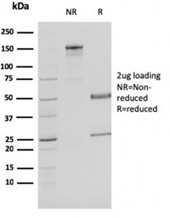
Biotium Malate dehydrogenase 1 NAD (soluble)(CPTC-MDH1-1), CF647 conjugate, 0.1mg/mL
Malate dehydrogenase catalyzes the reversible oxidation of malate to oxaloacetate, utilizing the NAD/NADH cofactor system in the citric acid cycle. The protein encoded by this gene is localized to the cytoplasm and may play pivotal roles in the malate-aspartate shuttle that operates in the metabolic coordination between cytosol and mitochondria. Alternatively spliced transcript variants encoding distinct isoforms have been found for this gene. Primary antibodies are available purified, or with a selection of fluorescent CF Dyes and other labels. CF Dyes offer exceptional brightness and photostability. Note: Conjugates of blue fluorescent dyes like CF405S and CF405M are not recommended for detecting low abundance targets, because blue dyes have lower fluorescence and can give higher non-specific background than other dye colors.
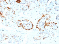
Biotium Crystallin Alpha B(CPTC-CRYAB-1), CF647 conjugate, 0.1mg/mL
Crystallins are the major proteins of the vertebrate eye lens, where they maintain the transparency and refractive index of the lens. Crystallins are divided into B-crystallin is associated with many neurological diseases, and a missense mutation in this gene has co-segregated in a family with a Desmin-related myopathy. Primary antibodies are available purified, or with a selection of fluorescent CF Dyes and other labels. CF Dyes offer exceptional brightness and photostability. Note: Conjugates of blue fluorescent dyes like CF405S and CF405M are not recommended for detecting low abundance targets, because blue dyes have lower fluorescence and can give higher non-specific background than other dye colors.
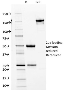
Biotium Cdk2 / p34cdc2 Serine-Threonine Kinase(AN4.3), CF647 conjugate, 0.1mg/mL
In vertebrates, as in yeast, multiple cyclins have been identified, including a total of eight such regulatory proteins in mammals. In contrast to the situation in yeast, the Cdc2 p34 kinase is not the only catalytic subunit identified in vertebrates that can interact with cyclins. While Cdc2 p34 is essential for the G2 to M transition in vertebrate cells, a second Cdc2-related kinase has also been implicated in cell cycle control. This protein, designated cyclindependent kinase 2 (Cdk2), also binds to cyclins and its kinase activity is temporally regulated during the cell cycle. Several additional Cdc2-related cyclin dependent kinases have been identified. These include Cdk3, Cdk4, Cdk5, PCTAIRE-1, PCTAIRE-2, PCTAIRE-3, Cdk6 Cdk7, Cdk8 and KKIALRE. Primary antibodies are available purified, or with a selection of fluorescent CF Dyes and other labels. CF Dyes offer exceptional brightness and photostability. Note: Conjugates of blue fluorescent dyes like CF405S and CF405M are
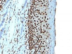
Biotium Histone H1 (Nuclear Marker) (r1415-1), CF647 conjugate, 0.1mg/mL
Eukaryotic histones are basic and water-soluble nuclear proteins that form hetero-octameric nucleosome particles by wrapping 146 base pairs of DNA in a left-handed supealpha-helicalturn sequentially to form chromosomal fiber. Two molecules of each of the four core histones (H2A, H2B, H3, and H4) form the octamer; formed of two H2A-H2B dimers and two H3-H4 dimers, forming two nearly symmetrical halves by tertiary structure. Over 80% of nucleosomes contain the linker Histone H1, derived from an intronless gene that interacts with linker DNA between nucleosomes and mediates compaction into higher order chromatin. Histones are subject to posttranslational modification by enzymes primarily on their N-terminal tails, but also in their globular domains. Such modifications include methylation, citrullination, acetylation, phosphorylation, sumoylation, ubiquitination and ADP-ribosylation.Primary antibodies are available purified, or with a selection of fluorescent CF Dyes and other l
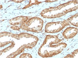
Biotium YBX1 / Y-box Binding Protein 1 / YB-1 (Tumor Biomarker)(YBX1/2430), CF647 conjugate, 0.1mg/mL
The specificity of this monoclonal antibody to its intended target was validated by HuProt™ Array, containing more than 19,000 full-length human proteins. Y-box binding protein-1 (YBX1) is the prototypic member of the cold shock protein family that fulfills numerous cellular functions. In the nucleus, YBX1 protein orchestrates transcription of proliferation-related genes, whereas in the cytoplasm it associates with mRNA and directs translation. In human tumor entities, such as breast, lung and prostate cancer, cellular YBX1 expression indicates poor clinical outcome, suggesting that YBX1 is an attractive marker to predict patients' prognosis. Primary antibodies are available purified, or with a selection of fluorescent CF Dyes and other labels. CF Dyes offer exceptional brightness and photostability. Note: Conjugates of blue fluorescent dyes like CF405S and CF405M are not recommended for detecting low abundance targets, because blue dyes have lower fluorescence an
Biotium Arginase 1 (Hepatocellular Carcinoma Marker) (ARG1/1126) , CF647 conjugate, 0.1mg/mL
This antibody recognizes a protein of 35-38 kDa, which is identified as Arginase 1 (ARG1). Arginase is a manganese metallo-enzyme that catalyzes the hydrolysis of arginine to generate ornithine and urea. Arginase I and II are isoenzymes which differ in subcellular localization, regulation, and possibly function. Arginase I is a cytosolic enzyme, which is expressed mainly in the liver as part of the urea cycle, whereas arginase II is a mitochondrial protein found in a variety of tissues. Antibodies to Arginase 1 label hepatocytes in normal tissues and granulocytes in peripheral blood. Arginase 1 is a sensitive and specific marker for identification of hepatocellular carcinoma.Primary antibodies are available purified, or with a selection of fluorescent CF Dyes and other labels. CF Dyes offer exceptional brightness and photostability. Note: Conjugates of blue fluorescent dyes like CF405S and CF405M are not recommended for detecting low abundance targets, because blue dyes have