Donkey Secondary Antibodies

Biotium EGFR (Epidermal Growth Factor Receptor) (GFR/1667), CF647 conjugate, 0.1mg/mL
This MAb recognizes a protein of 170 kDa, identified as EGFR. EGFR is type I receptor tyrosine kinase with sequence homology to erbB-1, -2, -3 -4 or HER-1, -2, -3 -4. It binds to Epidermal Growth Factor (EGF), Transforming Growth Factor-a (TGF-a), Heparin-binding EGF (HB-EGF), amphiregulin, betacellulin and epiregulin. EGFR is overexpressed in tumors of breast, brain, bladder, lung, gastric, head & neck, esophagus, cervix, vulva, ovary, and endometrium. It is predominantly present in squamous cell carcinomas.Primary antibodies are available purified, or with a selection of fluorescent CF Dyes and other labels. CF Dyes offer exceptional brightness and photostability. Note: Conjugates of blue fluorescent dyes like CF405S and CF405M are not recommended for detecting low abundance targets, because blue dyes have lower fluorescence and can give higher non-specific background than other dye colors.

Biotium CD40, Mouse(FGK45.5), CF647 conjugate, 0.1mg/mL
Upon binding to its ligand CD154, CD40 acts as a costimulatory molecule for the activation of B cells, dendritic cells, monocytes, and other APCs. CD40 plays roles in B cell activation, differentiation, proliferation and Ig isotype switching as well as dendritic cell maturation. FGK4. 5 can be used for blocking of CD40/CD154 interaction and forin vitroandin vivoactivation of CD40 expressing APCs. This MAb is shown to indirectly activate natural killer (NK) cells, producing significant antitumor and anti-metastatic effects. Effective in boosting immune responses against infectious agents and can potentially be used to treat chronic autoimmune inflammatory processes. Primary antibodies are available purified, or with a selection of fluorescent CF Dyes and other labels. CF Dyes offer exceptional brightness and photostability. Note: Conjugates of blue fluorescent dyes like CF405S and CF405M are not recommended for detecting low abundance targets, because blue dyes have lower
Biotium HSV1 (Herpes Simplex Virus Type I) (HSV1/1934), CF647 conjugate, 0.1mg/mL
The antibody reacts with HSV type 1 specific antigen. It is suitable for detection of HSV in human cellular material obtained from superficial lesions or biopsies and for the early identification of HSV in infected tissue cultures. The herpes simplex virus (HSV) (also known as cold sore, night fever or fever blister) is a virus that causes a contagious disease. There are two main types of Herpes Simplex Virus (HSV), 1 and 2. The HSV-1 strain generally appears in the orafacial organs. HSV2 usually resides in the sacral ganglion at the base of the spine. All herpes viruses are morphologically identical: they have a large double-stranded DNA genome and the virion consists of an icosahedral nucleo-capsid, which is surrounded by a lipid bilayer envelope.Primary antibodies are available purified, or with a selection of fluorescent CF Dyes and other labels. CF Dyes offer exceptional brightness and photostability. Note: Conjugates of blue fluorescent dyes like CF405S and CF405M
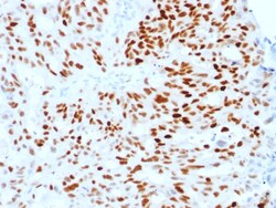
Biotium Estrogen Receptor, alpha (Marker of Estrogen Dependence)(ESR1/1904), CF647 conjugate, 0.1mg/mL
This monoclonal antibody is specific to estrogen receptor alpha (ER alpha) and shows minimal cross-reaction with other members of the family. ER is an important regulator of growth and differentiation in the mammary gland. Presence of ER in breast tumors indicates an increased likelihood of response to anti-estrogen (e. g. tamoxifen) therapy. It strongly stains nuclei of epithelial cells in breast carcinomas. Primary antibodies are available purified, or with a selection of fluorescent CF Dyes and other labels. CF Dyes offer exceptional brightness and photostability. Note: Conjugates of blue fluorescent dyes like CF405S and CF405M are not recommended for detecting low abundance targets, because blue dyes have lower fluorescence and can give higher non-specific background than other dye colors.
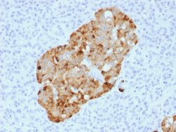
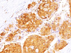
Biotium Chromogranin A / CHGA (Neuroendocrine Marker) (CHGA/1731R), CF647 conjugate, 0.1mg/mL
Chromogranin A is present in neuroendocrine cells throughout the body, including the neuroendocrine cells of the large and small intestine, adrenal medulla and pancreatic islets. It is an excellent marker for carcinoid tumors, pheochromocytomas, paragangliomas, and other neuroendocrine tumors. Co-expression of chromogranin A and neuron specific enolase (NSE) is common in neuroendocrine neoplasms. Reportedly, co-expression of certain keratins and chromogranin indicates neuroendocrine lineage. The presence of strong anti-chromogranin staining and absence of anti-keratin staining should raise the possibility of paraganglioma. The co-expression of chromogranin and NSE is typical of neuroendocrine neoplasms. Most pituitary adenomas and prolactinomas readily express chromogranin.Primary antibodies are available purified, or with a selection of fluorescent CF Dyes and other labels. CF Dyes offer exceptional brightness and photostability. Note: Conjugates of blue fluorescent dyes
Biotium PAX8 (Renal Cell Marker)(PAX8/1491 + PAX8/1492), CF647 conjugate, 0.1mg/mL
Recognizes a protein of 62 kDa, identified as PAX8. It is a member of the paired box (PAX) family of transcription factors. This nuclear protein is involved in thyroid follicular cell development and expression of thyroid-specific genes. Mutations in this gene have been associated with thyroid dysgenesis, thyroid follicular carcinomas, and atypical thyroid adenomas. PAX-8 is expressed in the thyroid (and associated carcinomas), non-ciliated mucosal cells of the fallopian tubes, and simple ovarian inclusion cysts, but not normal ovarian surface epithelial cells. PAX-8 is expressed in a high percentage of ovarian serous, endometrioid, and clear cell carcinomas, but only rarely in primary ovarian mucinous adenocarcinomas. PAX-8 expression is reported in renal tubules as well as renal cell carcinoma, nephroblastoma, and seminoma. PAX-8 antibody may be used as an additional immunohistochemical marker for renal epithelial tumors. Primary antibodies are available purified, or with a
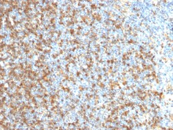
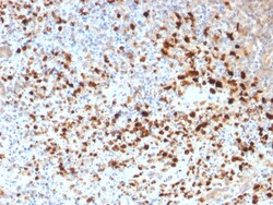
Biotium LMO2 (B-Cell Marker)(LMO2/1971), CF647 conjugate, 0.1mg/mL
The LMO2 protein has a central and crucial role in hematopoietic development and is highly conserved. It has a particular function in normal and lymphatic endothelial cells involving the regulation of angiogenesis and lymph-angiogenesis. Immunohistochemical studies have also demonstrated expression of LMO2 in both normal germinal center B-cells and germinal center-derived B-cell lymphomas, including follicular lymphoma and diffuse large B-cell lymphoma. The use of anti-LMO2 is valuable as a tool in the identification of lymphomas of B-cell origin. LMO2 is useful in differentiating follicular lymphoma (LMO2 ) from nodal marginal zone lymphoma (LM02-). It also is positive in Hodgkin's and Burkitt's lymphomas. Primary antibodies are available purified, or with a selection of fluorescent CF Dyes and other labels. CF Dyes offer exceptional brightness and photostability. Note: Conjugates of blue fluorescent dyes like CF405S and CF405M are not recommended for detecting low abun
Biotium Epstein-Barr Virus (LMP-1)(CS1), CF647 conjugate, 0.1mg/mL
This antibody is a mixture of four different monoclonal antibodies. This antibody is specific to 60 kDa latent membrane protein (LMP-1) encoded by the BNLF1 gene of the EBV. Each clone reacts with different epitopes on the hydrophilic C-terminus of the cytoplasmic domain of LMP-1. This antibody stains strongly with EBV-positive lymphoblastoid cell lines and EBV infected B cell immunoblasts in infectious mononucleosis. EBV, also designated human herpesvirus 4 (HHV-4), is a member of the herpesvirus family and is one of the most common human viruses. EBV infects B cells and, though often asymptomatic, it can cause infectious mononucleosis, a disease characterized by fatigue, fever, sore throat and muscle soreness. Primary antibodies are available purified, or with a selection of fluorescent CF Dyes and other labels. CF Dyes offer exceptional brightness and photostability. Note: Conjugates of blue fluorescent dyes like CF405S and CF405M are not recommended for detecting low
Biotium Cytokeratin 13 (Non-Keratinized Squamous Epithelial Marker)(KRT13/2213), CF594 conjugate, 0.1mg/mL
Cytokeratin 13 (KRT13) is the major acidic keratin, which together with KRT4, its basic partner, is expressed in the suprabasal layers of non-cornified stratified epithelia including tongue mucosa, esophagus, anal canal epithelium, tracheal epithelium, uterine cervix, and urothelium. Defects in the KRT13 gene are a cause of white sponge nevus of cannon (WSN), a rare autosomal dominant disorder, which predominantly affects non-cornified stratified squamous epithelia and is characterized by the presence of soft, white and spongy plaques in the oral mucosa. KRT13 has been used as a marker for non-keratinized squamous epithelium. It is also expressed in various squamous metaplasia, but it is down regulated in squamous dysplasia and squamous carcinoma. Primary antibodies are available purified, or with a selection of fluorescent CF Dyes and other labels. CF Dyes offer exceptional brightness and photostability. Note: Conjugates of blue fluorescent dyes like CF405S and CF405M a
Biotium MUC5AC (Mucin 5AC / Gastric Mucin)(2-11M1), CF647 conjugate, 0.1mg/mL
This MAb recognizes the peptide core of gastric mucin M1 (recently identified as Mucin 5AC). Its epitope is located in the N-terminal cysteine rich part of the peptide core of MUC5AC, which is heavily glycosylated. Its epitope is destroyed by beta-mercaptoethanol but not by periodate treatment. MAb 2-11M1 reacts with the protein backbone exclusively; it only reacts with fully deglycosylated MUC5AC. Therefore, the material under test should also be fully deglycosylated. This can be achieved with standard periodate oxidation method. The success of the deglycosylation can be checked with routine PAS (Periodic Acid Shiff) staining. After deglycosylation, the preparation should no longer be stainable with PAS reagent. Only then 2-11M1 will react should MUC5AC be present. This mucin is present in primary ovarian mucinous cancer but usually absent in colorectal adenocarcinoma, thus showing an expression pattern opposite to MUC2. Together with a panel of antibodies, Anti-MUC5AC may b
Biotium Spectrin beta III (SPTBN2) (SPTBN2/1247), CF647 conjugate, 0.1mg/mL
Spectrin is an actin binding protein that is a major component of the plasma membrane skeleton. Spectrins function as membrane organizers and stabilizers by forming dimers, tetramers and higher polymers. Vertebrate spectrins have two alpha-subunits (alpha-I/alpha-II), four beta-subunits (beta-I-beta-IV) and a beta-H subunit creating diversity and specialization of function. Spectrin alpha and spectrin beta are present in erythrocytes, whereas spectrin alpha II (also designated fodrin alpha) and spectrin beta I (also designated fodrin beta) are present in other somatic cells. The spectrin tetramers in erythrocytes act as barriers to lateral diffusion, but spectrin dimers seem to lack this function. Spectrin beta III is highly homologous to both spectrin beta I and spectrin beta II. Spectrin beta III is highly expressed in brain, kidney, pancreas and liver, and at lower levels in lung and placenta. Spectrin beta 3 is primarily expressed in nervous tissues with highest expressio
Biotium Arginase 1 (Hepatocellular Carcinoma Marker)(ARG1/1125+ ARG1/1126), CF647 conjugate, 0.1mg/mL
This antibody recognizes a protein of 35-38 kDa, which is identified as Arginase 1 (ARG1). Arginase is a manganese metallo-enzyme that catalyzes the hydrolysis of arginine to generate ornithine and urea. Arginase I and II are isoenzymes which differ in subcellular localization, regulation, and possibly function. Arginase I is a cytosolic enzyme, which is expressed mainly in the liver as part of the urea cycle, whereas arginase II is a mitochondrial protein found in a variety of tissues. Antibodies to Arginase 1 label hepatocytes in normal tissues and granulocytes in peripheral blood. Arginase 1 is a sensitive and specific marker for identification of hepatocellular carcinoma.Primary antibodies are available purified, or with a selection of fluorescent CF Dyes and other labels. CF Dyes offer exceptional brightness and photostability. Note: Conjugates of blue fluorescent dyes like CF405S and CF405M are not recommended for detecting low abundance targets, because blue dyes
Biotium NOX4 / NADPH Oxidase 4 (NOX4/1245), CF647 conjugate, 0.1mg/mL
The superoxide-generating NADPH oxidase includes a membrane-bound flavocytochrome containing two subunits, gp91-phox and p22-phox, and the cytosolic proteins p47-phox and p67-phox. During activation of the NADPH oxidase, p47-phox and p67-phox migrate to the plasma membrane where they associate with the flavocytochrome, cytochrome b558, to form the active enzyme complex. The p22 and gp91-phox subunits also function as surface O2 sensors that initiate cellular signaling in response to hypoxic conditions. NOX4 is a renal gp91-phox homolog highly expressed at the site of erythropoietin production in the proximal convoluted tubule epithelial cells of the renal cortex. It is also expressed in fetal tissues, placenta, glioblastoma and vascular cells.Primary antibodies are available purified, or with a selection of fluorescent CF Dyes and other labels. CF Dyes offer exceptional brightness and photostability. Note: Conjugates of blue fluorescent dyes like CF405S and CF405M are no
Biotium CD11c (Dendritic Cell Marker)(ITGAX/1242), CF647 conjugate, 0.1mg/mL
Recognizes a protein of 145 kDa, identified as CD11c. CD11c (ITGAX), a member of the leukointegrin family, shares the same beta subunit with other members of the leukocyte adhesion molecule family, which includes CD11a (LFA-1), CD11b (MAC-1) and CD11d (ITGAD), but has a unique alpha chain. CD11c has been shown to play a role in phagocytosis, cell migration, and cytokine production by monocytes/macrophages as well as induction of T-cell proliferation by Langerhans cells. CD11c is expressed prominently on the plasma membranes of monocytes, tissue macrophages, NK cells, and most dendritic cells (DCs). A lower level of expression is also observed on neutrophils as a result of its high level of expression on most DCs. An antibody to CD11c may aid in identification of lesions with histiocytic origin. It may also been used as a marker for hairy cell leukemia in paraffin-embedded tissues.