
Donkey Secondary Antibodies


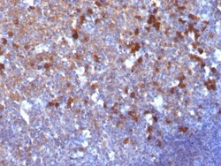
Biotium Human IgG Immunoglobulin(IG217), CF647 conjugate, 0.1mg/mL
Recognizes a protein of 75 kDa, identified as γ heavy chain of human immunoglobulins. It reacts with all sub-classes of γ chain of human immunoglobulins. It does not cross-react with α (IgA), μ (IgM), ε (IgE), or δ (IgD), heavy chains, T-cells, monocytes, granulocytes, or erythrocytes. This MAb is useful in the identification of leukemias, plasmacytomas, and certain non-Hodgkin's lymphomas. The most common feature of these malignancies is the restricted expression of a single heavy chain class. Demonstration of clonality in lymphoid infiltrates indicates that the infiltrate is clonal and therefore malignant.Primary antibodies are available purified, or with a selection of fluorescent CF Dyes and other labels. CF Dyes offer exceptional brightness and photostability. Note: Conjugates of blue fluorescent dyes like CF405S and CF405M are not recommended for detecting low abundance targets, because blue dyes have lower fluorescence and can give higher non-specific ba

Jackson Immuno Research Labs Rhodamine (TRITC)-AffiniPure Donkey Anti-Mouse IgM µ Chain Specific (min X Hu Bov Hrs Rat Sr Prot)
Rhodamine (TRITC)-AffiniPure Donkey Anti-Mouse IgM µ Chain Specific (min X Hu Bov Hrs Rat Sr Prot)
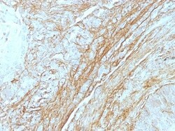
Biotium Tenascin(T2H5), CF647 conjugate, 0.1mg/mL
In Western blotting, this antibody reacts with two bands of ~MW of 210 kDa and 300 kDa, identified as two isoforms of Tenascin C. Specificity of this MAb is validated by sequential immunoprecipitation with a PAb against Tenascin C. Tenascin C is a multifunctional, disulfide-linkedhexameric extracellular matrix glycoprotein expressed in association with mesenchymal epithelial interactions during development and in the neo-vasculature and stroma of undifferentiated tumors. In adults, it is restricted to certain epithelial-stromal interfaces and increases markedly in hyper-proliferative diseases and in stroma of many neoplasms, including gliomas, breast, squamous and lung carcinomas.Primary antibodies are available purified, or with a selection of fluorescent CF Dyes and other labels. CF Dyes offer exceptional brightness and photostability. Note: Conjugates of blue fluorescent dyes like CF405S and CF405M are not recommended for detecting low abundance targets, because blue
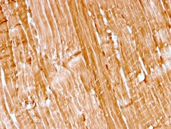
Biotium Myoglobin (Muscle Cell Marker)(rMB/2105), CF647 conjugate, 0.1mg/mL
Myoglobin is a cytosolic oxygen-binding protein responsible for the storage and diffusion of oxygen within myocytes. Expression of myoglobin is highest in skeletal and cardiac muscle. Myoglobin is well accepted as an O2-storage protein in muscle, capable of releasing O2 during periods of hypoxia or anoxia. In combination with other striated muscle markers such as vimentin and myogenin, myoglobin is helpful in the identification of rhabdomyosarcoma and tumors with skeletal muscle differentiation. Reportedly, myoglobin is expressed on epithelial cancer cells due to changed metabolic and environmental conditions. Primary antibodies are available purified, or with a selection of fluorescent CF Dyes and other labels. CF Dyes offer exceptional brightness and photostability. Note: Conjugates of blue fluorescent dyes like CF405S and CF405M are not recommended for detecting low abundance targets, because blue dyes have lower fluorescence and can give higher non-specific backgroun
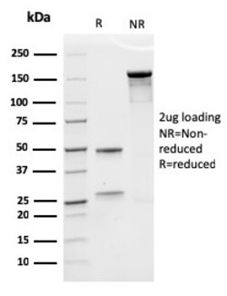
Biotium Erythropoietin (EPO) (Marker of Placentation Disorders)(rEPO/1367), CF647 conjugate, 0.1mg/mL
Recognizes a protein of about 37 kDa, which is identified as Erythropoietin (EPO). Erythropoietin is a secreted, glycosylated cytokine hormone composed of four alpha helical bundles. It is the primary factor responsible for regulating erythropoiesis during steady-state conditions and in response to blood loss and hemorrhage in the adult organism. Erythropoietin is synthesized by the kidney and stimulates the proliferation and maturation of bone marrow erythroid precursor cells. The protein is found in the plasma and regulates red cell production by promoting erythroid differentiation and initiating hemoglobin synthesis. Primary antibodies are available purified, or with a selection of fluorescent CF Dyes and other labels. CF Dyes offer exceptional brightness and photostability. Note: Conjugates of blue fluorescent dyes like CF405S and CF405M are not recommended for detecting low abundance targets, because blue dyes have lower fluorescence and can give higher non-specific
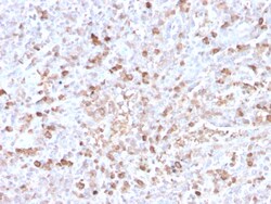
Biotium Human Lambda Light Chain (B-Cell Marker)(LLC/3778R), CF647 conjugate, 0.1mg/mL
This MAb is specific to lambda light chain of immunoglobulin and shows no cross-reaction with lambda light chain or any of the five heavy chains. In mammals, the two light chains in an antibody are always identical, with only one type of light chain, kappa or lambda. The ratio of Kappa to Lambda is 70:30. However, with the occurrence of multiple myeloma or other B-cell malignancies this ratio is disturbed. Antibody to the lambda light chain is reportedly useful in the identification of leukemias, plasmacytomas, and certain non-Hodgkin's lymphomas. Demonstration of clonality in lymphoid infiltrates indicates that the infiltrate is malignant. Primary antibodies are available purified, or with a selection of fluorescent CF Dyes and other labels. CF Dyes offer exceptional brightness and photostability. Note: Conjugates of blue fluorescent dyes like CF405S and CF405M are not recommended for detecting low abundance targets, because blue dyes have lower fluorescence and can giv
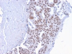
Biotium CHD4(3F2/4), CF647 conjugate, 0.1mg/mL
CHD4/Mi-2b is a component of the nucleosome remodeling and deacetylase (NuRD) complex which is a multi-subunit protein complex containing both histone deacetylase and nucleosome-dependent ATPase subunits. Current models predict that this complex function primarily in transcriptional repression. Accumulating evidence indicates that NuRD may regulate the transcription of specific genes by interacting with specific transcriptional factors. In addition, the NuRD complex may also participate in genome-wide transcriptional regulation through an association with histone tails. Primary antibodies are available purified, or with a selection of fluorescent CF Dyes and other labels. CF Dyes offer exceptional brightness and photostability. Note: Conjugates of blue fluorescent dyes like CF405S and CF405M are not recommended for detecting low abundance targets, because blue dyes have lower fluorescence and can give higher non-specific background than other dye colors.
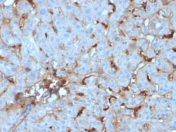
Biotium C1QA / Complement C1q A-Chain(C1QA/2955), CF647 conjugate, 0.1mg/mL
Complement C1q A Chain (C1qa) is a subunit of C1q. C1q is composed of 6 polypeptide chains each of C1qa, C1qb, and C1qc. C1q is a component of the classical complement pathway, and thereby plays an important role in the immune response. Each C1q subunit contains an N-terminal collagen-like region and a C-terminal C1q globular domain. The presence of receptors for C1q on effector cells modulates its activity, which may be antibody-dependent or independent. Macrophages are the primary source of C1q, while anti-inflammatory drugs as well as cytokines differentially regulate expression of the mRNA as well as the protein. C1q deficiency is associated with lupus erythematosus and glomerulonephritis. Primary antibodies are available purified, or with a selection of fluorescent CF Dyes and other labels. CF Dyes offer exceptional brightness and photostability. Note: Conjugates of blue fluorescent dyes like CF405S and CF405M are not recommended for detecting low abundance targets,
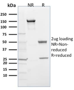
Biotium EGLN1 / PHD2(366G/76/3), CF647 conjugate, 0.1mg/mL
This antibody recognises human prolyl hydroxylase 2 (PHD2), a 46 kDa enzyme expressed abundantly in all tissues with the highest expression in testis. Hypoxia inducible factor-1 (HIF-1) is a transcriptional complex, consisting of an alpha and beta subunit, which plays a key role in coordinating the cellular response to hypoxia. During normal oxygen conditions, the alpha subunit of HIF-1 is rapidly degraded, however when hypoxia occurs this degradation is suppressed and HIF-1 activates the transcription of various genes important for survival and adaptation to hypoxia. Prolyl hydroxylase 2 catalyses the hydroxylation of specific prolyl residues within the HIF-1 alpha subunit, thereby targeting this subunit for degradation. Primary antibodies are available purified, or with a selection of fluorescent CF Dyes and other labels. CF Dyes offer exceptional brightness and photostability. Note: Conjugates of blue fluorescent dyes like CF405S and CF405M are not recommended for det
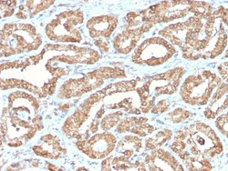
Biotium Ornithine Decarboxylase-1 (ODC-1)(ODC1/2878R), CF647 conjugate, 0.1mg/mL
Recognizes a 53 kDa protein, identified as the Ornithine Decarboxylase (ODC-1). ODC is the initial and rate-limiting enzyme in the biosynthetic pathway of polyamines and is involved in the conversion of ornithine to putrescine. The biological activity of ODC-1 is rapidly induced in response to virtually all agents known to promote cell proliferation including hormones, drugs, growth factors, mitogens, and tumor promoters. Reportedly, ODC mRNA levels are elevated in lung carcinomas as well as in colon adenomas and carcinomas. ODC activity in colorectal carcinomas is greater than those in adenomas and normal mucosa. Primary antibodies are available purified, or with a selection of fluorescent CF Dyes and other labels. CF Dyes offer exceptional brightness and photostability. Note: Conjugates of blue fluorescent dyes like CF405S and CF405M are not recommended for detecting low abundance targets, because blue dyes have lower fluorescence and can give higher non-specific backg
Biotium CD8A (Cytotoxic- & Suppressor T-Cell Marker)(UCHT4), CF647 conjugate, 0.1mg/mL
CD8 is a cell surface receptor expressed either as a heterodimer with the CD8 beta chain (CD8 alpha/beta) or as a homodimer (CD8 alpha/alpha). A majority of thymocytes and a subpopulation of mature T cells and NK cells express CD8a. CD8 binds to MHC class 1 and through its association with protein tyrosine kinase p56lck plays a role in T cell development and activation of mature T cells. For mature T-cells, CD4 and CD8 are mutually exclusive, so anti-CD8, generally used in conjunction with anti-CD4. It is a useful marker for distinguishing helper/inducer T-lymphocytes, and most peripheral T-cell lymphomas are CD4 /CD8-. Anaplastic large cell lymphoma is usually CD4 and CD8-, and in T-lymphoblastic lymphoma/leukemia, CD4 and CD8 are often co-expressed. CD8 is also found in littoral cell angioma of the spleen. This MAb binds to the CD8 alpha chain, at an epitope distinct from that recognized by UCHT14.Primary antibodies are available purified, or with a selection of fluorescent
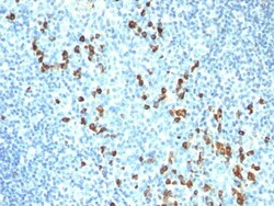
Biotium Human IgM Immunoglobulin(ICO-30), CF647 conjugate, 0.1mg/mL
Recognizes a protein of 75 kDa, identified as mu heavy chain of human immunoglobulins. It does not cross-react with alpha (IgA), gamma (IgG), epsilon (IgE), or delta (IgD), heavy chains, T-cells, monocytes, granulocytes, or erythrocytes. Monomeric IgM is expressed as a membrane bound antibody on the surface of B cells and as a pentamer when secreted by plasma cells. IgM antibody is prominent in early immune responses to most antigens. Aberrant levels are associated with immune deficiency states, hereditary deficiencies, myeloma, Waldenstrom's macroglobulinemia, chronic infection and hepatocellular disease. This MAb is useful in the identification of leukemias, plasmacytomas, and certain non-Hodgkin's lymphomas. The most common feature of these malignancies is the restricted expression of a single heavy chain class. Demonstration of clonality in lymphoid infiltrates indicates that the infiltrate is clonal and therefore malignant.Primary antibodies are available purified, o