
Donkey Secondary Antibodies


Biotium NOX4 / NADPH Oxidase 4 (NOX4/1245), CF647 conjugate, 0.1mg/mL
The superoxide-generating NADPH oxidase includes a membrane-bound flavocytochrome containing two subunits, gp91-phox and p22-phox, and the cytosolic proteins p47-phox and p67-phox. During activation of the NADPH oxidase, p47-phox and p67-phox migrate to the plasma membrane where they associate with the flavocytochrome, cytochrome b558, to form the active enzyme complex. The p22 and gp91-phox subunits also function as surface O2 sensors that initiate cellular signaling in response to hypoxic conditions. NOX4 is a renal gp91-phox homolog highly expressed at the site of erythropoietin production in the proximal convoluted tubule epithelial cells of the renal cortex. It is also expressed in fetal tissues, placenta, glioblastoma and vascular cells.Primary antibodies are available purified, or with a selection of fluorescent CF Dyes and other labels. CF Dyes offer exceptional brightness and photostability. Note: Conjugates of blue fluorescent dyes like CF405S and CF405M are no

Biotium IDH1(IDH1/1152), CF647 conjugate, 0.1mg/mL
This antibody recognizes a 45 kDa protein, which is identified as isocitrate dehydrogenase (IDH1). It belongs to the isocitrate and isopropylmalate dehydrogenases family. IDH1 catalyzes the third step of the citric acid cycle, which involves the oxidative decarboxylation of isocitrate, forming alpha-ketoglutarate and CO2 in a two-step reaction. The first step involves the oxidation of isocitrate to the intermediate oxalosuccinate, while the second step involves the production of alpha-ketoglutarate. During this process, either NADH or NADPH is produced along with CO2. Recently, an inactivating mutation of IDH1 has been implicated in glioblastoma. IDH1 appears to function as a tumor suppressor that, when mutationally inactivated, contributes to tumorigenesis in part through induction of the HIF-1 pathway.Primary antibodies are available purified, or with a selection of fluorescent CF Dyes and other labels. CF Dyes offer exceptional brightness and photostability. Note: Conjugat
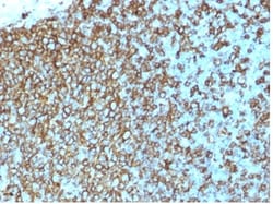
Biotium HLA-Pan (MHC II)(rHLA-Pan/3475), CF647 conjugate, 0.1mg/mL
Reacts with a common epitope of human major histocompatibility (MHC) class II antigens, HLA-DP, -DQ and -DR. Human MHC class II antigens are transmembrane glycoproteins composed of an alpha chain (36 kDa) and a beta chain (27 kDa). They are expressed primarily on antigen presenting cells such as B lymphocytes, monocytes, macrophages, and thymic epithelial cells and are also present on activated T lymphocytes. Human MHC class II genes are located in the HLA-D region that encodes at least six alpha and ten beta chain genes. Three loci, DR, DQ and DP, encode the major expressed products of the human class II region. The human MHC class II molecules bind intracellularly processed peptides and present them to T-helper cells. They, therefore, have a critical role in the initiation of the immune response. Primary antibodies are available purified, or with a selection of fluorescent CF Dyes and other labels. CF Dyes offer exceptional brightness and photostability. Note: Conjugates
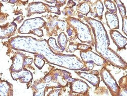
Biotium EGFR(GFR1195 + 31G7), CF647 conjugate, 0.1mg/mL
This MAb recognizes a protein of 170 kDa, identified as EGFR. EGFR is type I receptor tyrosine kinase with sequence homology to erbB-1, -2, -3 -4 or HER-1, -2, -3 -4. It binds to Epidermal Growth Factor (EGF), Transforming Growth Factor-a (TGF-a), Heparin-binding EGF (HB-EGF), amphiregulin, βcellulin and epiregulin. EGFR is overexpressed in tumors of breast, brain, bladder, lung, gastric, head & neck, esophagus, cervix, vulva, ovary, and endometrium. It is predominantly present in squamous cell carcinomas.Primary antibodies are available purified, or with a selection of fluorescent CF Dyes and other labels. CF Dyes offer exceptional brightness and photostability. Note: Conjugates of blue fluorescent dyes like CF405S and CF405M are not recommended for detecting low abundance targets, because blue dyes have lower fluorescence and can give higher non-specific background than other dye colors.
Biotium CD30 / TNFRSF8 (Hodgkin & Reed-Sternberg Cell Marker) (rKi-1/779), CF647 conjugate, 0.1mg/mL
This antibody recognizes a single chain glycoprotein of 105/120 kDa, identified as CD30/Ki-1. CD30 is synthesized as a 90 kDa precursor, which is processed in the Golgi complex into a membrane-bound phosphorylated mature 105/120 kDa glycoprotein. In Hodgkin's disease, CD30/Ki-1 antigen is expressed by mononuclear-Hodgkin and multinucleated Reed-Sternberg cells. It is also expressed by the tumor cells of a majority of anaplastic large cell lymphomas as well as by a varying proportion of activated T and B cells. This MAb distinguishes large cell lymphomas derived from activated lymphoid cells from histiocytic malignancies and lymphomas derived from resting and precursor lymphoid cells or from anaplastic carcinomas. About one third of the Ki-1 positive lymphomas lack the leukocyte common antigen (CD45).Primary antibodies are available purified, or with a selection of fluorescent CF Dyes and other labels. CF Dyes offer exceptional brightness and photostability. Note: Conjugate

Biotium Primary Antibody Neurofilament H rRmdO-20 CF647 1/EA
Neurofilament-H (rRmdO-20) is a recombinant mouse monoclonal antibody that recognizes Neurofilament-H This CF647 antibody conjugate has been validated in Immunofluorescence CF and reg dyes are Biotiums line of next-generation fluorescent dyes with advantages in brightness and photostability
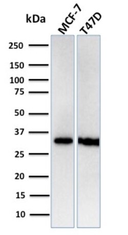
Biotium Replication Protein A2 (RPA2)(RPA2/3140R), CF647 conjugate, 0.1mg/mL
Recognizes a protein of 34 kDa, identified as a subunit of Replication Protein A (RPA) (also known as human single-stranded DNA binding protein, or HSSB). RPA from human cells is a stable heterotrimer of 70 kDa, 32-34 kDa, and 11-14 kDa subunits (RPA70, RPA32, and RPA14 respectively). It is involved in DNA replication, repair, and recombination. RPA is required for the SV40 large tumor antigen-catalyzed unwinding of SV40 DNA and stimulates DNA polymerase (pol) alpha and delta. RPA34 is phosphorylated at the G1/S boundary of the cell cycle or upon exposure of cells to DNA damage-inducing agents including ionizing and UV radiation. Primary antibodies are available purified, or with a selection of fluorescent CF Dyes and other labels. CF Dyes offer exceptional brightness and photostability. Note: Conjugates of blue fluorescent dyes like CF405S and CF405M are not recommended for detecting low abundance targets, because blue dyes have lower fluorescence and can give higher no

Biotium Primary Antibody Mouse IgG1 Fc N2504.M1FC CF647 1/EA
Anti-Mouse IgG1 (Fc) rVHH (N2504 M1FC) is a recombinant Alpaca VHH antibody that recognizes the Fc fragment of mouse IgG1 and is validated for IF flow and WB Far-red fluorescent STORM-compatible CF647 dye has Ex/Em at 650/665 nm Unit size 500 uL at 0 1 mg/mL
Biotium FOXP3(FXP3/197), CF647 conjugate, 0.1mg/mL
Recognizes a protein of 47-55 kDa, which is identified as FOXP3. Its precise epitope is not known, but it has been mapped to the N-terminal portion of the protein. The FOX family of transcription factors is a large group of proteins that share a common DNA binding domain termed a winged-helix or forkhead domain. During early development, FOXP1 and FOXP2 are expressed abundantly in the lung, with lower levels of expression in neural, intestinal and cardiovascular tissues, where they act as transcription repressors. FOXP1 is widely expressed in adult tissues, while neoplastic cells often exhibit a dramatic change in expression level or localization of FOXP1. Mutations in FOXP3 gene cause IPEX, a fatal, X-linked inherited disorder characterized by immune dysregulation. The FOXP3 protein is essential for normal immune homeostasis. Specifically, FOXP3 represses transcription through a DNA binding forkhead domain, thereby regulating T cell activation.Primary antibodies are availabl
Biotium Tyrosinase (Melanoma Marker) (TYR/2024R), CF647 conjugate, 0.1mg/mL
This antibody recognizes a cluster of proteins between 70-80 kDa, identified as tyrosinase. Occasionally a minor band at 55 kDa is also detected. This MAb shows no cross-reaction with MAGE-1 and tyrosinase-related protein 1, TRP-1/gp75. Tyrosinase is a copper-containing metalloglycoprotein that catalyzes several steps in the melanin pigment biosynthetic pathway; the hydroxylation of tyrosine to L-3,4-dihydroxy-phenylalanine (dopa), and the subsequent oxidation of dopa to dopaquinone. Mutations of the tyrosinase gene occur in various forms of albinism. Tyrosinase is one of the targets for cytotoxic T-cell recognition in melanoma patients. Staining of melanomas with this MAb shows tyrosinase in melanotic as well as amelanotic variants. This MAb is a useful marker for melanocytes and melanomas.Primary antibodies are available purified, or with a selection of fluorescent CF Dyes and other labels. CF Dyes offer exceptional brightness and photostability. Note: Conjugates of blue fluo
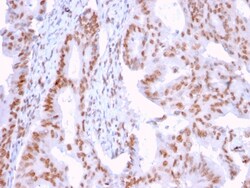
Biotium DMC1(2H12/4), CF647 conjugate, 0.1mg/mL
DNA repair proteins are necessary for the maintenance of chromosome integrity and are involved in the elimination of premutagenic lesions from DNA. The DNA repair proteins Rad51 and Rad52 are key components of the doublestrand-break repair (DSBR) pathway. Rad51 is essential for mitotic and meiotic recombination, and its mutation in yeast and mammalian cells results in chromosome loss. Overexpression of Rad52 confers resistance to ionizing radiation and induces homologous intrachromosomal recombination. Rad52 is thought to be involved in an early stage of Rad51-mediated recombination. Additional proteins involved in the pathway include Nibrin and Dmc1. Nibrin, which complexes with Mre11 and Rad50, is absent in Nijemegen breakage syndrome (NBS) patients. Dmc1 is specifically involved in meiotic recombination. An alternative spliced form of Dmc1, designated Dmc1-D, is deleted for a region between the two motifs involved in nucleotide binding. The alternatively spliced Dmc1-D transcri
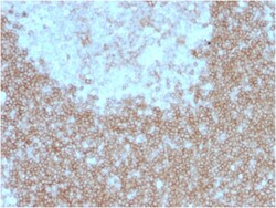
Biotium CD268 / BAFFR / TNFRSF13C(BAFFR/1557), CF647 conjugate, 0.1mg/mL
Defects in TNFRSF13C are the cause of immunodeficiency common variable type 4 (CVID4) ; also called antibody deficiency due to BAFFR defect. CVID4 is a primary immunodeficiency characterized by antibody deficiency, hypogammaglobulinemia, recurrent bacterial infections and an inability to mount an antibody response to antigen. The defect results from a failure of B-cell differentiation and impaired secretion of immunoglobulins; the numbers of circulating B cells is usually in the normal range, but can be low. Primary antibodies are available purified, or with a selection of fluorescent CF Dyes and other labels. CF Dyes offer exceptional brightness and photostability. Note: Conjugates of blue fluorescent dyes like CF405S and CF405M are not recommended for detecting low abundance targets, because blue dyes have lower fluorescence and can give higher non-specific background than other dye colors.
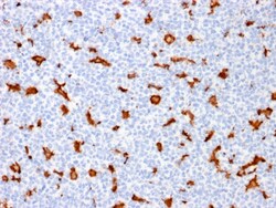
Biotium AIF1 / Iba1 (Microglia Marker) (AIF1/1909), CF647 conjugate, 0.1mg/mL
Allograft Inflammatory Factor 1 (also known as AIF1 or IBA1) is a cytoplasmic, calcium-binding protein that is thought to play a role in macrophage activation and function. AIF1, containing two EF domains, is induced by cytokines and Interferons. In an unstimulated state, AIF1 colocalizes with actin, and upon stimulation, translocates to lamellipodia. It is also a marker of human microglia and is expressed by macrophages in injured skeletal muscle. The gene encoding AIF1 resides in the tumor necrosis factor (TNF) cluster of genes, located in the region represented by the human major histocompatibility complex (MHC).Primary antibodies are available purified, or with a selection of fluorescent CF Dyes and other labels. CF Dyes offer exceptional brightness and photostability. Note: Conjugates of blue fluorescent dyes like CF405S and CF405M are not recommended for detecting low abundance targets, because blue dyes have lower fluorescence and can give higher non-specific
Biotium TSH-Receptor, B-Chain (Thyroid Marker) (TSHRB/1405), CF647 conjugate, 0.1mg/mL
Thyroid-stimulating hormone (TSH, also known as thyrotropin) is a glycoprotein involved in the control of thyroid structure and metabolism, which stimulates the release of the thyroid hormones. TSH is regulated by thyroid hormone (T3) and various retinoid compounds. TSH binds to the thyroid-stimulating hormone receptor (TSHR), which is cleaved into two subunits, A and B, and plays a major role in regulating thyroid function. The third cytoplasmic loop of TSHR has been identified as critical for its role in regulating inositol phosphate and cAMP formation. In Grave s disease, an autoimmune disorder, TSHR is activated by autoantibodies, which may be stimulated by the cleavage of the A and B subunits.Primary antibodies are available purified, or with a selection of fluorescent CF Dyes and other labels. CF Dyes offer exceptional brightness and photostability. Note: Conjugates of blue fluorescent dyes like CF405S and CF405M are not recommended for detecting low abundance targ
Biotium Spectrin beta III (SPTBN2) (SPTBN2/1584), CF647 conjugate, 0.1mg/mL
Spectrin is an actin binding protein that is a major component of the plasma membrane skeleton. Spectrins function as membrane organizers and stabilizers by forming dimers, tetramers and higher polymers. Vertebrate spectrins have two alpha-subunits (alpha-I/alpha-II), four beta-subunits (beta-I-beta-IV) and a beta-H subunit creating diversity and specialization of function. Spectrin alpha and spectrin beta are present in erythrocytes, whereas spectrin alpha II (also designated fodrin alpha) and spectrin beta I (also designated fodrin beta) are present in other somatic cells. The spectrin tetramers in erythrocytes act as barriers to lateral diffusion, but spectrin dimers seem to lack this function. Spectrin beta III is highly homologous to both spectrin beta I and spectrin beta II. Spectrin beta III is highly expressed in brain, kidney, pancreas and liver, and at lower levels in lung and placenta. Spectrin beta 3 is primarily expressed in nervous tissues with highest expressio