
Donkey Secondary Antibodies


Abcam Donkey Anti-Mouse IgG H&L (Alexa Fluor 647) preadsorbed
Donkey Anti-Mouse IgG H&L (Alexa Fluor 647) preadsorbed
The product is subject to the following: Abcam Restricted Use Statement


Biotium Cytokeratin 7 (Glandular and Transitional Epithelial Marker) (KRT7/1499R), CF647 conjugate, 0.1mg/mL
This antibody recognizes an intermediate filament protein (IFP) of 55 kDa, which is identified as cytokeratin 7. This MAb is highly specific to cytokeratin 7 and shows no cross-reaction with other IFPs. Cytokeratin 7 is a basic cytokeratin, which is found in most glandular and transitional epithelia but not in the stratified squamous epithelia. Keratin 7 is expressed in the epithelial cells of ovary, lung, and breast but not of colon, prostate, or gastrointestinal tract. This MAb is highly useful in distinguishing ovarian carcinomas (keratin 7 ) from colon carcinomas (keratin 7-).Primary antibodies are available purified, or with a selection of fluorescent CF Dyes and other labels. CF Dyes offer exceptional brightness and photostability. Note: Conjugates of blue fluorescent dyes like CF405S and CF405M are not recommended for detecting low abundance targets, because blue dyes have lower fluorescence and can give higher non-specific background than other dye colors.
Biotium Cdk1 / p34cdc2 Serine-Threonine Kinase(A17.1.1), CF647 conjugate, 0.1mg/mL
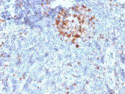
Recognizes a 34 kDa protein (cdk1), identified as p34cdc2 (a catalytic subunit of Maturation Promoting Factor). Its epitope maps near the C-terminus of the protein and its core is thought to be LGTPNNEV (aa220-227 in murine cdc2). It shows no cross reaction with cdk2 p32. It supports the kinase activity. p34cdc2 plays a crucial role during cell division and is most active during mitosis. It is a serine/threonine kinase, which is activated by cyclin, by dephosphorylation of tyrosine residues. p34cdc2 is inactivated by a tyrosine kinase. This MAb reportedly inhibits the activation of p34cdc2 kinase by cyclins. Primary antibodies are available purified, or with a selection of fluorescent CF Dyes and other labels. CF Dyes offer exceptional brightness and photostability. Note: Conjugates of blue fluorescent dyes like CF405S and CF405M are not recommended for detecting low abundance targets, because blue dyes have lower fluorescence and can give higher non-specific background
Biotium Ksp-Cadherin / CDH16 (Renal Cell Marker)(CDH16/2125), CF647 conjugate, 0.1mg/mL
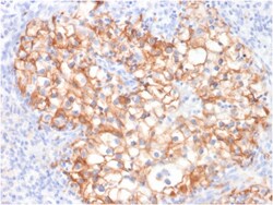
This MAb recognizes a protein of 130 kDa, identified as Ksp-cadherin. Cadherins form a superfamily of related glycoproteins that mediate calcium-dependent cell adhesion and transmit signals from the extracellular matrix to the cytoplasm. Cadherins have been implicated in embryogenesis, tissue morphogenesis, tissue structure maintenance, cell polarization, neoplastic invasiveness and metastasis, and membrane transport. It is suggested that Ksp-cadherin is a marker for terminal differentiation of the basolateral membranes of renal tubular epithelial cells. Within the kidney, Ksp-Cadherin is found exclusively in the basolateral membrane of renal tubular epithelial cells and collecting duct cells, and not in glomeruli, renal interstitial cells, or blood vessels. Ksp-Cadherin has been suggested to distinguish Chromophobe Renal-Cell Carcinoma from Oncocytoma. Primary antibodies are available purified, or with a selection of fluorescent CF Dyes and other labels. CF Dyes offer exception
Biotium MMP3 (Marker of Metastasis and Rheumatoid Arthritis)(MMP3/1994R), CF647 conjugate, 0.1mg/mL
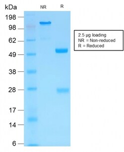
The matrix metalloproteinases (MMP) are a family of peptidase enzymes responsible for the degradation of extracellular matrix components, including collagen, gelatin, fibronectin, laminin and proteoglycan. Transcription of MMP genes is differentially activated by phorbol ester, lipopolysaccharide (LPS) or staphylococcal enterotoxin B (SEB). MMP catalysis requires both calcium and zinc. MMP-3, MMP-10 and MMP-11 (also designated stromelysin-1, 2 and 3, respectively) activate procollagenase. MMP-3 activation of procollagenase can occur via two pathways. Direct activation by MMP-3 is slow and activation by MMP-3 in conjunction with tissue or plasma proteinases is rapid. MMP-10 is expressed in small intestine, and at lower levels in lung and heart. MMP-11 is specifically expressed in stromal cells of breast carcinomas and contributes to epithelial cell malignancies. Primary antibodies are available purified, or with a selection of fluorescent CF Dyes and other labels. CF Dyes of
Biotium CD257 / BAFF / TNFSF13B (C257/1638), CF647 conjugate, 0.1mg/mL
BAFF/CD257 is a type II transmembrane protein, and a member of the tumor necrosis factor ligand superfamily. It is proteolytically cleaved to form a soluble protein. BAFF, like other TNF ligand family members, forms homo-trimers. The predicted molecular weight of a BAFF monomer is approximately 31 kDa. It is expressed on peripheral blood B and T lymphocytes, monocytes, macrophages, and dendritic cells and is upregulated by IFN-gamma and down-regulated by PMA/ionomycin treatment. BAFF stimulates B and T cell immunity and regulates humoral immunity by binding TACI and BCMA receptors; BAFF prevents apoptosis and promotes B cell survival by binding the BAFF-specific receptor (BAFFR/BR3).Primary antibodies are available purified, or with a selection of fluorescent CF Dyes and other labels. CF Dyes offer exceptional brightness and photostability. Note: Conjugates of blue fluorescent dyes like CF405S and CF405M are not recommended for detecting low abundance targets, because
Biotium Cytokeratin 10 (Suprabasal Epithelial Marker)(AE20), CF647 conjugate, 0.1mg/mL
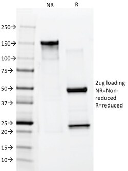
This MAb recognizes a protein of 56. 5 kDa, identified as cytokeratin 10 (CK10). CK10 is expressed in all suprabasal layers of the epidermis. In the epidermis, expression of CK10 strictly parallels the extent of differentiation; it is absent in the basal layer, appears in the first suprabasal layers and increases in concentration towards the granular layer. However, CK10 is rarely detected in early stages of vulvar squamous carcinomas (tumors less than 2 cm, clinical stage I) regardless of the tumor grade. In larger and more advanced tumors (greater than 2 cm, clinical stages II and III), CK10 is detected very frequently. Expression of CK10 is related to maturation of malignant keratinocytes, being preferentially detected in more-differentiated parts. Primary antibodies are available purified, or with a selection of fluorescent CF Dyes and other labels. CF Dyes offer exceptional brightness and photostability. Note: Conjugates of blue fluorescent dyes like CF405S and CF40
Biotium PTH / Parathyroid Hormone (N-Terminal)(PTH/1173), CF647 conjugate, 0.1mg/mL
The epitope of this MAb maps in the C-terminus of PTH, a hormone produced by the parathyroid gland that regulates the concentration of calcium and phosphorus in extracellular fluid. This hormone elevates blood calcium levels by dissolving the salts in bone and preventing their renal excretion.It is produced in the parathyroid gland as an 84 amino acid single chain polypeptide. It can also be secreted as N-terminal truncated fragments or C-terminal fragments after intracellular degradation, as in case of hypercalcemia. Defects in this gene are a cause of familial isolated hypoparathyroidism (FIH); also called autosomal dominant hypoparathyroidism or autosomal dominant hypocalcemia. FIH is characterized by hypocalcemia and hyperphosphatemia due to inadequate secretion of parathyroid hormone. Symptoms are seizures, tetany and cramps. FIH exist both as autosomal dominant and recessive forms of hypoparathyroidism.Primary antibodies are available purified, or with a selection of fluore
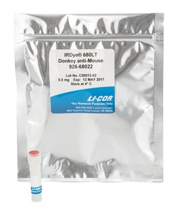
LICOR IRDye 680LT Donkey anti-Mouse IgG Secondary Antibody (0.5 mg)
Immunogen: Mouse IgG | Applications: IRDye 680LT Donkey anti-Mouse IgG Secondary Antibody is recommended for: Western Blot, Protein Array, Immunohistochemistry, Microscopy, 2D Gel Detection, and Tissue Section Imaging. IRDye 680LT Donkey anti-Mouse IgG Secondary Antibody is not recommended for Small Animal Imaging, In-Cell Western Assay, On-Cell Western Assay | Formulation: IRDye 680LT Secondary Antibodies are supplied as purified immunoglobulin conjugates, lyophilized in phosphate-buffered saline, pH 7.4. Protect from light. Store at 4 °C prior to reconstitution. Each vial contains 10 mg/mL BSA (free of IgG and protease) as a stabilizer and 0.01% sodium azide as a preservative, after reconstitution. Concentration is 1.0 mg/mL when reconstituted as directed. Refer to the pack insert for details on reconstitution.
Biotium Prostate Specific Antigen (PSA)(KLK3/1248), CF647 conjugate, 0.1mg/mL
Recognizes a single protein of 33-34 kDa, identified as the prostate specific antigen (PSA). This MAb is highly specific to PSA and stains prostatic secretory and ductal epithelium in both normal and neoplastic tissues. PSA is a chymotrypsin-like serine protease (kallikrein family) exclusively produced by the prostate epithelium, and abundant in seminal fluid. PSA can be detected in the sera of patients with prostatic carcinoma. It is predominantly complexed to a liver-derived serine protease inhibitor, alpha-1-antichymotrypsin (ACT). A higher proportion of serum PSA is complexed to ACT in prostate cancer than in benign prostate hyperplasia. This MAb makes an excellent pair with MAb A67-B/E3 for PSA tests.
Biotium pS2 / pNR-2 / TFF1 (Estrogen-Regulated Protein) (TFF1/2969R), CF647 conjugate, 0.1mg/mL
This antibody recognizes a polypeptide of 6.5 kDa, identified as pS2 estrogen-regulated protein. Its epitope is localized between aa57-84 of human pS2 protein. pS2 is a trefoil peptide. Trefoil peptides are protease resistant molecules secreted throughout the gut that play a role in mucosal healing. These peptides contain three intra-chain disulfide bonds, forming the trefoil motif, or P-domain. pS2 is known to form dimers and this dimerization is thought to play a role in its protective and healing properties. About 60% of breast carcinomas are positive for pS2. Staining is cytoplasmic, often with localization to the Golgi apparatus. pS2 is shown to be localized in normal stomach mucosa, gastric fluid, goblet cells in the colon and small intestine, and in ulcerations of the gastrointestinal tract. Several studies have shown that pS2 is primarily expressed in estrogen receptor-positive breast tumors and it may define a subset of estrogen-dependent tumors that displays an increased
Biotium CD10 (Membrane Metalloendopeptidase)(MME/2580), CF647 conjugate, 0.1mg/mL
CD10 (also known as Common Acute Lymphocytic Leukemia Antigen (CALLA)) is a type II transmembrane protein. It is a cell surface enzyme with neutral metalloendopeptidase activity, which inactivates a variety of biologically active peptides. CD10 is expressed on the cells of lymphoblastic, Burkitt's, and follicular germinal center lymphomas, and on cells from patients with chronic myelocytic leukemia (CML). It is also expressed on the surface of normal early lymphoid progenitor cells, immature B cells within adult bone marrow and germinal center B cells within lymphoid tissue.Primary antibodies are available purified, or with a selection of fluorescent CF Dyes and other labels. CF Dyes offer exceptional brightness and photostability. Note: Conjugates of blue fluorescent dyes like CF405S and CF405M are not recommended for detecting low abundance targets, because blue dyes have lower fluorescence and can give higher non-specific background than other dye colors.
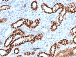
Biotium Ksp-Cadherin (Kidney-Specific Cadherin) / CDH16 (rCDH16/1071), CF647 conjugate, 0.1mg/mL
This MAb recognizes a protein of 130 kDa, identified as Ksp-cadherin. Cadherins form a superfamily of related glycoproteins that mediate calcium-dependent cell adhesion and transmit signals from the extracellular matrix to the cytoplasm. Cadherins have been implicated in embryogenesis, tissue morphogenesis, tissue structure maintenance, cell polarization, neoplastic invasiveness and metastasis, and membrane transport. It is suggested that Ksp-cadherin is a marker for terminal differentiation of the basolateral membranes of renal tubular epithelial cells. Within the kidney, Ksp-Cadherin is found exclusively in the basolateral membrane of renal tubular epithelial cells and collecting duct cells, and not in glomeruli, renal interstitial cells, or blood vessels. Ksp-Cadherin has been suggested to distinguish Chromophobe Renal-Cell Carcinoma from Oncocytoma.Primary antibodies are available purified, or with a selection of fluorescent CF Dyes and other labels. CF Dyes offer exc
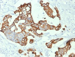
Biotium Cytokeratin 20 (KRT20) (Colorectal Epithelial Marker) (KRT20/1992), CF647 conjugate, 0.1mg/mL
This MAb recognizes an intermediate filament protein of 46 kDa, identified as cytokeratin 20 (KRT20). KRT is abundantly expressed in goblet cells and enterocytes of the gastrointestinal tract. It is a useful marker of pancreatic and colorectal cancer. KRT20 is expressed under normal, hyperplastic and neoplastic conditions. It has been detected in adenocarcinomas of the colon, stomach and biliary tract. Breast carcinomas are generally non-reactive.Primary antibodies are available purified, or with a selection of fluorescent CF Dyes and other labels. CF Dyes offer exceptional brightness and photostability. Note: Conjugates of blue fluorescent dyes like CF405S and CF405M are not recommended for detecting low abundance targets, because blue dyes have lower fluorescence and can give higher non-specific background than other dye colors.
Biotium Connexin 32 (Gap Junction Protein) (R5.21C), CF647 conjugate, 0.1mg/mL
This antibody recognizes a protein of 27-32 kDa, identified as Connexin 32. The connexin family of proteins forms hexameric complexes called connexons that facilitate movement of low molecular weight proteins between cells via gap junctions. Connexin proteins share a common topology of four transmembrane alphalpha-helicaldomains, two extracellular loops, a cytoplasmic loop and cytoplasmic N- and C-termini. Many of the key functional differences arise from specific amino-acid substitutions in the most highly conserved domains, the transmembrane and extracellular regions. Each of the approximately 20-connexin isoforms produces channels with distinct permeability and electrical and chemical sensitivities; therefore, one connexin usually cannot fully substitute for another.Primary antibodies are available purified, or with a selection of fluorescent CF Dyes and other labels. CF Dyes offer exceptional brightness and photostability. Note: Conjugates of blue fluorescent dyes like