
Mouse Secondary Antibodies

Biotium Melanoma Associated Antigen (MAA)(MAA/1414), CF568 conjugate, 0.1mg/mL
The antibody reacts strongly with melanoma cells derived from cell lines and short-term cultures and reacts preferentially with melanoma cells in frozen tissue sections. The antibody can also be used to detect melanoma lesions in vivo. Primary antibodies are available purified, or with a selection of fluorescent CF Dyes and other labels. CF Dyes offer exceptional brightness and photostability. Note: Conjugates of blue fluorescent dyes like CF405S and CF405M are not recommended for detecting low abundance targets, because blue dyes have lower fluorescence and can give higher non-specific background than other dye colors.


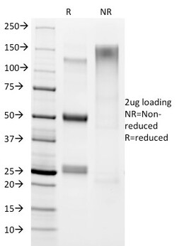
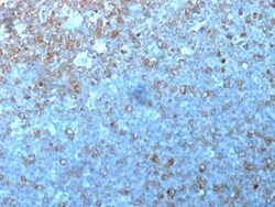
Biotium CD20 / MS4A1 (B-Cell Marker) (IGEL/1497R), Biotin conjugate, 0.1mg/mL
This antibody recognizes a protein of 30-33 kDa, which is identified as CD20. It is a non-Ig differentiation antigen of B-cells and its expression is restricted to normal and neoplastic B-cells, being absent from all other leukocytes and tissues. CD20 is expressed by pre B-cells and persists during all stages of B-cell maturation but is lost upon terminal differentiation into plasma cells. This MAb can be used for immunophenotyping of leukemia and malignant cells, B lymphocyte detection in peripheral blood and B cell localization in tissues. It reacts with the majority of B-cells present in peripheral blood and lymphoid tissues and their derived lymphomas. In lymphoid tissue, germinal center blasts and B-immunoblasts are particularly reactive. It is a reliable antibody for ascribing a B-cell phenotype in known lymphoid tissues. Rarely, CD20-positive T-cell lymphomas have been reported. Reactivity has also been noted with Reed-Sternberg cells in cases of Hodgkin's disease, par
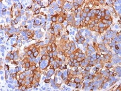
Biotium VLDL-Receptor (Very Low Density Lipoprotein Receptor) (VLDLR/1337), Biotin conjugate, 0.1mg/mL
VLDLR (very low density lipoprotein receptor) is a member of the LDL receptor gene family, which includes LDL receptor, LRP, megalin, VLDLR and ApoER2. The LDL receptor family is characterized by a cluster of cysteine-rich class A repeats, epidermal growth factor (EGF)-like repeats, YWTD repeats and an O-linked sugar domain. VLDLR associates with RAP (receptor associated protein) during receptor folding, and RAP facilitates the secretion of the extracellular region of VLDLR. VLDLR is thought to mediate the interaction of extracellular Reelin and cytosolic mDab1 (mammalian disabled protein), which activates a tyrosine kinase. This pathway regulates the migration of neurons along the radial glial fiber network during brain development.Primary antibodies are available purified, or with a selection of fluorescent CF Dyes and other labels. CF Dyes offer exceptional brightness and photostability. Note: Conjugates of blue fluorescent dyes like CF405S and CF405M are not recommen
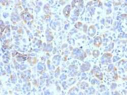
Biotium TRP1 (Tyrosinase Related Protein 1)(TYRP1/807), CF740 conjugate, 0.1mg/mL
This antibody reacts with a 75 kDa melanocyte-specific gene product, identified as Tyrosinase-related protein-1 (TRP-1). It is involved in melanin synthesis. TRP1 is present on the melanosomal membranes of melanoma, normal melanocytes and nevi.Recent evidence suggests that TRP-1 is involved in maintaining stability of tyrosinase protein and modulating its catalytic activity. TRP-1 is also involved in maintenance of melanosome ultrastructure and affects melanocyte proliferation and cell death.Primary antibodies are available purified, or with a selection of fluorescent CF Dyes and other labels. CF Dyes offer exceptional brightness and photostability. Note: Conjugates of blue fluorescent dyes like CF405S and CF405M are not recommended for detecting low abundance targets, because blue dyes have lower fluorescence and can give higher non-specific background than other dye colors.
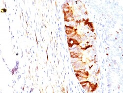
Biotium Helicobacter pylori(HP/1336), CF740 conjugate, 0.1mg/mL
The spiral shaped bacterium Helicobacter pyloriis strongly associated with inflammation of the stomach and is also implicated in the development of gastric malignancy. H. pyloriis known to cause peptic ulcers and chronic gastritis in human. It is associated with duodenal ulcers and may be involved in development of adenocarcinoma and low-grade lymphoma of mucosa associated lymphoid tissue in the stomach. This antibody stains the individual H. pylori bacterium when it presents on the surface of the epithelium or in the cytoplasm of the epithelial cells in biopsy tissue sections from the antrum and body of the stomach. Primary antibodies are available purified, or with a selection of fluorescent CF Dyes and other labels. CF Dyes offer exceptional brightness and photostability. Note: Conjugates of blue fluorescent dyes like CF405S and CF405M are not recommended for detecting low abundance targets, because blue dyes have lower fluorescence and can give higher non-specific ba
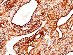
Biotium Cytokeratin 8(TS1), CF740 conjugate, 0.1mg/mL
Cytokeratin 8 (CK8) belongs to the type II (or B or basic) subfamily of high molecular weight cytokeratins, and exists in combination with cytokeratin 18 (CK18). CK8 is primarily found in the non-squamous epithelia and is present in majority of adenocarcinomas and ductal carcinomas. It is absent in squamous cell carcinomas. Hepatocellular carcinomas are defined by the use of antibodies that recognize only cytokeratin 8 and 18. CK8 exists on several types of normal and neoplastic epithelia, including many ductal and glandular epithelia such as colon, stomach, small intestine, trachea, and esophagus as well as in transitional epithelium. Anti-CK8 does not react with skeletal muscle or nerve cells. Epithelioid sarcoma, chordoma, and adamantinoma show strong positivity corresponding to that of simple epithelia (with antibodies against CK8, CK18 and CK19). Reportedly, anti-CK8 is useful for the differentiation of lobular (ring-like, perinuclear) from ductal (peripheral-predominant
Biotium TIA1 (T-Cell-Restricted Intracellular Antigen-1) (TIA1/1313), CF488A conjugate, 0.1mg/mL
TIA-1 (T-cell intracytoplasmic antigen) is a cytoplasmic granule-associated protein expressed in lymphocytes processing cytolytic potential. TIA-1 is a member of an RNA-binding protein family and possesses nucleolytic activity against cytotoxic lymphocyte (CTL) target cells. It has been suggested that this protein may be involved in the induction of apoptosis as it preferentially recognizes poly(A) homopolymers and induces DNA fragmentation in CTL targets. The major granule-associated species is a 15 kDa protein thought to be derived from the carboxyl terminus of the 40 kDa product by proteolytic processing. TIA1 antibody labels cytotoxic T cells and natural killer cells (NK cells). It is also expressed in T-cell lymphoma, large granular lymphocyte (LGL) leukemia and hairy cell leukemia. TIA1 expression in T-cell malignancies may help in differentiating LGL leukemia (high expression) from T-cell lymphocytosis and other T-cell diseases (low expression). TIA1 may also be used t
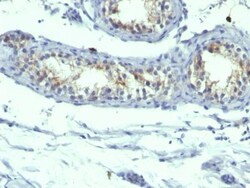
Biotium ASRGL1 (Asparaginase Like 1) (CRASH/1289), CF568 conjugate, 0.1mg/mL
ASRGL1 (Asparaginase-like protein 1), also known as CRASH, is a 308 amino acid protein belonging to the Ntn-hydrolase family. Asparaginases utilize asparagine as a substrate to produce aspartic acid and ammonia. ASRGL1 has been identified as a autoantigenic protein that is present in the mid-piece of sperm after obstruction of the male reproductive tract. ASRGL1 is expressed highly in testis, but is also expressed in brain, kidney and gastrointestinal tissues. High levels of ASRGL1 have also been identified in ovarian, uterine and mammary tumors in comparison with normal tissues of the same origin.Primary antibodies are available purified, or with a selection of fluorescent CF Dyes and other labels. CF Dyes offer exceptional brightness and photostability. Note: Conjugates of blue fluorescent dyes like CF405S and CF405M are not recommended for detecting low abundance targets, because blue dyes have lower fluorescence and can give higher non-specific background than other
Biotium Alpha-1-Antitrypsin (SERPINA1) (Hepatocellular & Histiocytic Marker) (AAT/1379), CF568 conjugate, 0.1mg/mL
This antibody recognizes a protein of 54 kDa, which is identified as Alpha-1 antitrypsin (AAT). AAT is secreted and is a serine protease inhibitor whose targets include elastase, plasmin, thrombin, trypsin, chymotrypsin, and plasminogen activator. Defects in this gene can cause emphysema or liver disease.Primary antibodies are available purified, or with a selection of fluorescent CF Dyes and other labels. CF Dyes offer exceptional brightness and photostability. Note: Conjugates of blue fluorescent dyes like CF405S and CF405M are not recommended for detecting low abundance targets, because blue dyes have lower fluorescence and can give higher non-specific background than other dye colors.
Biotium Prolactin Receptor(B6.2 + PRLR742), CF740 conjugate, 0.1mg/mL
This antibody recognizes a protein of 70 kDa, identified as prolactin receptor. Prolactin is a pituitary hormone involved in the stimulation of milk production, salt and water regulation, growth, development and reproduction. The initial step in its action is the binding to a specific membrane receptor (prolactin receptor), which belongs to the superfamily of class 1 cytokine receptors. The function of the prolactin receptor is mediated, at least in part, by two families of signaling molecules: Janus kinases and signal transducers and activators of transcription.Primary antibodies are available purified, or with a selection of fluorescent CF Dyes and other labels. CF Dyes offer exceptional brightness and photostability. Note: Conjugates of blue fluorescent dyes like CF405S and CF405M are not recommended for detecting low abundance targets, because blue dyes have lower fluorescence and can give higher non-specific background than other dye colors.
Biotium ALDH1A1 (Aldehyde Dehydrogenase 1A1)(ALDH1A1/1382), CF488A conjugate, 0.1mg/mL
Aldehyde dehydrogenase 1 family member A1 (ALDH1A1), also known as retinal dehydrogenase 1, belongs to the aldehyde dehydrogenase enzyme family, which is involved in the metabolism of alcohol. ALDH1A1 is predominantly expressed in the epithelium of testis, brain, eye, liver, kidney, as well as neural and hematopoietic stem cells. Reportedly, high ALDH1A1 expression is found in solitary fibrous tumor (SFT) and hemangiopericytoma (HPC), compared to meningiomas and synovial sarcomas. In combination with CD34, ALDH1A1 may be useful for the differentiation among SFT, HPC, meningioma, and synovial sarcoma. Primary antibodies are available purified, or with a selection of fluorescent CF Dyes and other labels. CF Dyes offer exceptional brightness and photostability. Note: Conjugates of blue fluorescent dyes like CF405S and CF405M are not recommended for detecting low abundance targets, because blue dyes have lower fluorescence and can give higher non-specific background than oth

Biotium TRP1 (Tyrosinase Related Protein 1)(TYRP1/807), CF740 conjugate, 0.1mg/mL
This antibody reacts with a 75 kDa melanocyte-specific gene product, identified as Tyrosinase-related protein-1 (TRP-1). It is involved in melanin synthesis. TRP1 is present on the melanosomal membranes of melanoma, normal melanocytes and nevi.Recent evidence suggests that TRP-1 is involved in maintaining stability of tyrosinase protein and modulating its catalytic activity. TRP-1 is also involved in maintenance of melanosome ultrastructure and affects melanocyte proliferation and cell death.Primary antibodies are available purified, or with a selection of fluorescent CF Dyes and other labels. CF Dyes offer exceptional brightness and photostability. Note: Conjugates of blue fluorescent dyes like CF405S and CF405M are not recommended for detecting low abundance targets, because blue dyes have lower fluorescence and can give higher non-specific background than other dye colors.
Biotium p34 / cdk1(POH-1), CF740 conjugate, 0.1mg/mL
Recognizes a 34 kDa protein, which is identified as cyclin dependent kinase 1 (cdk1) or p34cdc2 protein kinase. cdk1 / p34cdc2 plays a crucial role during cell division and is most active during mitosis. It is predominantly localized in the nucleus. It is a serine/threonine kinase, which is activated by cyclin, presumably by de-phosphorylation of tyrosine residues. Activated cdk1 / p34cdc2 performs specific functions during mitosis, including nuclear envelope breakdown and chromosome condensation.Primary antibodies are available purified, or with a selection of fluorescent CF Dyes and other labels. CF Dyes offer exceptional brightness and photostability. Note: Conjugates of blue fluorescent dyes like CF405S and CF405M are not recommended for detecting low abundance targets, because blue dyes have lower fluorescence and can give higher non-specific background than other dye colors.
Biotium MART-1 / Melan-A(A103), CF740 conjugate, 0.1mg/mL
This antibody recognizes a protein doublet of 20-22 kDa, identified as MART-1 (Melanoma Antigen Recognized by T cells 1) or Melan-A. MART-1 is a newly identified melanocyte differentiation antigen recognized by autologous cytotoxic T lymphocytes. Seven other melanoma associated antigens recognized by autologous cytotoxic T cells include MAGE-1, MAGE-3, tyrosinase, gp100, gp75, BAGE-1, and GAGE-1. Subcellular fractionation shows that MART-1 is present in melanosomes and endoplasmic reticulum. This MAb labels melanomas and other tumors showing melanocytic differentiation. It is also a useful positive-marker for angiomyolipomas. It does not stain tumor cells of epithelial, lymphoid, glial, or mesenchymal origin.Primary antibodies are available purified, or with a selection of fluorescent CF Dyes and other labels. CF Dyes offer exceptional brightness and photostability. Note: Conjugates of blue fluorescent dyes like CF405S and CF405M are not recommended for detecting low abu
Biotium Alpha-1-Antichymotrypsin (SERPINA3) (Histiocytoma Marker) (AACT/1451), CF488A conjugate, 0.1mg/mL
Alpha-1 Antichymotrypsin (AACT) is a plasma protease inhibitor synthesized in the liver as a single glycopeptide chain. In human, the normal serum level of AACT is about one-tenth that of alpha-1-antitrypsin (AAT), with which it shares nucleic acid and protein sequence homology. Both are major acute phase reactants; their concentrations in plasma increase in response to trauma, surgery and infection. Elevated levels of AACT are widely, but not universally, reported in the cerebrospinal fluid and plasma of AD patients. Prostate-specific antigen (PSA) and its SDS-stable complex with AACT are in widespread use as markers for the diagnosis of prostate cancer. AACT deficiency may also be a possible cause of chronic liver disease. AACT antibody reacts with histiocytes and histiocytic neoplasms. It is widely used to identify histiocytes and tumors derived from them. Acinar tumors of the pancreas and salivary gland may also exhibit AACT positivity. Primary antibodies are available