
Mouse Secondary Antibodies

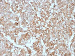
Biotium DOG-1 / TMEM16A (Gastrointestinal Stromal Tumor Marker)(rDG1/447), Biotin conjugate, 0.1mg/mL
Expression of DOG-1 protein is elevated in the gastrointestinal stromal tumors (GIST's), c-kit signaling-driven mesenchymal tumors of the GI tract. DOG-1 is rarely expressed in other soft tissue tumors, which, due to appearance, may be difficult to diagnose. Immunoreactivity for DOG-1 has been reported in 97. 8 percent of scorable GIST's, including all c-kit negative GIST's. Overexpression of DOG-1 has been suggested to aid in the identification of GISTs, including Platelet-Derived Growth Factor Receptor Alpha mutants that fail to express c-kit antigen. The overall sensitivity of DOG1 and c-kit in GIST's is nearly identical: 94. 4% vs. 94. 7%. Primary antibodies are available purified, or with a selection of fluorescent CF Dyes and other labels. CF Dyes offer exceptional brightness and photostability. Note: Conjugates of blue fluorescent dyes like CF405S and CF405M are not recommended for detecting low abundance targets, because blue dyes have lower fluorescence and can

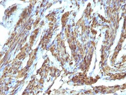
Biotium Calponin-1 (Smooth Muscle Marker), CF488A conjugate, 0.1mg/mL
Calponin is a calmodulin, F-actin and tropomyosin binding protein, which is thought to be involved in the regulation of smooth muscle contraction. Calponin expression is restricted to smooth muscle cells and has been shown to be a marker of the differentiated (contractile) phenotype of developing smooth muscle. Multiple isoelectric variants of calponin have been identified, however only two molecular weight isoforms exist, a 34 kDa form and a 29 kDa form. Expression of the 29 kDa form, I-calponin, is primarily restricted to muscle of the urogenital tract, whereas the higher molecular weight variant has been demonstrated in vascular and visceral smooth muscle. In Western blotting, this MAb reacts with only the 34 kDa form of calponin in extracts of human aortic medial smooth muscle and is unreactive with fibroblast extracts of cultivated human foreskin.Primary antibodies are available purified, or with a selection of fluorescent CF Dyes and other labels. CF Dyes offer except
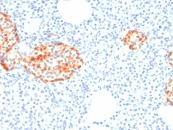
Biotium Chromogranin A / CHGA (Neuroendocrine Marker) (rCHGA/798), Biotin conjugate, 0.1mg/mL
Chromogranin A is present in neuroendocrine cells throughout the body, including the neuroendocrine cells of the large and small intestine, adrenal medulla and pancreatic islets. It is an excellent marker for carcinoid tumors, pheochromocytomas, paragangliomas, and other neuroendocrine tumors. Co-expression of chromogranin A and neuron specific enolase (NSE) is common in neuroendocrine neoplasms. Reportedly, co-expression of certain keratins and chromogranin indicates neuroendocrine lineage. The presence of strong anti-chromogranin staining and absence of anti-keratin staining should raise the possibility of paraganglioma. The co-expression of chromogranin and NSE is typical of neuroendocrine neoplasms. Most pituitary adenomas and prolactinomas readily express chromogranin.Primary antibodies are available purified, or with a selection of fluorescent CF Dyes and other labels. CF Dyes offer exceptional brightness and photostability. Note: Conjugates of blue fluorescent dyes
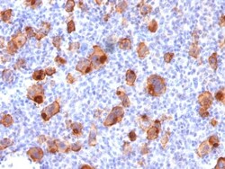
Biotium CD30 / TNFRSF8 (Hodgkin & Reed-Sternberg Cell Marker) (rKi-1/779), CF568 conjugate, 0.1mg/mL
This antibody recognizes a single chain glycoprotein of 105/120 kDa, identified as CD30/Ki-1. CD30 is synthesized as a 90 kDa precursor, which is processed in the Golgi complex into a membrane-bound phosphorylated mature 105/120 kDa glycoprotein. In Hodgkin's disease, CD30/Ki-1 antigen is expressed by mononuclear-Hodgkin and multinucleated Reed-Sternberg cells. It is also expressed by the tumor cells of a majority of anaplastic large cell lymphomas as well as by a varying proportion of activated T and B cells. This MAb distinguishes large cell lymphomas derived from activated lymphoid cells from histiocytic malignancies and lymphomas derived from resting and precursor lymphoid cells or from anaplastic carcinomas. About one third of the Ki-1 positive lymphomas lack the leukocyte common antigen (CD45).Primary antibodies are available purified, or with a selection of fluorescent CF Dyes and other labels. CF Dyes offer exceptional brightness and photostability. Note: Conjugate
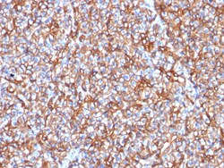
Biotium Spectrin beta III (SPTBN2) (SPTBN2/1778), Biotin conjugate, 0.1mg/mL
Spectrin is an actin binding protein that is a major component of the plasma membrane skeleton. Spectrins function as membrane organizers and stabilizers by forming dimers, tetramers and higher polymers. Vertebrate spectrins have two alpha-subunits (alpha-I/alpha-II), four beta-subunits (beta-I-beta-IV) and a beta-H subunit creating diversity and specialization of function. Spectrin α and spectrinβ are present in erythrocytes, whereas spectrin α II (also designated fodrin α) and spectrinβ I (also designated fodrinβ) are present in other somatic cells. The spectrin tetramers in erythrocytes act as barriers to lateral diffusion, but spectrin dimers seem to lack this function. Spectrinβ III is highly homologous to both spectrinβ I and spectrinβ II. Spectrinβ III is highly expressed in brain, kidney, pancreas and liver, and at lower levels in lung and placenta. Spectrin beta 3 is primarily expressed in nervous tissues with high

Biotium MUC5AC (Mucin 5AC / Gastric Mucin)(9-13M1), CF568 conjugate, 0.1mg/mL
This MAb recognizes the peptide core of gastric mucin M1 (recently identified as Mucin 5AC). Its epitope is located in the N-terminal cysteine rich part of the peptide core of MUC5AC, which is heavily glycosylated. Its epitope is destroyed by beta-mercaptoethanol but not by periodate treatment. MAb 2-11M1 reacts with the protein backbone exclusively; it only reacts with fully deglycosylated MUC5AC. Therefore, the material under test should also be fully deglycosylated. This can be achieved with standard periodate oxidation method. The success of the deglycosylation can be checked with routine PAS (Periodic Acid Shiff) staining. After deglycosylation, the preparation should no longer be stainable with PAS reagent. Only then 2-11M1 will react should MUC5AC be present. This mucin is present in primary ovarian mucinous cancer but usually absent in colorectal adenocarcinoma, thus showing an expression pattern opposite to MUC2. Together with a panel of antibodies, Anti-MUC5AC may b
Biotium CD50 / ICAM3(ICO-60), CF740 conjugate, 0.1mg/mL
Recognizes the N-glycosylated glycoprotein of 120 kDa with intra-chain disulfide bonds, identified as CD50 or ICAM-3. CD50 is the major ligand for LFA-1 (CD11a/CD18) and may have signaling role to increase adhesion. It is expressed on thymocytes and T lymphocytes and is resistant to treatment with phosphatidylinositol (PI) phospholipase C.
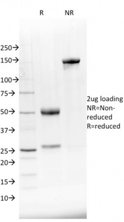
Biotium NKX2.8(NKX28/2548), 1mg/mL
The protein encoded by this gene is a homeobox-containing developmental regulator associated with liver development. The encoded protein binds to the alpha-fetoprotein (AFP) gene promoter and increases the expression of AFP. This gene is overexpressed in some lung cancers and is linked to poor patient survival, possibly due to its resistance to cisplatin. This gene is aberrantly methylated in pancreatic cancer, deleted in squamous cell lung carcinomas, and acts as a tumor suppressor in esophageal cancer. Mutations in this gene may also be a cause of neural tube defects. Primary antibodies are available purified, or with a selection of fluorescent CF Dyes and other labels. CF Dyes offer exceptional brightness and photostability. Note: Conjugates of blue fluorescent dyes like CF405S and CF405M are not recommended for detecting low abundance targets, because blue dyes have lower fluorescence and can give higher non-specific background than other dye colors.
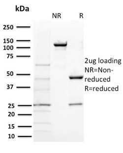
Biotium BAP1 (BRCA1 Associated Protein 1)(BAP1/2431), CF488A conjugate, 0.1mg/mL
The BRCA 1 Associated Protein 1 (BAP1) protein belongs to the ubiquitin C-terminal hydrolase subfamily of de-ubiquitination enzymes that are involved in the removal of ubiquitin from proteins. The encoded enzyme binds to the breast cancer type 1 susceptibility protein (BRCA1) via the RING finger domain of the latter and acts as a tumor suppressor. In addition, the enzyme may be involved in regulation of transcription, regulation of cell cycle and growth, response to DNA damage and chromatin dynamics. Germ line mutations in this gene may be associated with tumor predisposition syndrome (TPDS), which involves increased risk of cancers including malignant mesothelioma, Uveal melanoma and cutaneous melanoma. Primary antibodies are available purified, or with a selection of fluorescent CF Dyes and other labels. CF Dyes offer exceptional brightness and photostability. Note: Conjugates of blue fluorescent dyes like CF405S and CF405M are not recommended for detecting low abundan
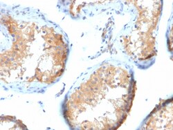
Biotium Vinculin (Marker of Age-related Macular Degeneration)(VCL/2573), CF488A conjugate, 0.1mg/mL
Focal adhesions are identified as areas within the plasma membrane of tissue culture cells that adhere tightly to the underlying substrate. In vivo, these regions are involved in the adhesion of cells to the extracellular matrix. Paxillin and vinculin are cytoskeletal, focal adhesion proteins that are components of a protein complex which links the Actin network to the plasma membrane. Vinculin binding sites have been identified on other cytoskeletal proteins, including talin and alpha-actinin each contain actin binding sites. Reportedly, vinculin is a potential plasma biomarker for Age-related Macular Degeneration (AMD). The early detection of AMD using novel plasma biomarkers with genetic modeling may enable timely treatment and vision preservation in the elderly. Primary antibodies are available purified, or with a selection of fluorescent CF Dyes and other labels. CF Dyes offer exceptional brightness and photostability. Note: Conjugates of blue fluorescent dyes like CF
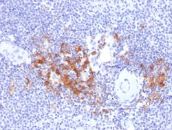
Biotium TRAF1 (TNFR-Associated Factor 1) (Lymphomatoid Papulosis Marker)(TRAF1/2770), CF740 conjugate, 0.1mg/mL
This MAb recognizes a protein of 52 kDa, which is identified as TNFR1 (TNFR-associated factor 1). CD30-positive lymphoproliferations of the skin comprise 30% of all primary cutaneous T-cell lymphomas (CTCLs). Besides borderline cases this group includes lymphomatoid papulosis (LyP) and primary cutaneous anaplastic large T-cell lymphoma (cALCL). Although the two entities overlap clinically, histopathologically, immunopathologically and genetically, they differ considerably in their prognosis. In particular, common feature of both cases is histologically the presence of atypical lymphoid CD30-positive T blasts and genetically a clonal T-cell-receptor rearrangement. However, both cases differ considerably in their clinical course: Lesions of LyP regress spontaneously, whereas those of cALCL persist and may progress and spread. Moreover, LyP patients do not benefit from an aggressive radio- and/or chemotherapeutic approach, in contrast to patients with cALCL. Besides, LyP and cAL
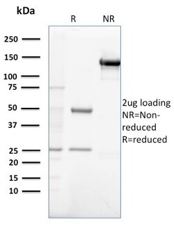
Biotium Gastrin(GAST/2632), CF488A conjugate, 0.1mg/mL
Gastrin, is a hormone that normally formed by mucosal cells in the gastric antrum and by the D cells of the pancreatic islets. Its primary function is to stimulate secretion of HCl by the gastric mucosa. HCl, in turn, inhibits gastrin formation. It also responsible for stimulating smooth muscle contraction and increasing blood circulation and water secretion in the stomach and intestine. Gastrin is regulated by epidermal growth factor in both mice and humans. Gastrin is excreted in excess by pancreatic tumors in the Zollinger-Ellison syndrome. Gastrin-Releasing Peptide (GRP) stimulates the release of gastrin as well as other gastrointestinal hormones and also acts as an autocrine growth factor for certain cell types. High levels of GRP are found in the human lung just after birth and levels decrease thereafter in parallel with the observed disease in a number of pulmonary neuroendocrine cells. GRP is known to promote lung tumorigenesis in model systems. Primary antibodies are
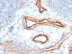
Biotium MTF1(MTF1/2649), CF740 conjugate, 0.1mg/mL
The metal-responsive element (MRE)-binding transcription factor (MTF-1) stimulates the expression of metallothioneins in response to the exposure of cells to heavy metals. MTF-1 contains six zinc fingers in the DNA binding domain. The phosphorylation of MTF-1 in response to metal exposure appears to play a significant role in the ability of MTF-1 to activate metallothionein transcription. In addition to its role in metallothionein activation, MTF-1 is involved in a post-transcription regulatory complex for ribosomal protein S25. MTF-1, La and p53 inhibit the nuclear export of S25 mRNA in response to nutrient depravation. Furthermore, MTF-1 acts as a chromatin insulator on integrated transgenes in cultured cells to insulate active loci against chromatin silencing. Primary antibodies are available purified, or with a selection of fluorescent CF Dyes and other labels. CF Dyes offer exceptional brightness and photostability. Note: Conjugates of blue fluorescent dyes like CF40
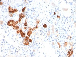
Biotium Calbindin 1 (CALB1)(CALB1/2782), CF568 conjugate, 0.1mg/mL
Calbindin 1, also known as calbindin D28K, is a member of a family of calcium binding proteins. This protein family also includes calmodulin, calbindin 2, S-100, and troponin C. Calbindin 1 is a 261-amino acid protein with 6 EF-hand domains, 4 of which are active calcium-binding domains. Expressed in brain, ovary, uterus, testis, pancreas, liver, kidney and intestine, calbindin 1 acts as a calcium-buffering agent and alters the activity of the plasma membrane ATPase. In neuronal cells, calbindin 1 modulates calcium channel activity, calcium transients and intrinsic neuronal firing activity. Also, calbindin 1 has been implicated to play a role in apoptosis and microtubule function. Primary antibodies are available purified, or with a selection of fluorescent CF Dyes and other labels. CF Dyes offer exceptional brightness and photostability. Note: Conjugates of blue fluorescent dyes like CF405S and CF405M are not recommended for detecting low abundance targets, because blue
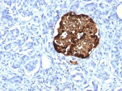
Biotium Chromogranin A / CHGA (Neuroendocrine Marker) (CHGA/1815R), 0.2mg/mL
Chromogranin A is present in neuroendocrine cells throughout the body, including the neuroendocrine cells of the large and small intestine, adrenal medulla and pancreatic islets. It is an excellent marker for carcinoid tumors, pheochromocytomas, paragangliomas, and other neuroendocrine tumors. Co-expression of chromogranin A and neuron specific enolase (NSE) is common in neuroendocrine neoplasms. Reportedly, co-expression of certain keratins and chromogranin indicates neuroendocrine lineage. The presence of strong anti-chromogranin staining and absence of anti-keratin staining should raise the possibility of paraganglioma. The co-expression of chromogranin and NSE is typical of neuroendocrine neoplasms. Most pituitary adenomas and prolactinomas readily express chromogranin.Primary antibodies are available purified, or with a selection of fluorescent CF Dyes and other labels. CF Dyes offer exceptional brightness and photostability. Note: Conjugates of blue fluorescent dyes