
Mouse Secondary Antibodies


BIOTIUM INC Cytokeratin 10 (Suprabasal Epithelial Marker) (DE-K10), Biotin conjugate, 0.1mg/mL
This MAb recognizes a protein of 56.5 kDa, identified as cytokeratin 10 (CK10). CK10 is expressed in all suprabasal layers of the epidermis. In the epidermis, expression of CK10 strictly parallels the extent of differentiation; it is absent in the basal layer, appears in the first suprabasal layers and increases in concentration towards the granular layer. However, CK10 is rarely detected in early stages of vulvar squamous carcinomas (tumors less than 2 cm, clinical stage I) regardless of the tumor grade. In larger and more advanced tumors (greater than 2 cm, clinical stages II and III), CK10 is detected very frequently. Expression of CK10 is related to maturation of malignant keratinocytes, being preferentially detected in more-differentiated parts.Primary antibodies are available purified, or with a selection of fluorescent CF Dyes and other labels. CF Dyes offer exceptional brightness and photostability. Note: Conjugates of blue fluorescent dyes like CF405S and CF405M


BIOTIUM INC Ksp-Cadherin (Kidney-Specific Cadherin) / CDH16, 0.2mg/mL
This antibody recognizes a protein of 130 kDa, identified as Ksp-cadherin. Cadherins form a superfamily of related glycoproteins that mediate calcium-dependent cell adhesion and transmit signals from the extracellular matrix to the cytoplasm. Cadherins have been implicated in embryogenesis, tissue morphogenesis, tissue structure maintenance, cell polarization, neoplastic invasiveness and metastasis, and membrane transport. It is suggested that Ksp-cadherin is a marker for terminal differentiation of the basolateral membranes of renal tubular epithelial cells. Within the kidney, Ksp-Cadherin is found exclusively in the basolateral membrane of renal tubular epithelial cells and collecting duct cells, and not in glomeruli, renal interstitial cells, or blood vessels. Ksp-Cadherin has been suggested to distinguish Chromophobe Renal-Cell Carcinoma from Oncocytoma.Primary antibodies are available purified, or with a selection of fluorescent CF Dyes and other labels. CF Dyes of

BIOTIUM INC Topoisomerase II alpha (Proliferation & Drug-Resistance Marker) (TOP2A/1362), 1mg/mL
This antibody recognizes a 170 kDa protein, which is identified as topoisomerase IIα. It shows no cross-reaction with topoisomerase IIβ or topoisomerase I. Topo IIα plays important roles in synthesis and transcription of DNA as well as chromosomal segregation during mitosis. It is reported to be a sensitive and specific marker of late S-, G2- & M-phases in transformed and developmentally regulated normal cells. Topo IIα is also implicated in drug resistance of tumor cells.Primary antibodies are available purified, or with a selection of fluorescent CF Dyes and other labels. CF Dyes offer exceptional brightness and photostability. Note: Conjugates of blue fluorescent dyes like CF405S and CF405M are not recommended for detecting low abundance targets, because blue dyes have lower fluorescence and can give higher non-specific background than other dye colors.

BIOTIUM INC CELA3B / ELA3B (Pancreatic Function Marker) (CELA3B/1374), 1mg/mL
This MAb recognizes a protein of ~30 kDa, identified as CELA3B (Chymotrypsin like elastase family member 3B). Elastases form a subfamily of serine proteases that hydrolyze many proteins in addition to elastin. Humans have six elastase genes which encode the structurally similar proteins elastase 1, 2, 2A, 2B, 3A, and 3B. Unlike other elastases, elastase 3B has little elastolytic activity. Like most of the human elastases, elastase 3B is secreted from the pancreas as a zymogen and, like other serine proteases such as trypsin, chymotrypsin and kallikrein; it has a digestive function in the intestine. Elastase 3B preferentially cleaves proteins after alanine residues. Elastase 3B may also function in the intestinal transport and metabolism of cholesterol. Both elastase 3A and elastase 3B have been referred to as protease E and as elastase 1, and excretion of this protein in fecal material is frequently used as a measure of pancreatic function in clinical assays.Primary antibodie
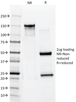
BIOTIUM INC Cytokeratin 10 (Suprabasal Epithelial Marker)(AE20), 1mg/mL
This MAb recognizes a protein of 56. 5 kDa, identified as cytokeratin 10 (CK10). CK10 is expressed in all suprabasal layers of the epidermis. In the epidermis, expression of CK10 strictly parallels the extent of differentiation; it is absent in the basal layer, appears in the first suprabasal layers and increases in concentration towards the granular layer. However, CK10 is rarely detected in early stages of vulvar squamous carcinomas (tumors less than 2 cm, clinical stage I) regardless of the tumor grade. In larger and more advanced tumors (greater than 2 cm, clinical stages II and III), CK10 is detected very frequently. Expression of CK10 is related to maturation of malignant keratinocytes, being preferentially detected in more-differentiated parts. Primary antibodies are available purified, or with a selection of fluorescent CF Dyes and other labels. CF Dyes offer exceptional brightness and photostability. Note: Conjugates of blue fluorescent dyes like CF405S and CF40
BIOTIUM INC Cytokeratin 8 (KRT8)(B22.1), 1mg/mL
Cytokeratin 8 (CK8) belongs to the type II (or B or basic) subfamily of high molecular weight cytokeratins and exists in combination with cytokeratin 18 (CK18). CK8 is primarily found in the non-squamous epithelia and is present in majority of adenocarcinomas and ductal carcinomas. It is absent in squamous cell carcinomas. Hepatocellular carcinomas are defined by the use of antibodies that recognize only cytokeratin 8 and 18. CK8 exists on several types of normal and neoplastic epithelia, including many ductal and glandular epithelia such as colon, stomach, small intestine, trachea, and esophagus as well as in transitional epithelium. Anti-CK8 does not react with skeletal muscle or nerve cells. Epithelioid sarcoma, chordoma, and adamantinoma show strong positivity corresponding to that of simple epithelia (with antibodies against CK8, CK18 and CK19). Reportedly, anti-CK8 is useful for the differentiation of lobular ('ring-like, perinuclear') from ductal ('peripheral-predomina

BIOTIUM INC Spectrin beta III (SPTBN2) (SPTBN2/1582), 0.2mg/mL
Spectrin is an actin binding protein that is a major component of the plasma membrane skeleton. Spectrins function as membrane organizers and stabilizers by forming dimers, tetramers and higher polymers. Vertebrate spectrins have two alpha-subunits (alpha-I/alpha-II), four beta-subunits (beta-I-beta-IV) and a beta-H subunit creating diversity and specialization of function. Spectrin alpha and spectrin beta are present in erythrocytes, whereas spectrin alpha II (also designated fodrin alpha) and spectrin beta I (also designated fodrin beta) are present in other somatic cells. The spectrin tetramers in erythrocytes act as barriers to lateral diffusion, but spectrin dimers seem to lack this function. Spectrin beta III is highly homologous to both spectrin beta I and spectrin beta II. Spectrin beta III is highly expressed in brain, kidney, pancreas and liver, and at lower levels in lung and placenta. Spectrin beta 3 is primarily expressed in nervous tissues with highest expressio

BIOTIUM INC p57Kip2 (Mitotic Inhibitor/Suppressor Protein)(57P06), 1mg/mL
Recognizes a protein of 57 kDa, identified as p57Kip2. It shows no cross-reaction with p27Kip1. p57Kip2 is a potent tight-binding inhibitor of several G1 cyclin complexes, and is a negative regulator of cell proliferation. Anti-p57 has been used as an aide in identification of complete hydatidiform mole (CHM) (no nuclear labeling of cytotrophoblasts and stromal cells) from partial hydatidiform mole (PHM) in which both cytotrophoblasts and stromal cells stain. The histological differentiation of complete mole, partial mole, and hydropic spontaneous abortion is problematic. Most complete hydatidiform moles are diploid, whereas most partial moles are triploid. Ploidy studies will identify partial moles, but will not differentiate complete moles from non-molar gestations. Complete moles carry a high risk of persistent disease and choriocarcinoma, while partial moles have a very low risk. In normal placenta, many cytotrophoblast nuclei and stromal cells are labeled with this antib
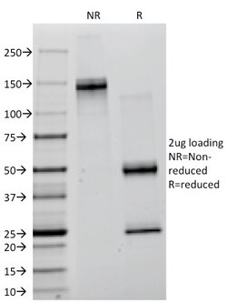
BIOTIUM INC Creatine Kinase-BB (CK-BB) (CKBB/1268), Biotin conjugate, 0.1mg/mL
Creatine kinases (CK) are a large family of isoenzymes that regulate levels of ATP in subcellular compartments, where they provide ATP at sites of fluctuating energy demand by the transfer of phosphates between creatine and adenine nucleotides. CKs provide the energy of phosphate hydrolysis necessary to drive the normal function of many cellular systems. In cells, the cytosolic CK enzymes consist of two subunits, which can be either B (brain type) or M (muscle type). There are three different isoenzymes: CKMM, CKBB and CKMB. This MAb recognizes the CKBB isoenzyme and does not react with the B subunit in CKMB. It shows minimal reactivity with other human serum proteins.Primary antibodies are available purified, or with a selection of fluorescent CF Dyes and other labels. CF Dyes offer exceptional brightness and photostability. Note: Conjugates of blue fluorescent dyes like CF405S and CF405M are not recommended for detecting low abundance targets, because blue dyes have lo
BIOTIUM INC HPV-16 (Human Papilloma Virus 16) (HPV16/1296), Biotin conjugate, 0.1mg/mL
This antibody reacts with E6 protein of human papilloma virus type 16 (HPV-16). Infection with specific types of HPV has been associated with an increased risk of developing cervical neoplasia. HPV types 6 and 11 have been associated with relatively benign diseases such as genital warts but types 16 and 18 are strongly associated with cervical, vaginal, and vulvar malignancies. The antibody reacts very strongly with formalin-fixed, paraffin-embedded tissues containing HPV-16.Primary antibodies are available purified, or with a selection of fluorescent CF Dyes and other labels. CF Dyes offer exceptional brightness and photostability. Note: Conjugates of blue fluorescent dyes like CF405S and CF405M are not recommended for detecting low abundance targets, because blue dyes have lower fluorescence and can give higher non-specific background than other dye colors.

BIOTIUM INC ASRGL1 (Asparaginase Like 1) (CRASH/1289), 1mg/mL
ASRGL1 (Asparaginase-like protein 1), also known as CRASH, is a 308 amino acid protein belonging to the Ntn-hydrolase family. Asparaginases utilize asparagine as a substrate to produce aspartic acid and ammonia. ASRGL1 has been identified as a autoantigenic protein that is present in the mid-piece of sperm after obstruction of the male reproductive tract. ASRGL1 is expressed highly in testis, but is also expressed in brain, kidney and gastrointestinal tissues. High levels of ASRGL1 have also been identified in ovarian, uterine and mammary tumors in comparison with normal tissues of the same origin.Primary antibodies are available purified, or with a selection of fluorescent CF Dyes and other labels. CF Dyes offer exceptional brightness and photostability. Note: Conjugates of blue fluorescent dyes like CF405S and CF405M are not recommended for detecting low abundance targets, because blue dyes have lower fluorescence and can give higher non-specific background than other

BIOTIUM INC Topoisomerase II alpha (Proliferation & Drug-Resistance Marker) (TOP2A/1362), Biotin conjugate, 0.1mg/mL
This antibody recognizes a 170 kDa protein, which is identified as topoisomerase IIα. It shows no cross-reaction with topoisomerase IIβ or topoisomerase I. Topo IIα plays important roles in synthesis and transcription of DNA as well as chromosomal segregation during mitosis. It is reported to be a sensitive and specific marker of late S-, G2- & M-phases in transformed and developmentally regulated normal cells. Topo IIα is also implicated in drug resistance of tumor cells.Primary antibodies are available purified, or with a selection of fluorescent CF Dyes and other labels. CF Dyes offer exceptional brightness and photostability. Note: Conjugates of blue fluorescent dyes like CF405S and CF405M are not recommended for detecting low abundance targets, because blue dyes have lower fluorescence and can give higher non-specific background than other dye colors.
BIOTIUM INC Thomsen-Friedenreich Antigen / CD176 (Pan Carcinoma Marker)(A84-A/F10), 1mg/mL
Recognizes a disaccharide epitope, Gal 1-3GalNAc, of Thomsen-Friedenreich (TF) antigen. It is specific for both anomeric forms of the disaccharide (TF and TF , including related structures on the glycolipid) and shows no cross-reactivity with sialylated glycophorin. The TF antigen acts as an oncofetal antigen, with low expression in normal adult tissues but increasing to fetal levels of expression in hyperplasia or malignancy. Considered to be a pan-carcinoma marker. It is capable to agglutinate desialylated red blood cells. During metastasis, the ability of malignant cells to form multicellular aggregates via homotypic or heterotypic aggregation and their adhesion to the endothelium are critical. It is involved in tumor cell adhesion and tissue invasion. It also causes an immune response, and overexpression of the antigen causes cancer cells to be more sensitive to natural killer cell lysis. TF antigen is suppressed in normal healthy cells and represents one of the few chemi