
Mouse Secondary Antibodies

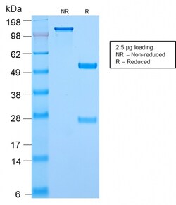
Biotium c-Myc Oncoprotein(MYC2895R), CF568 conjugate, 0.1mg/mL
The epitope of this antibody spans the Myc tag epitope, aa 410-419 (EQKLISEEDL), which is a specific portion of an alpha helical region of human c-myc protein. Because the epitope is located at the dimerization site of the protein, the antibody is not sensitive for detecting endogenous c-myc, making it suitable for detecting Myc tagged proteins or myc overexpression. This MAb shows no cross-reaction with v-myc. c-myc is involved in the control of cell proliferation and differentiation and is amplified and/or overexpressed in a variety of tumors. Over-expression of c-myc protein occurs frequently in luminal cells of prostate intraepithelial neoplasia as well as in most primary carcinomas and metastatic disease.Primary antibodies are available purified, or with a selection of fluorescent CF Dyes and other labels. CF Dyes offer exceptional brightness and photostability. Note: Conjugates of blue fluorescent dyes like CF405S and CF405M are not recommended for detecting lo


Biotium BCL10 (MALT-Lymphoma Marker) (BL10/2988R), CF568 conjugate, 0.1mg/mL
B-cell lymphoma/leukemia 10 (BCL10) is a signaling protein that promotes apoptosis, pro-caspase-9 maturation, and activation of NF-kappa-B via NIK and IKK. BCL10 contains an N-terminal caspase recruitment domain (CARD), which is found in a number of apoptotic regulatory molecules. BCL10 is involved in the adaptive immune response, and may be an adapter protein between upstream TNFR1-TRADD-RIP complex and the downstream NIK-IKK-IKAP complex. A BCL10 translocation is recurrent in low-grade mucosa-associated lymphoid tissue (MALT lymphoma).Primary antibodies are available purified, or with a selection of fluorescent CF Dyes and other labels. CF Dyes offer exceptional brightness and photostability. Note: Conjugates of blue fluorescent dyes like CF405S and CF405M are not recommended for detecting low abundance targets, because blue dyes have lower fluorescence and can give higher non-specific background than other dye colors.
Biotium Renal Cell Carcinoma (Carbonic Anhydrase IX) (CA9/2993R), CF568 conjugate, 0.1mg/mL
Carbonic anhydrase IX (carbonic anhydrase 9) is one of several carbolic anhydrases that vary in tissue distribution and localization. Carbonic anhydrases catalyze the interconversion of carbon dioxide and water into carbonic acid and bicarbonate and are found in all mammalian tissues. Carbonic anhydrase IX is a transmembrane protein that is not expressed in most healthy tissues, but is expressed in most renal cell carcinomas.Primary antibodies are available purified, or with a selection of fluorescent CF Dyes and other labels. CF Dyes offer exceptional brightness and photostability. Note: Conjugates of blue fluorescent dyes like CF405S and CF405M are not recommended for detecting low abundance targets, because blue dyes have lower fluorescence and can give higher non-specific background than other dye colors.
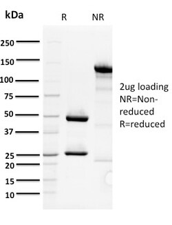
Biotium ORC1(7F6/1), CF568 conjugate, 0.1mg/mL
The initiation of DNA replication is a multi-step process that depends on the formation of pre-replication complexes, which trigger initiation. Among the proteins required for establishing these complexes are the origin recognition complex (ORC) proteins. ORC proteins bind specifically to origins of replication where they serve as scaffold for the assembly of additional initiation factors. Human ORC subunits 1-6 are expressed in the nucleus of proliferating cells and tissues, such as the testis. ORC1 and ORC2 are both expressed at equivalent concentrations throughout the cell cycle; however, only ORC2 remains stably bound to chromatin. ORC4 and ORC6 are also expressed constantly throughout the cell cycle. ORC2, ORC3, ORC4 and ORC5 form a core complex upon which ORC6 and ORC1 assemble. The formation of this core complex suggests that ORC proteins play a crucial role in the G1-S transition in mammalian cells. Primary antibodies are available purified, or with a selection of flu
Biotium CDX2 (GI Epithelial Marker) (CDX2/2951R), CF568 conjugate, 0.1mg/mL
The intestine-specific transcription factors CDX1 and CDX2 are important for directing intestinal development, differentiation, proliferation and maintenance of the intestinal phenotype. CDX2 protein expression has been seen in GI carcinomas. Anti-CDX2 has been useful to establish GI origin of metastatic adenocarcinomas and carcinoids and is especially useful to distinguish metastatic colorectal adenocarcinoma from lung adenocarcinoma. However, mucinous carcinomas of the ovary also express CDX2 protein. It limits the usefulness of this marker in the distinction of metastatic colorectal adenocarcinoma from mucinous carcinoma of the ovary.Primary antibodies are available purified, or with a selection of fluorescent CF Dyes and other labels. CF Dyes offer exceptional brightness and photostability. Note: Conjugates of blue fluorescent dyes like CF405S and CF405M are not recommended for detecting low abundance targets, because blue dyes have lower fluorescence and can give higher
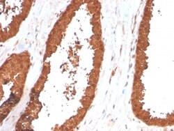
Biotium TIGIT / VSTM3 / VSIG9 (Immune Checkpoint for Cancer)(TIGIT/3017), CF568 conjugate, 0.1mg/mL
TIGIT is a checkpoint inhibitor which binds with high affinity to the poliovirus receptor (PVR), causing increased IL10 secretion, decreased IL12B secretion. TIGIT binding to PVR also causes the suppression of T cell activation by promoting the generation of mature immuno-regulatory dendritic cells. It is expressed at low levels on natural killer (NK) cells, as well as peripheral memory and regulatory CD4 T cells. At the protein level, it is upregulated following the activation of these cells. Functionally, TIGIT is similar to CTLA4. The ligands for TIGIT include CD155 (signal abrogation) and CD226 (signal stimulation). It has been demonstrated to be upregulated on T cells in many cancers and is an immuno-oncology target for therapy. Primary antibodies are available purified, or with a selection of fluorescent CF Dyes and other labels. CF Dyes offer exceptional brightness and photostability. Note: Conjugates of blue fluorescent dyes like CF405S and CF405M are not recomm
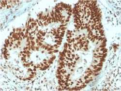
Biotium MSH6 (DNA Mismatch Repair Protein)(MSH6/3091), CF568 conjugate, 0.1mg/mL
The finding that mutations in DNA mismatch repair genes are associated with hereditary nonpolyposis colorectal cancer (HNPCC) has resulted in considerable interest in the understanding of the mechanism of DNA mismatch repair. Initially, inherited mutations in the MSH2 and MLH1 homologs of the bacterial DNA mismatch repair genes mutS and mutL were demonstrated at high frequency in HNPCC and were shown to be associated with microsatellite instability. A member of the mismatch repair family, GTBP (also designated MSH6), is an MSH2-related protein that binds to DNA containing G/T mismatches. Findings suggest that the mismatch-binding factor in human cells is composed of a heterodimer of GTBP and MSH2. Primary antibodies are available purified, or with a selection of fluorescent CF Dyes and other labels. CF Dyes offer exceptional brightness and photostability. Note: Conjugates of blue fluorescent dyes like CF405S and CF405M are not recommended for detecting low abundance targ
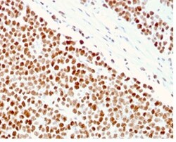
Biotium MSH6 (DNA Mismatch Repair Protein)(MSH6/3086), CF568 conjugate, 0.1mg/mL
The finding that mutations in DNA mismatch repair genes are associated with hereditary nonpolyposis colorectal cancer (HNPCC) has resulted in considerable interest in the understanding of the mechanism of DNA mismatch repair. Initially, inherited mutations in the MSH2 and MLH1 homologs of the bacterial DNA mismatch repair genes mutS and mutL were demonstrated at high frequency in HNPCC and were shown to be associated with microsatellite instability. A member of the mismatch repair family, GTBP (also designated MSH6), is an MSH2-related protein that binds to DNA containing G/T mismatches. Findings suggest that the mismatch-binding factor in human cells is composed of a heterodimer of GTBP and MSH2. Primary antibodies are available purified, or with a selection of fluorescent CF Dyes and other labels. CF Dyes offer exceptional brightness and photostability. Note: Conjugates of blue fluorescent dyes like CF405S and CF405M are not recommended for detecting low abundance targ
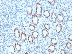
Biotium MSH6 (DNA Mismatch Repair Protein)(MSH6/3089), CF568 conjugate, 0.1mg/mL
The finding that mutations in DNA mismatch repair genes are associated with hereditary nonpolyposis colorectal cancer (HNPCC) has resulted in considerable interest in the understanding of the mechanism of DNA mismatch repair. Initially, inherited mutations in the MSH2 and MLH1 homologs of the bacterial DNA mismatch repair genes mutS and mutL were demonstrated at high frequency in HNPCC and were shown to be associated with microsatellite instability. A member of the mismatch repair family, GTBP (also designated MSH6), is an MSH2-related protein that binds to DNA containing G/T mismatches. Findings suggest that the mismatch-binding factor in human cells is composed of a heterodimer of GTBP and MSH2. Primary antibodies are available purified, or with a selection of fluorescent CF Dyes and other labels. CF Dyes offer exceptional brightness and photostability. Note: Conjugates of blue fluorescent dyes like CF405S and CF405M are not recommended for detecting low abundance targ
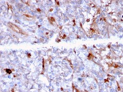
Biotium C1QA / Complement C1q A-Chain(C1QA/2956), CF568 conjugate, 0.1mg/mL
Complement C1q A Chain (C1qa) is a subunit of C1q. C1q is composed of 6 polypeptide chains each of C1qa, C1qb, and C1qc. C1q is a component of the classical complement pathway, and thereby plays an important role in the immune response. Each C1q subunit contains an N-terminal collagen-like region and a C-terminal C1q globular domain. The presence of receptors for C1q on effector cells modulates its activity, which may be antibody-dependent or independent. Macrophages are the primary source of C1q, while anti-inflammatory drugs as well as cytokines differentially regulate expression of the mRNA as well as the protein. C1q deficiency is associated with lupus erythematosus and glomerulonephritis. Primary antibodies are available purified, or with a selection of fluorescent CF Dyes and other labels. CF Dyes offer exceptional brightness and photostability. Note: Conjugates of blue fluorescent dyes like CF405S and CF405M are not recommended for detecting low abundance targets,
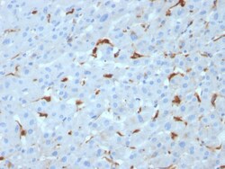
Biotium C1QA / Complement C1q A-Chain(C1QA/2953), CF568 conjugate, 0.1mg/mL
Complement C1q A Chain (C1qa) is a subunit of C1q. C1q is composed of 6 polypeptide chains each of C1qa, C1qb, and C1qc. C1q is a component of the classical complement pathway, and thereby plays an important role in the immune response. Each C1q subunit contains an N-terminal collagen-like region and a C-terminal C1q globular domain. The presence of receptors for C1q on effector cells modulates its activity, which may be antibody-dependent or independent. Macrophages are the primary source of C1q, while anti-inflammatory drugs as well as cytokines differentially regulate expression of the mRNA as well as the protein. C1q deficiency is associated with lupus erythematosus and glomerulonephritis. Primary antibodies are available purified, or with a selection of fluorescent CF Dyes and other labels. CF Dyes offer exceptional brightness and photostability. Note: Conjugates of blue fluorescent dyes like CF405S and CF405M are not recommended for detecting low abundance targets,
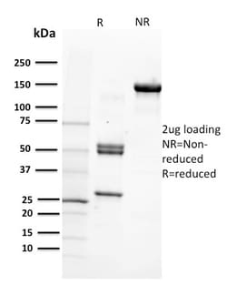
Biotium CD29 (Stem Cell Marker)(12G10), CF568 conjugate, 0.1mg/mL
Recognizes CD29. Binds to the cell membrane of T- and B- lymphocytes, monocytes, platelets, fibroblasts, endothelial cells, mast cells and smooth muscle cells. CD29 is an adhesion receptor and co-stimulatory molecule. This antigen was initially characterized as GPIIa on platelets and as the common b1 subunit of the very late antigen (VLA) protein family. CD29 forms a non-covalent heterodimeric complex with integrin alpha subunits. It also mediates adhesion to invasion and thrombospondin. Primary antibodies are available purified, or with a selection of fluorescent CF Dyes and other labels. CF Dyes offer exceptional brightness and photostability. Note: Conjugates of blue fluorescent dyes like CF405S and CF405M are not recommended for detecting low abundance targets, because blue dyes have lower fluorescence and can give higher non-specific background than other dye colors.
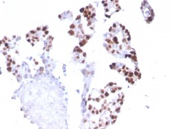
Biotium RNA Polymerase II(CTD4H8), CF568 conjugate, 0.1mg/mL
RNA polymerase II (Pol II) is an enzyme that is composed of 12 subunits and is responsible for the transcription of protein-coding genes. Transcription initiation requires Pol II-mediated recruitment of transcription machinery to a target promoter, thereby allowing transcription to begin. The largest subunit of Pol II (referred to as RPB1 or RPB205) is a 1, 840 amino acid protein that contains one C2H2-type zinc finger and a C-terminal domain comprised of several heptapeptide repeats. Although Pol II function requires the cooperation of all twelve subunits, the largest subunit conveys Pol II catalytic activity and, together with the second largest subunit, forms the active center of the Pol II enzyme. Additionally, the large subunit participates in forming the DNA-binding domain of Pol II, a groove that is necessary for transcription of the DNA template. Without proper function of the large subunit, mRNA synthesis and subsequent transcription elongation cannot occur. Primary
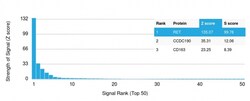
Biotium RET Proto-oncogene(RET/2976), CF568 conjugate, 0.1mg/mL
The Ret proto-oncogene is structurally related to the growing family of tyrosine kinase transmembrane receptors and is involved in GDNF signaling. RET expression is reported in several regions of the central nervous system; in the developing cranial nerve ganglia and a subset of cells within dorsal root ganglia, in motor neurons in the spinal cord and hindbrain, in neuro-retina and the growing tips of the renal collecting ducts in developing kidney. Alterations in RETgene are associated with diseases including papillary thyroid carcinoma, multiple endocrine neoplasia (type 2A and 2B), familial medullary thyroid carcinoma, and a congenital developmental disorder known as Hirschsprung's disease. Primary antibodies are available purified, or with a selection of fluorescent CF Dyes and other labels. CF Dyes offer exceptional brightness and photostability. Note: Conjugates of blue fluorescent dyes like CF405S and CF405M are not recommended for detecting low abundance targets,
Biotium Tenascin C (Stromal Marker For Epithelial Malignancy) (TNC/2981R), CF740 conjugate, 0.1mg/mL
In Western blotting, this antibody reacts with two bands of ~MW of 210 kDa and 300 kDa, identified as two isoforms of Tenascin C. Specificity of this MAb is validated by sequential immunoprecipitation with a PAb against Tenascin C. Tenascin C is a multifunctional, disulfide-linked hexameric extracellular matrix glycoprotein expressed in association with mesenchymal epithelial interactions during development and in the neo-vasculature and stroma of undifferentiated tumors. In adults, it is restricted to certain epithelial-stromal interfaces and increases markedly in hyper-proliferative diseases and in stroma of many neoplasms, including gliomas, breast, squamous and lung carcinomas.Primary antibodies are available purified, or with a selection of fluorescent CF Dyes and other labels. CF Dyes offer exceptional brightness and photostability. Note: Conjugates of blue fluorescent dyes like CF405S and CF405M are not recommended for detecting low abundance targets, because blue dyes