
Mouse Secondary Antibodies

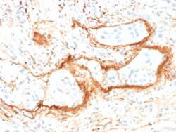
Biotium Elastin (ELN) (Marker of Arterial Stiffness and Atherosclerosis) (ELN/1981), CF488A conjugate, 0.1mg/mL
Elastin is a polymeric protein and is the main component of the extracellular matrix of arteries. It is synthesized and secreted as a soluble, single-chain protein (tropoelastin), which undergoes a number of post-translational modifications prior to its organization into an elastic fiber in the extracellular space. Elastin performs a regulatory function during arterial development by controlling proliferation of smooth muscle and stabilizing arterial structure. It imparts elasticity to the connective tissue. During aging, the elasticity of connective tissue is reduced because of the cross-linking of collagenous fibers with elastin. The abnormal accumulation of elastic tissue in blood vessels is found in atherosclerosis and hypertension.Primary antibodies are available purified, or with a selection of fluorescent CF Dyes and other labels. CF Dyes offer exceptional brightness and photostability. Note: Conjugates of blue fluorescent dyes like CF405S and CF405M are not recom


Biotium Villin (GI-Mucosal & Urogenital Brush Border Marker) (VIL1/1314), Biotin conjugate, 0.1mg/mL
This antibody recognizes a protein of 95 kDa, which is identified as villin. It is a major constituent in the microvilli, which compose the brush border of epithelial cells forming absorptive surfaces of the intestinal and renal proximal tubular epithelia. Anti-Villin labels the brush border area in the gastrointestinal mucosal epithelium and urogenital tract. Among neoplasms, villin is predominantly expressed in tumors of colorectal origin. Antibody to villin is useful in identifying malignant cells from primary and metastatic colorectal carcinomas. This antibody also labels Merkel cells of the skin.Primary antibodies are available purified, or with a selection of fluorescent CF Dyes and other labels. CF Dyes offer exceptional brightness and photostability. Note: Conjugates of blue fluorescent dyes like CF405S and CF405M are not recommended for detecting low abundance targets, because blue dyes have lower fluorescence and can give higher non-specific background than oth
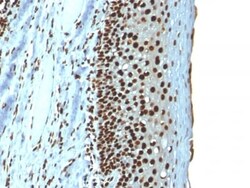
Biotium Histone H1 (Nuclear Marker) (r1415-1), 0.2mg/mL
Eukaryotic histones are basic and water-soluble nuclear proteins that form hetero-octameric nucleosome particles by wrapping 146 base pairs of DNA in a left-handed supealpha-helicalturn sequentially to form chromosomal fiber. Two molecules of each of the four core histones (H2A, H2B, H3, and H4) form the octamer; formed of two H2A-H2B dimers and two H3-H4 dimers, forming two nearly symmetrical halves by tertiary structure. Over 80% of nucleosomes contain the linker Histone H1, derived from an intronless gene that interacts with linker DNA between nucleosomes and mediates compaction into higher order chromatin. Histones are subject to posttranslational modification by enzymes primarily on their N-terminal tails, but also in their globular domains. Such modifications include methylation, citrullination, acetylation, phosphorylation, sumoylation, ubiquitination and ADP-ribosylation.Primary antibodies are available purified, or with a selection of fluorescent CF Dyes and other l
Biotium DOG-1 / TMEM16A (Gastrointestinal Stromal Tumor Marker) (DG1/1484), CF488A conjugate, 0.1mg/mL
Expression of DOG-1 protein is elevated in gastrointestinal stromal tumors (GISTs), c-kit signaling-driven mesenchymal tumors of the GI tract. DOG-1 is rarely expressed in other soft tissue tumors, which, due to appearance, may be difficult to diagnose. Immunoreactivity for DOG-1 has been reported in 97.8 percent of scorable GISTs, including all c-kit negative GISTs. Overexpression of DOG-1 has been suggested to aid in the identification of GISTs, including Platelet-Derived Growth Factor Receptor Alpha mutants that fail to express c-kit antigen. The overall sensitivity of DOG1 and c-kit in GISTs is nearly identical: 94.4% vs. 94.7%.Primary antibodies are available purified, or with a selection of fluorescent CF Dyes and other labels. CF Dyes offer exceptional brightness and photostability. Note: Conjugates of blue fluorescent dyes like CF405S and CF405M are not recommended for detecting low abundance targets, because blue dyes have lower fluorescence and can give higher non-s
Biotium Neurofilament (H+L)(NF421 + NFL736), CF740 conjugate, 0.1mg/mL
This MAb reacts with a 200 kDa and 68 kDa protein, identified as heavy and light sub-units of neurofilaments (NF-H & NF-L). Neurofilaments make up the main structural elements of axons and dendrites and are found in neurons, peripheral nerves, and sympathetic ganglion cells. Neurofilaments consist of three major subunits with molecular weights of 68 kDa (NF-L), 160 kDa (NF-M) and 200 kDa (NF-H). Anti-neurofilament stains a number of neural, neuroendocrine, and endocrine tumors. Neuromas, ganglioneuromas, gangliogliomas, ganglioneuroblastomas, and neuroblastomas stain positively for anti-neurofilament. Neurofilaments are also present in paragangliomas as well as adrenal and extra-adrenal pheochromocytomas. Carcinoids, neuroendocrine carcinomas of the skin, and oat cell carcinomas of the lung also express neurofilament.Primary antibodies are available purified, or with a selection of fluorescent CF Dyes and other labels. CF Dyes offer exceptional brightness and photos
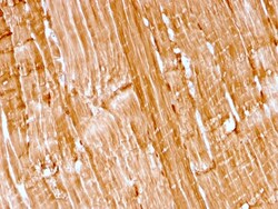
Biotium Myoglobin (Muscle Cell Marker)(rMB/2105), CF568 conjugate, 0.1mg/mL
Myoglobin is a cytosolic oxygen-binding protein responsible for the storage and diffusion of oxygen within myocytes. Expression of myoglobin is highest in skeletal and cardiac muscle. Myoglobin is well accepted as an O2-storage protein in muscle, capable of releasing O2 during periods of hypoxia or anoxia. In combination with other striated muscle markers such as vimentin and myogenin, myoglobin is helpful in the identification of rhabdomyosarcoma and tumors with skeletal muscle differentiation. Reportedly, myoglobin is expressed on epithelial cancer cells due to changed metabolic and environmental conditions. Primary antibodies are available purified, or with a selection of fluorescent CF Dyes and other labels. CF Dyes offer exceptional brightness and photostability. Note: Conjugates of blue fluorescent dyes like CF405S and CF405M are not recommended for detecting low abundance targets, because blue dyes have lower fluorescence and can give higher non-specific backgroun
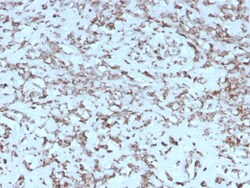
Biotium Cytochrome C (Mitochondrial Marker)(rCYCS/1010), CF568 conjugate, 0.1mg/mL
Recognizes an epitope within amino acids 93-104 of pigeon Cytochrome C, a well-characterized mobile electron transport protein that is essential to energy conversion in all aerobic organisms. In mammalian cells, this highly conserved protein is normally localized to the mitochondrial inter-membrane space. More recent studies have identified cytosolic cytochrome c as a factor necessary for activation of apoptosis. During apoptosis, cytochrome c is trans-located from the mitochondrial membrane to the cytosol, where it is required for activation of caspase-3 (CPP32). Overexpression of Bcl-2 has been shown to prevent the translocation of cytochrome c, thereby blocking the apoptotic process. Overexpression of Bax has been shown to induce the release of cytochrome c and to induce cell death. The release of cytochrome c from the mitochondria is thought to trigger an apoptotic cascade, whereby Apaf-1 binds to Apaf-3 (caspase-9) in a cytochrome c-dependent manner, leading to caspase-9
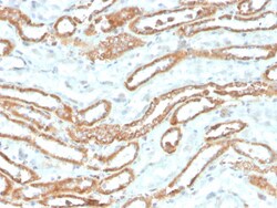
Biotium Aurora B (Proliferation Marker)(rAURKB/1592), 0.2mg/mL
This antibody recognizes a protein of 39 kDa, which is identified as Aurora kinase B (or Aurora B). Aurora B is a serine/threonine protein kinase and component of the chromosomal passenger complex (CPC), a complex that acts as a key regulator of mitosis. It is important for accurate chromosome segregation, cytokinesis, protein localization to the centromere and kinetochore, correct microtubule-kinetochore attachment, and regulation of the mitotic checkpoint. Aurora B forms a tight complex with inner centrosome protein and survivin. Inactivation of any of these proteins causes similar defects in chromosome segregation. A significant overexpression of Aurora B has been found in a variety of human tumors including non-small cell lung carcinoma, astrocytoma, seminoma and carcinomas of the colon, prostate, endometrium and thyroid. The expression level of Aurora B is associated with cell proliferation and prognosis in these tumors. Primary antibodies are available purified, or with
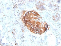
Biotium MTAP (Tumor Suppressor Marker)(rMTAP/1813), Biotin conjugate, 0.1mg/mL
Recognizes a protein of 31 kDa, which is identified as MTAP (5'-deoxy-5'-methylthioadenosine phosphorylase). It catalyzes the reversible phosphorolysis of methylthioadenosine, which is important in polyamine metabolism and for the salvage of adenine and methionine. The gene encoding MTAP is linked to the tumor suppressor gene, p16INK4A. Deficient levels of MTAP can occur in cancers primarily through co-deletion of the MTAP gene and the p16INK4A gene. Cells expressing MTAP and possessing adenine salvage pathway activity may be less susceptible to malignancy due to growth-inhibitory actions of agents (e. g. antifolates), whose mechanism of action, in part, involves this de novo purine pathway. Primary antibodies are available purified, or with a selection of fluorescent CF Dyes and other labels. CF Dyes offer exceptional brightness and photostability. Note: Conjugates of blue fluorescent dyes like CF405S and CF405M are not recommended for detecting low abundance targets, b
Biotium CELA3B / ELA3B (Pancreatic Function Marker) (CELA3B/2809R), CF488A conjugate, 0.1mg/mL
This MAb recognizes a protein of ~30 kDa, identified as CELA3B (Chymotrypsin like elastase family member 3B). Elastases form a subfamily of serine proteases that hydrolyze many proteins in addition to elastin. Humans have six elastase genes which encode the structurally similar proteins elastase 1, 2, 2A, 2B, 3A, and 3B. Unlike other elastases, elastase 3B has little elastolytic activity. Like most of the human elastases, elastase 3B is secreted from the pancreas as a zymogen and, like other serine proteases such as trypsin, chymotrypsin and kallikrein; it has a digestive function in the intestine. Elastase 3B preferentially cleaves proteins after alanine residues. Elastase 3B may also function in the intestinal transport and metabolism of cholesterol. Both elastase 3A and elastase 3B have been referred to as protease E and as elastase 1, and excretion of this protein in fecal material is frequently used as a measure of pancreatic function.Primary antibodies are available purified
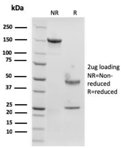
Biotium Langerin / CD207 (Marker of Langerhans Cells)(rLGRN/1821), CF740 conjugate, 0.1mg/mL
Langerhans cells (LCs) are a subset of immature dendritic cells (DCs) that specifically localize in the epidermis and other mucosal epithelia. Epidermal LCs possess strong immuno-stimulatory capacity and play a central role in the initiation and regulation of immune responses. Langerin (CD207) is a Ca2 -dependent, C-type lectin domain containing, type II transmembrane protein that induces epidermal LCs to differentiate into Birbeck granules (BG). BGs are organelles with superimposing and zippering membranes that influence proper class I type antigen presentation to the circulating T cells. Human spleen, lymph node, thymus, liver, lung and heart express Langerin protein. Primary antibodies are available purified, or with a selection of fluorescent CF Dyes and other labels. CF Dyes offer exceptional brightness and photostability. Note: Conjugates of blue fluorescent dyes like CF405S and CF405M are not recommended for detecting low abundance targets, because blue dyes have
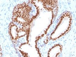
Biotium NKX3.1 (Metastatic Prostate Adenocarcinoma Marker)(NKX3.1/2576), CF568 conjugate, 0.1mg/mL
NKX3. 1 is a prostate specific gene encoding a transcription factor that plays an important role in normal prostate development and carcinogenesis. It is a prostatic tumor suppressor gene located on chromosome 8p21. 2, which frequently undergoes a loss of heterozygosity. NKX3. 1 expression is highly restricted in prostate epithelial cells and therefore can be used as a diagnostic biomarker for prostate cancer and other metastatic lesions of prostatic origin. Furthermore, NKX3. 1 shows better sensitivity than Prostate Specific Antigen (PSA) for identifying metastatic prostatic adenocarcinoma. This suggests that immunohistochemical staining of NKX3. 1, along with other prostate-restricted markers, may be valuable for the definitive determination of prostatic origin in poorly differentiated metastatic carcinomas. Primary antibodies are available purified, or with a selection of fluorescent CF Dyes and other labels. CF Dyes offer exceptional brightness and photostability. Note
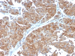
Biotium PAI-RBP1 / SERBP1 / SERPINE1(SERBP1/3498), 1mg/mL
PAI-RBP1 (plasminogen activator inhibitor 1 RNA-binding protein), also known as SERBP1 (SERPINE1 mRNA-binding protein 1), CGI-55, CHD3IP (chromodomain helicase DNA binding protein 3 interacting protein), HABP4L or PAI-RBP1, is a membrane-associated protein that localizes to the nucleus, the perinuclear region of the cytoplasm and the plasma membrane. PAI-RBP1 is believed to play a role in the regulation of mRNA stability, as it specifically binds to the CRS (cyclic nucleotide-responsive sequence) motif of the PAI-1 mRNA and acts to stabilize the mRNA and regulate its expression. In addition, PAI-RBP1 interacts with Mi2- and may be involved in chromatin remodeling. PAI-RBP1 also interacts with PGRMC1 and participates in the transduction of Progesterone's antiapoptotic action in ovarian cell types. The gene encoding PAI-RBP1 is overexpressed in ovarian cancer, suggesting a possible role for PAI-RBP1 in tumorigenesis and tumor metastasis. Primary antibodies are available purifie
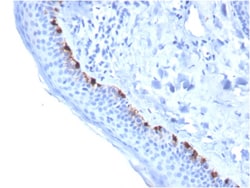
Biotium Tyrosinase-Related Protein-1 (TYRP-1) (Melanoma Marker)(TYRP1/3284), Biotin conjugate, 0.1mg/mL
Reacts with a 75 kDa melanocyte-specific gene product, identified as Tyrosinase-related protein-1 (TRP-1). It is involved in melanin synthesis. TRP1 is present on the melanosomal membranes of melanoma, normal melanocytes and nevi. Recent evidence suggests that TRP-1 is involved in maintaining stability of tyrosinase protein and modulating its catalytic activity. TRP-1 is also involved in maintenance of melanosome ultrastructure and affects melanocyte proliferation and cell death. Primary antibodies are available purified, or with a selection of fluorescent CF Dyes and other labels. CF Dyes offer exceptional brightness and photostability. Note: Conjugates of blue fluorescent dyes like CF405S and CF405M are not recommended for detecting low abundance targets, because blue dyes have lower fluorescence and can give higher non-specific background than other dye colors.

Biotium Sarcomeric Actinin Alpha 2 / ACTN2(ACTN2/3294), 0.2mg/mL
Alpha actinins belong to the spectrin gene superfamily which represents a diverse group of cytoskeletal proteins, including the alpha and beta spectrins and dystrophins. Alpha actinin is an actin-binding protein with multiple roles in different cell types. In non-muscle cells, the cytoskeletal isoform is found along microfilament bundles and adherens-type junctions, where it is involved in binding actin to the membrane. In contrast, skeletal, cardiac, and smooth muscle isoforms are localized to the Z-disc and analogous dense bodies, where they help anchor the myofibrillar actin filaments. This gene encodes a muscle-specific, alpha actinin isoform that is expressed in both skeletal and cardiac muscles. Several transcript variants encoding different isoforms have been found for this gene. Primary antibodies are available purified, or with a selection of fluorescent CF Dyes and other labels. CF Dyes offer exceptional brightness and photostability. Note: Conjugates of blue flu