
Mouse Secondary Antibodies

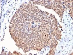
Biotium Fas (TNFRSF6) associated factor 1(CPTC-FAF1-2), CF488A conjugate, 0.1mg/mL
In contrast to growth factors which promote cell proliferation, FAS ligand (FAS-L) and the tumor necrosis factors (TNFs) rapidly induce apoptosis. Cellular response to FAS-L and TNF is mediated by structurally related receptors containing a conserved 'death domain' and belonging to the TNF receptor superfamily. TRADD, FADD and RIP are FAS/TNF-RI interacting proteins that contain a death domain homologous region (DDH). TRADD (TNF-RI-associated death domain) and FADD (FAS-associated death domain) associate with the death domains of both FAS and TNF-RI via their DDH regions, while RIP associates exclusively with FAS. An additional FAS interacting protein designated FAF1, for FAS-associated protein factor-1, binds with the cytoplasmic tail of wildtype but not LPR mutant FAS. When overexpressed in cells, FAF1 enhances the efficiency of FAS-mediated apoptosis. In contrast to TRADD, FADD and RIP, FAF1 lacks a DDH and cannot induce apoptosis independently of FAS activation. Primary antibo

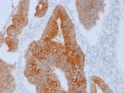
Biotium Aurora B (Proliferation Marker)(AURKB/1592), CF568 conjugate, 0.1mg/mL
This antibody recognizes a protein of 39 kDa, which is identified as Aurora kinase B (or Aurora B). Aurora B is a serine/threonine protein kinase and component of the chromosomal passenger complex (CPC), a complex that acts as a key regulator of mitosis. It is important for accurate chromosome segregation, cytokinesis, protein localization to the centromere and kinetochore, correct microtubule-kinetochore attachment, and regulation of the mitotic checkpoint. Aurora B forms a tight complex with inner centrosome protein and survivin. Inactivation of any of these proteins causes similar defects in chromosome segregation. A significant overexpression of Aurora B has been found in a variety of human tumors including non-small cell lung carcinoma, astrocytoma, seminoma and carcinomas of the colon, prostate, endometrium and thyroid. The expression level of Aurora B is associated with cell proliferation and prognosis in these tumors. Primary antibodies are available purified, or with
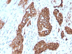
Biotium PDZ and LIM Domain 1(CPTC-PDLIM1-1), CF568 conjugate, 0.1mg/mL
CLP36, also known as PDLIM1, CLIM1 or Elfin, is a 329 amino acid cytoplasmic protein that associates with actin stress fibers at the cytoskeleton. Expressed at high levels in skeletal muscle and heart and at lower levels in colon, small intestine, spleen, lung, placenta, kidney, liver, thymus and pancreas, CLP36 functions as a cytoskeletal protein that is thought to act as an adaptor, bringing target proteins to the cytoskeleton. Specifically, CLP36 interacts with Clik1 (-actinin-1, thereby facilitating the association of Clik1 with actin stress fibers. CLP36 contains one PDZ domain and one LIM zinc-binding domain through which it conveys its protein-protein binding capabilities. Human CLP36 shares 88% sequence similarity with its rat counterpart, suggesting a conserved function between species. Primary antibodies are available purified, or with a selection of fluorescent CF Dyes and other labels. CF Dyes offer exceptional brightness and photostability. Note: Conjugates of blue

Biotium CD99 / MIC2 (Ewing s Sarcoma Marker) (MIC2/1495R), CF488A conjugate, 0.1mg/mL
This antibody recognizes a sialoglycoprotein of 27-32 kDa, identified as CD99, or MIC2 gene product, or E2 antigen. MIC2 gene is located in the pseudo-autosomal region of the human X and Y chromosome. MIC2 gene encodes two distinct proteins, which are produced by alternative splicing of the CD99 gene transcript and are identified as bands of 30 and 32 kDa (p30/32). Although its function is not fully understood, CD99 is implicated in various cellular processes including homotypic aggregation of T cells, upregulation of T cell receptor and MHS molecules, apoptosis of immature thymocytes and leukocyte diapedesis. CD99 is expressed on the cell membrane of some lymphocytes, cortical thymocytes, and granulosa cells of the ovary. Most pancreatic islet cells, Sertoli cells of the testis, and some endothelial cells express this antigen. Mature granulocytes express very little or no CD99. MIC2 is strongly expressed on Ewing s sarcoma cells and primitive peripheral neuroectodermal tumor

Biotium Desmoglein-1 (DSG1) (18D4), 0.2mg/mL
This antibody recognizes a protein of ~150 kDa, identified as Desmoglein-1 (DSG1). Desmoglein-1 is a member of the desmosomal cadherin family. Desmosomes are intercellular adhering junctions that represent cell surface attachment sites for intermediate filament. Desmocollins and desmogleins are the main desmosomal transmembrane proteins. Desmogleins consist of Dsg1, Dsg2, Dsg3, and Dsg4 isoforms. Within the desmosome, the extracellular domain of desmoglein is essential for calcium dependent heterophilic binding to the desmocollins, whereas the intracellular domain is essential for binding to the desmosomal plaque protein, plakoglobin. Desmoglein 1 is synthesized exclusively in the suprabasal layers. Intact and functionally active desmoglein 1 is essential to epidermal integrity.Primary antibodies are available purified, or with a selection of fluorescent CF Dyes and other labels. CF Dyes offer exceptional brightness and photostability. Note: Conjugates of blue fluorescent
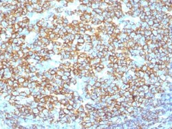
Biotium CD20 / MS4A1 (B-Cell Marker) (IGEL/1497R), CF568 conjugate, 0.1mg/mL
This antibody recognizes a protein of 30-33 kDa, which is identified as CD20. It is a non-Ig differentiation antigen of B-cells and its expression is restricted to normal and neoplastic B-cells, being absent from all other leukocytes and tissues. CD20 is expressed by pre B-cells and persists during all stages of B-cell maturation but is lost upon terminal differentiation into plasma cells. This MAb can be used for immunophenotyping of leukemia and malignant cells, B lymphocyte detection in peripheral blood and B cell localization in tissues. It reacts with the majority of B-cells present in peripheral blood and lymphoid tissues and their derived lymphomas. In lymphoid tissue, germinal center blasts and B-immunoblasts are particularly reactive. It is a reliable antibody for ascribing a B-cell phenotype in known lymphoid tissues. Rarely, CD20-positive T-cell lymphomas have been reported. Reactivity has also been noted with Reed-Sternberg cells in cases of Hodgkin's disease, par

Biotium Cytochrome C(CYCS/1010), CF488A conjugate, 0.1mg/mL
Cytochrome c is a well-characterized mobile electron transport protein that is essential to energy conversion in all aerobic organisms. In mammalian cells, this highly conserved protein is normally localized to the mitochondrial inter-membrane space. More recent studies have identified cytosolic cytochrome c as a factor necessary for activation of apoptosis. During apoptosis, cytochrome c is trans-located from the mitochondrial membrane to the cytosol, where it is required for activation of caspase-3 (CPP32). Overexpression of Bcl-2 has been shown to prevent the translocation of cytochrome c, thereby blocking the apoptotic process. Overexpression of Bax has been shown to induce the release of cytochrome c and to induce cell death. The release of cytochrome c from the mitochondria is thought to trigger an apoptotic cascade, whereby Apaf-1 binds to Apaf-3 (caspase-9) in a cytochrome c-dependent manner, leading to caspase-9 cleavage of caspase-3. This MAb recognizes total cytochrome
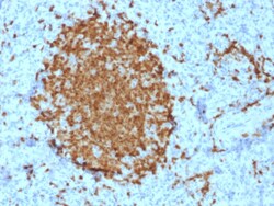
Biotium BOB.1 (B-Cell Marker)(BOB1/2421), 1mg/mL
BOB-1 is a transcriptional coactivator that specifically associates with either POU2F1/OCT1 or POU2F2/OCT2, boosting the promoter activity of those complexes. It is essential for the response of B-cells to antigens, required for the formation of germinal centers, and regilates IL-6 expression. BOB-1 expression in a variety of established B-cell lines, representing different stages of B-cell development, has suggested a constitutive, B-cell-specific expression pattern. LP cells in nodular lymphocyte predominant Hodgkin lymphoma, because they are germinal center-derived, are consistently immuno-positive for BOB-1. Conversely, only some cases of classical Hodgkin lymphoma show BOB-1 immuno-reactivity within the Hodgkin and Reed-Sternberg cells. Expression of BOB-1 has been reported in follicular center cell lymphoma, diffuse large B-cell lymphoma and some cases of acute myeloid leukemia. B-CLL, marginal zone lymphoma, and mantle cell lymphoma may show weak to moderate immunoreactivit
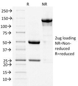
Biotium Cycloxygenase-2 (COX-2)(COX2/1941), CF568 conjugate, 0.1mg/mL
Prostaglandins are a diverse group of autocrine and paracrine hormones that mediate many cellular and physiologic processes. Prostaglandin H2 (PGH2) is an intermediate molecule in formation of the prostaglandins. Cyclooxygenase-1 (Cox-1) and cyclooxygenase-2 (Cox-2) are prostaglandin synthases that catalyze the formation of PGH2 from arachidonic acid (AA). Cox-1 and Cox-2 are isozymes of prostaglandin-endoperoxidase synthase (PTGS). Cox-1 is constitutively expressed in most tissues and is thought to serve in general housekeeping functions. Cox-2 is efficiently induced in migratory cells responding to pro-inflammatory stimuli and is considered to be an important mediator of inflammation. Both enzymes are targets for the nonsteroidal therapeutic anti-inflammatory drugs, NSAIDs. COX2 expression is significantly increased in 85-90% of human colorectal adenocarcinomas whereas levels of COX-1 are not changed. Primary antibodies are available purified, or with a selection of fluorescent
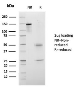
Biotium CELA3B / ELA3B (Pancreatic Function Marker)(rCELA3B/1811), 1mg/mL
This MAb recognizes a protein of ~30 kDa, identified as CELA3B (Chymotrypsin like elastase family member 3B). Elastases form a subfamily of serine proteases that hydrolyze many proteins in addition to elastin. Humans have six elastase genes which encode the structurally similar proteins elastase 1, 2, 2A, 2B, 3A, and 3B. Unlike other elastases, elastase 3B has little elastolytic activity. Like most of the human elastases, elastase 3B is secreted from the pancreas as a zymogen and, like other serine proteases such as trypsin, chymotrypsin and kallikrein; it has a digestive function in the intestine. Elastase 3B preferentially cleaves proteins after alanine residues. Elastase 3B may also function in the intestinal transport and metabolism of cholesterol. Both elastase 3A and elastase 3B have been referred to as protease E and as elastase 1, and excretion of this protein in fecal material is frequently used as a measure of pancreatic function. Primary antibodies are available pu
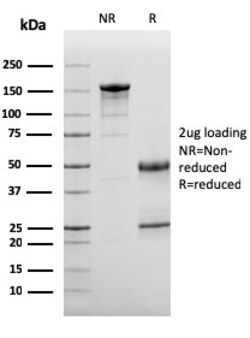
Biotium Complement 3d (C3d) (Acute Humoral Rejection Marker)(C3D/2891), Biotin conjugate, 0.1mg/mL
The complement component proteins, C2, C3, C4 and C5, are potent anaphylatoxins that are released during complement activation. Binding of these proteins to their respective G protein-coupled receptors, C3aR, C1R and C5aR, induces proinflammatory events, such as cellular degranulation, smooth muscle contraction, arachidonic acid metabolism, cytokine release, leukocyte activation and cellular chemotaxis. C3d is a terminal degradation product of C3 that plays an important role in modulation of the adaptive immune response through the interaction with complement receptor type 2 (CR2). CR2 is important in the switched-isotype, high-affinity and memory humoral immune responses to T-dependent foreign antigens, as well as in the development of the natural antibody repertoire. This pH- and ionic strength-dependent association of C3d with CR2 represents a link between innate and adaptive immunity. Primary antibodies are available purified, or with a selection of fluorescent CF Dyes a
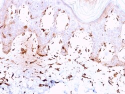
Biotium Collagen VII(LH7.2), 0.2mg/mL
Recognizes an epitope located on collagenase digested Type VII collagen (150 kDa), i. e. the non-helical carboxyl terminal region of the dimer. Binding is localized at the inferior border of the lamina densa as demonstrated by immunoelectron microscopy. Type VII is also found in the retina, where its function is unknown. It interacts with Laminin 5 and Fibronectin. Collagen VII is a stratified squamous epithelial basement membrane protein that forms anchoring fibrils which may contribute to epithelial basement membrane organization and adherence by interacting with extracellular matrix (ECM) proteins such as type IV collagen. Primary antibodies are available purified, or with a selection of fluorescent CF Dyes and other labels. CF Dyes offer exceptional brightness and photostability. Note: Conjugates of blue fluorescent dyes like CF405S and CF405M are not recommended for detecting low abundance targets, because blue dyes have lower fluorescence and can give higher non-specifi

Biotium Desmoglein-1 (DSG1) (18D4), CF568 conjugate, 0.1mg/mL
This antibody recognizes a protein of ~150 kDa, identified as Desmoglein-1 (DSG1). Desmoglein-1 is a member of the desmosomal cadherin family. Desmosomes are intercellular adhering junctions that represent cell surface attachment sites for intermediate filament. Desmocollins and desmogleins are the main desmosomal transmembrane proteins. Desmogleins consist of Dsg1, Dsg2, Dsg3, and Dsg4 isoforms. Within the desmosome, the extracellular domain of desmoglein is essential for calcium dependent heterophilic binding to the desmocollins, whereas the intracellular domain is essential for binding to the desmosomal plaque protein, plakoglobin. Desmoglein 1 is synthesized exclusively in the suprabasal layers. Intact and functionally active desmoglein 1 is essential to epidermal integrity.Primary antibodies are available purified, or with a selection of fluorescent CF Dyes and other labels. CF Dyes offer exceptional brightness and photostability. Note: Conjugates of blue fluorescent
Biotium CD11c (Dendritic Cell Marker) ( ITGAX/1243) , CF647 conjugate, 0.1mg/mL
This antibody recognizes a protein of 145 kDa, identified as CD11c. CD11c (ITGAX), a member of the leukointegrin family, shares the same beta subunit with other members of the leukocyte adhesion molecule family, which includes CD11a (LFA-1), CD11b (MAC-1) and CD11d (ITGAD), but has a unique alpha chain. CD11c has been shown to play a role in phagocytosis, cell migration, and cytokine production by monocytes/macrophages as well as induction of T-cell proliferation by Langerhans cells. CD11c is expressed prominently on the plasma membranes of monocytes, tissue macrophages, NK cells, and most dendritic cells (DCs). A lower level of expression is also observed on neutrophils as a result of its high level of expression on most DCs. An antibody to CD11c may aid in identification of lesions with histiocytic origin. It may also been used as a marker for hairy cell leukemia in paraffin-embedded tissues.Primary antibodies are available purified, or with a selection of fluorescent CF Dyes a
Biotium N-Cadherin / Cadherin-2 / CD325 (NCAD) (13A9), CF647 conjugate, 0.1mg/mL
This antibody recognizes a protein of ~140 kDa, identified as N-Cadherin (NCAD), also known as CD325. NCAD is a member of the Cadherin superfamily, and consists of five extracellular repeats, a transmembrane domain and a cytoplasmic domain. CD325 deficient mice die at day 10 of gestation and embryos display major heart defects and malformed neural tubes and somites. Consistent with this, CD325 has been implicated in several aspects of cardiac development including the precardiac mesoderm, establishment of left-right symmetry and cardiac looping morphogenesis. Furthermore, CD325 is normally involved in inducing cell cycle arrest and its expression is frequently deregulated in cancer cells. Studies have linked N-cadherin to cancer metastasis by showing the aggressive tumor cells had preferentially turned on N-cadherin as opposed to E- or P-cadherin.Primary antibodies are available purified, or with a selection of fluorescent CF Dyes and other labels. CF Dyes offer exceptional