
Mouse Secondary Antibodies

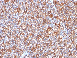
Biotium Spectrin beta III (SPTBN2) (SPTBN2/1778), CF740 conjugate, 0.1mg/mL
Spectrin is an actin binding protein that is a major component of the plasma membrane skeleton. Spectrins function as membrane organizers and stabilizers by forming dimers, tetramers and higher polymers. Vertebrate spectrins have two alpha-subunits (alpha-I/alpha-II), four beta-subunits (beta-I-beta-IV) and a beta-H subunit creating diversity and specialization of function. Spectrin α and spectrinβ are present in erythrocytes, whereas spectrin α II (also designated fodrin α) and spectrinβ I (also designated fodrinβ) are present in other somatic cells. The spectrin tetramers in erythrocytes act as barriers to lateral diffusion, but spectrin dimers seem to lack this function. Spectrinβ III is highly homologous to both spectrinβ I and spectrinβ II. Spectrinβ III is highly expressed in brain, kidney, pancreas and liver, and at lower levels in lung and placenta. Spectrin beta 3 is primarily expressed in nervous tissues with high

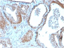
Biotium TIGIT / VSTM3 / VSIG9 (Immune Checkpoint for Cancer)(TIGIT/3106), CF568 conjugate, 0.1mg/mL
TIGIT is a checkpoint inhibitor which binds with high affinity to the poliovirus receptor (PVR), causing increased IL10 secretion, decreased IL12B secretion. TIGIT binding to PVR also causes the suppression of T cell activation by promoting the generation of mature immuno-regulatory dendritic cells. It is expressed at low levels on natural killer (NK) cells, as well as peripheral memory and regulatory CD4 T cells. At the protein level, it is upregulated following the activation of these cells. Functionally, TIGIT is similar to CTLA4. The ligands for TIGIT include CD155 (signal abrogation) and CD226 (signal stimulation). It has been demonstrated to be upregulated on T cells in many cancers and is a immuno-oncology target for therapy. Primary antibodies are available purified, or with a selection of fluorescent CF Dyes and other labels. CF Dyes offer exceptional brightness and photostability. Note: Conjugates of blue fluorescent dyes like CF405S and CF405M are not recomme
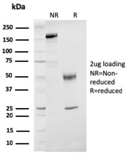
Biotium ER-beta-1 (Estrogen Receptor beta-1)(ESR2/3207), CF647 conjugate, 0.1mg/mL
Estrogen receptors (ER) are members of the steroid/thyroid hormone receptor superfamily of ligand-activated transcription factors. Estrogen receptors, including ER-alpha and ER-beta, contain DNA binding and ligand binding domains and are critically involved in regulating the normal function of reproductive tissues. They are located in the nucleus, though some estrogen receptors associate with the cell surface membrane and can be rapidly activated by exposure of cells to estrogen. ER-alpha and ER-beta are differentially activated by various ligands. Receptor-ligand interactions trigger a cascade of events, including dissociation from heat shock proteins, receptor dimerization, phosphorylation and the association of the hormone activated receptor with specific regulatory elements in target genes. Evidence suggests that ER-alpha and ER-beta may be regulated by distinct mechanisms even though they share many functional characteristics. Primary antibodies are available purified, o

Biotium CD20 / MS4A1 (B-Cell Marker)(MS4A1/3409), CF568 conjugate, 0.1mg/mL
Recognizes a protein of 30-33 kDa, which is identified as CD20. It is a non-Ig differentiation antigen of B-cells and its expression is restricted to normal and neoplastic B-cells, being absent from all other leukocytes and tissues. CD20 is expressed by pre-B-cells and persists during all stages of B-cell maturation but is lost upon terminal differentiation into plasma cells. This MAb can be used for immunophenotyping of leukemia and malignant cells, B lymphocyte detection in peripheral blood and B cell localization in tissues. It reacts with the majority of B-cells present in peripheral blood and lymphoid tissues and their derived lymphomas. In lymphoid tissue, germinal center blasts and B-immunoblasts are particularly reactive. It is a reliable antibody for ascribing a B-cell phenotype in known lymphoid tissues. Rarely, CD20-positive T-cell lymphomas have been reported. Reactivity has also been noted with Reed-Sternberg cells in cases of Hodgkin's disease, particularly of l
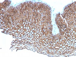
Biotium Desmoglein-3 (Squamous Cell Marker)(DSG3/2837), CF740 conjugate, 0.1mg/mL
Recognizes a protein of 130 kDa, identified as Desmoglein-3 (DSG3). This MAb is highly specific to Desmoglein-3 and does not cross-react with other members of the Desmoglein-family. DSG3 is a calcium-binding transmembrane glycoprotein component of desmosomes in vertebrate epithelial cells. Research has shown that DSG3 has a very high sensitivity (80%) and specificity (100%) in recognizing squamous cell carcinoma (SqCC). Therefore, DSG3 is considered a very important marker for lung SqCC and can be a useful ancillary marker to separate SqCC from other subtypes of lung cancer. Moreover, studies have shown that DSG3 expression in lung SqCC may indicate a poor prognosis. Primary antibodies are available purified, or with a selection of fluorescent CF Dyes and other labels. CF Dyes offer exceptional brightness and photostability. Note: Conjugates of blue fluorescent dyes like CF405S and CF405M are not recommended for detecting low abundance targets, because blue dyes have low
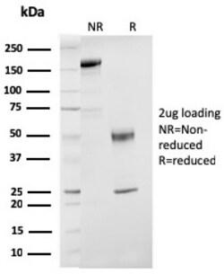
Biotium ER-beta-1 (Estrogen Receptor beta-1)(ESR2/3207), 1mg/mL
Estrogen receptors (ER) are members of the steroid/thyroid hormone receptor superfamily of ligand-activated transcription factors. Estrogen receptors, including ER-alpha and ER-beta, contain DNA binding and ligand binding domains and are critically involved in regulating the normal function of reproductive tissues. They are located in the nucleus, though some estrogen receptors associate with the cell surface membrane and can be rapidly activated by exposure of cells to estrogen. ER-alpha and ER-beta are differentially activated by various ligands. Receptor-ligand interactions trigger a cascade of events, including dissociation from heat shock proteins, receptor dimerization, phosphorylation and the association of the hormone activated receptor with specific regulatory elements in target genes. Evidence suggests that ER-alpha and ER-beta may be regulated by distinct mechanisms even though they share many functional characteristics. Primary antibodies are available purified, o

Cayman Chemical EPA Oxylipin MaxSpec LC-MS M
A mixture that contains PGE3, PGF3α, 5(S)-HEPE, (±)8-HEPE, (±)9-HEPE, (±)11-HEPE, 12(S)-HEPE, 15(S)-HEPE, and (±)18-HEPE

Biotium ATRX / RAD54 (Alpha Thalassemia Mental Retardation) (ATRX/2900R), CF740 conjugate, 0.1mg/mL
ATRX is a member of the Snf2 family of helicase/ATPases, which contribute to the remodeling of the nucelosome structure in an ATP-dependent manner, and facilitate the initiation of transcription and replication. Structurally, ATRX contains a PHD zinc finger motif. ATRX is regulated throughout the cell cycle where it is differentially distributed within the nucleus. During interphase, ATRX predominately associates with the nuclear matrix, while during mitosis, ATRX localizes with condensed chromatin. At the onset of M phase, phosphorylation rapidly induces this redistribution of ATRX to the short arms of human acrocentric chromosomes, where it then specifically complexes with heterochromatin protein 1 alpha to mediate chromosomal segregation. Mutations in the ATRX gene correlate with a high incidence of severe X-linked form of syndromal mental retardation associated with alpha-thalassemia or ATRX syndrome. Primary antibodies are available purified, or with a selection of fluorescen
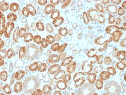
Biotium Occludin (OCLN) (Tight Junctions Marker)(OCLN/2183), 1mg/mL
Occludin is a tetraspan integral membrane protein in epithelial and endothelial tight junction (TJ) structures that can contain two extracellular loops. The protein exists in a variety of phosphorylated forms. Phosphorylation is involved in regulating both the localization and the function of Occludin. Expression of Occludin is upregulated by polyunsaturated fatty acids, increasing trans-endothelial cell resistance and reducing cellular permeability to large molecules. The level of Occludin varies greatly depending on tissue; in brain tissue, Occludin is highly expressed at cell-cell contact sites. Non-neural tissues show lower expression and discontinuous distribution. Up-regulation of epithelial Occludin may play a role in enhancing paracellular permeability and be related to the damage to the tight junction. Primary antibodies are available purified, or with a selection of fluorescent CF Dyes and other labels. CF Dyes offer exceptional brightness and photostability. Note: Co
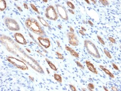
Biotium Occludin (Marker of Early Blood Brain Barrier Damage)(OCLN/2181), CF647 conjugate, 0.1mg/mL
Occludin is a tetraspan integral membrane protein in epithelial and endothelial tight junction (TJ) structures that can contain two extracellular loops. The protein exists in a variety of phosphorylated forms. Phosphorylation is involved in regulating both the localization and the function of Occludin. Expression of Occludin is upregulated by polyunsaturated fatty acids, increasing trans-endothelial cell resistance and reducing cellular permeability to large molecules. The level of Occludin varies greatly depending on tissue; in brain tissue, Occludin is highly expressed at cell-cell contact sites. Non-neural tissues show lower expression and discontinuous distribution. Occludin is a tight junction protein that is a key structural component of the blood-brain barrier (BBB). Degradation of Occludin is frequently seen in ischemic stroke and contributes to BBB disruption. Primary antibodies are available purified, or with a selection of fluorescent CF Dyes and other labels. CF Dyes
Biotium Alkaline Phosphatase (Placental) / PLAP (Germ Cell Tumor Marker) (ALPP/2899R), CF568 conjugate, 0.1mg/mL
This antibody reacts with a 70 kDa membrane-bound isozyme (Regan and Nagao type) of Placental Alkaline Phosphatase (PLAP) occurring in the placenta during the 3rd trimester of gestation. It is highly specific for PLAP and shows no cross-reaction with other isozymes of alkaline phosphatase. Anti-PLAP This antibody reacts with germ cell tumors and can discriminate between these and other neoplasms. Somatic neoplasms e.g. breast, gastrointestinal, prostatic, and urinary cancers may also immunoreact with antibodies to PLAP. Anti-PLAP positivity in conjunction with anti-keratin negativity favors seminoma over carcinoma. Germ cell tumors are usually anti-keratin positive, but they regularly fail to stain with anti-EMA, whereas most carcinomas stain with anti-EMA. Anti-PLAP has been useful in the diagnosis of gestational trophoblastic disease.Primary antibodies are available purified, or with a selection of fluorescent CF Dyes and other labels. CF Dyes offer exceptional brightness
Biotium Ki-67 (Proliferating Cell Marker) (MKI67/2465), CF740 conjugate, 0.1mg/mL
Ki-67 antigen is a nuclear, non-histone protein that is present in all stages of the cell cycle except G0. This characteristic makes Ki-67 an excellent marker for proliferating cells and is commonly used as one of the prognostic factors in cancer studies. A correlation has been demonstrated between Ki-67 index and the histo-pathological grade of neoplasms. Assessment of Ki-67 expression in renal and ureter tumors shows a correlation between tumor proliferation and disease progression, thus making it possible to differentiate high-risk patients. Ki-67 expression may also prove to be important for distinguishing between malignant and benign peripheral nerve sheath tumors. Ki-67 labeling index has been shown to be a prognostic marker in a number of neoplasms including grade II astrocytoma, oligodendroglioma, colon carcinoma, and breast carcinoma. In general, Ki-67 is a good marker of proliferating cell populations.Primary antibodies are available purified, or with a selection of

Biotium Small Cell Lung Cancer (SCLC)(MOC-52), CF740 conjugate, 0.1mg/mL
This MAb reacts with a membrane-associated protein present in normal and malignant neuroendocrine tissues including small cell lung cancer (SCLC). It stains neural and a variable number of endocrine tissues and in the lung it reacts preferentially with SCLC and carcinoids. Its epitope is destroyed during formalin fixation. This antibody was categorized during the First International Workshop on Small Cell Lung Cancer Antigens held in London in April 1987. There are two major types of Lung Carcinoma: non-small cell, which accounts for 80% of all cases; and small cell, which accounts for roughly 20% of all lung cancers reported. The lung continues to be a customary place for cancer migration from tumors elsewhere in the body. Treatment depends on the specific cell type of the cancer, level of progression and status of the individual patient.Primary antibodies are available purified, or with a selection of fluorescent CF Dyes and other labels. CF Dyes offer exceptional brightn
Biotium CELA3B / ELA3B (Pancreatic Function Marker) (CELA3B/1757), 0.2mg/mL
This MAb recognizes a protein of ~30 kDa, identified as CELA3B (Chymotrypsin like elastase family member 3B). Elastases form a subfamily of serine proteases that hydrolyze many proteins in addition to elastin. Humans have six elastase genes which encode the structurally similar proteins elastase 1, 2, 2A, 2B, 3A, and 3B. Unlike other elastases, elastase 3B has little elastolytic activity. Like most of the human elastases, elastase 3B is secreted from the pancreas as a zymogen and, like other serine proteases such as trypsin, chymotrypsin and kallikrein; it has a digestive function in the intestine. Elastase 3B preferentially cleaves proteins after alanine residues. Elastase 3B may also function in the intestinal transport and metabolism of cholesterol. Both elastase 3A and elastase 3B have been referred to as protease E and as elastase 1, and excretion of this protein in fecal material is frequently used as a measure of pancreatic function.Primary antibodies are available purified
Biotium Tyrosinase (Melanoma Marker) (TYR/2024R), Biotin conjugate, 0.1mg/mL
This antibody recognizes a cluster of proteins between 70-80 kDa, identified as tyrosinase. Occasionally a minor band at 55 kDa is also detected. This MAb shows no cross-reaction with MAGE-1 and tyrosinase-related protein 1, TRP-1/gp75. Tyrosinase is a copper-containing metalloglycoprotein that catalyzes several steps in the melanin pigment biosynthetic pathway; the hydroxylation of tyrosine to L-3,4-dihydroxy-phenylalanine (dopa), and the subsequent oxidation of dopa to dopaquinone. Mutations of the tyrosinase gene occur in various forms of albinism. Tyrosinase is one of the targets for cytotoxic T-cell recognition in melanoma patients. Staining of melanomas with this MAb shows tyrosinase in melanotic as well as amelanotic variants. This MAb is a useful marker for melanocytes and melanomas.Primary antibodies are available purified, or with a selection of fluorescent CF Dyes and other labels. CF Dyes offer exceptional brightness and photostability. Note: Conjugates of blue fluo