
Mouse Secondary Antibodies

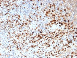
Biotium HSV1 (Herpes Simplex Virus Type I)(N/A), CF740 conjugate, 0.1mg/mL
The antibody reacts with HSV type 1 specific antigen. It is suitable for detection of HSV in human cellular material obtained from superficial lesions or biopsies and for the early identification of HSV in infected tissue cultures. The herpes simplex virus (HSV) (also known as cold sore, night fever or fever blister) is a virus that causes a contagious disease. There are two main types of Herpes Simplex Virus (HSV), 1 and 2. The HSV-1 strain generally appears in the orafacial organs. HSV2 usually resides in the sacral ganglion at the base of the spine. All herpes viruses are morphologically identical: they have a large double-stranded DNA genome and the virion consists of an icosahedral nucleo-capsid, which is surrounded by a lipid bilayer envelope. Primary antibodies are available purified, or with a selection of fluorescent CF Dyes and other labels. CF Dyes offer exceptional brightness and photostability. Note: Conjugates of blue fluorescent dyes like CF405S and CF405M


Biotium Parathyroid Hormone (PTH) (N-Terminal), CF568 conjugate, 0.1mg/mL
Epitope of this PAb maps in the N-terminus of PTH, a hormone produced by the parathyroid gland that regulates the concentration of calcium and phosphorus in extracellular fluid. This hormone elevates blood Ca2 levels by dissolving the salts in bone and preventing their renal excretion. It is produced in the parathyroid gland as an 84 amino acid single chain polypeptide. It can also be secreted as N-terminal truncated fragments or C-terminal fragments after intracellular degradation, as in case of hypercalcemia. Defects in this gene are a cause of familial isolated hypoparathyroidism (FIH); also called autosomal dominant hypoparathyroidism or autosomal dominant hypocalcemia. FIH is characterized by hypocalcemia and hyperphosphatemia due to inadequate secretion of parathyroid hormone. Symptoms are seizures, tetany and cramps. FIH exist both as autosomal dominant and recessive forms of hypoparathyroidism. Primary antibodies are available purified, or with a selection of fluores
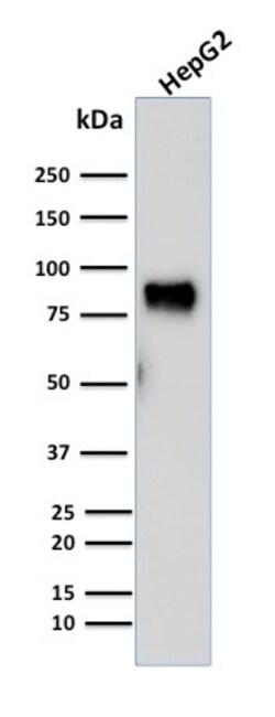
Biotium MDM2(SMP14), CF740 conjugate, 0.1mg/mL
MDM2 is a nuclear phosphoprotein that binds and inhibits transactivation by tumor protein p53. It can promote tumor formation by targeting tumor suppressor proteins, such as p53, for proteasomal degradation. Overexpression of MDM2 can result in excessive inactivation of tumor protein p53, diminishing its tumor suppressor function. This protein also affects the cell cycle, apoptosis, and tumorigenesis through interactions with other proteins, including retinoblastoma 1 and ribosomal protein L5. Overexpression of MDM2 protein is detected in a variety of cancers. Primary antibodies are available purified, or with a selection of fluorescent CF Dyes and other labels. CF Dyes offer exceptional brightness and photostability. Note: Conjugates of blue fluorescent dyes like CF405S and CF405M are not recommended for detecting low abundance targets, because blue dyes have lower fluorescence and can give higher non-specific background than other dye colors.
Biotium TIMP2 (Tissue Inhibitor of Metalloproteinase 2) (3A4), CF568 conjugate, 0.1mg/mL
This antibody recognizes a protein of 21 kDa, identified as tissue inhibitor of metalloproteinases-2 (TIMP-2). It is closely related to TIMP-1 and shows the highest binding affinity to both the latent (pro) and active forms of 72 kDa Type IV collagenase (also known as MMP-2 or gelatinase A). It also has affinity for the active form of 92 kDa Type IV collagenase (also known as MMP-9 or gelatinase B). TIMP-2 inhibits the proteolytic invasiveness of tumor cells and normal placental trophoblast cells.Primary antibodies are available purified, or with a selection of fluorescent CF Dyes and other labels. CF Dyes offer exceptional brightness and photostability. Note: Conjugates of blue fluorescent dyes like CF405S and CF405M are not recommended for detecting low abundance targets, because blue dyes have lower fluorescence and can give higher non-specific background than other dye colors.
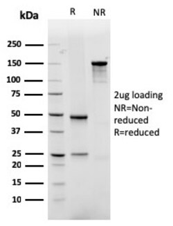
Biotium GLUT-1 (Tumor Progression and Mesothelioma Marker)(rGLUT1/2476), 1mg/mL
Recognizes a protein of 55 kDa, which is identified as GLUT-1. Glucose transporters are integral membrane glycoproteins involved in transporting glucose into most cells. There are many types of glucose transport carrier proteins, designated as Glut-1 to Glut-12. Glut-1 is a major glucose transporter in the mammalian blood-brain barrier. It is expressed in high density on the membranes of human erythrocytes and the brain capillaries that comprise the blood-brain barrier. Glut-1 is expressed at variable levels in many human tissues. Overexpression of Glut-1 has been linked to tumor progression or poor survival of patients with carcinomas of the colon, breast, cervical, lung, bladder and mesothelioma. Glut-1 is a sensitive and specific marker for the differentiation of malignant mesothelioma (positive) from reactive mesothelium (negative). Primary antibodies are available purified, or with a selection of fluorescent CF Dyes and other labels. CF Dyes offer exceptional brightnes
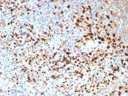
Biotium HSV1 (Herpes Simplex Virus Type I)(HSV1/1934), 1mg/mL
The antibody reacts with HSV type 1 specific antigen. It is suitable for detection of HSV in human cellular material obtained from superficial lesions or biopsies and for the early identification of HSV in infected tissue cultures. The herpes simplex virus (HSV) (also known as cold sore, night fever or fever blister) is a virus that causes a contagious disease. There are two main types of Herpes Simplex Virus (HSV), 1 and 2. The HSV-1 strain generally appears in the orafacial organs. HSV2 usually resides in the sacral ganglion at the base of the spine. All herpes viruses are morphologically identical: they have a large double-stranded DNA genome and the virion consists of an icosahedral nucleo-capsid, which is surrounded by a lipid bilayer envelope.Primary antibodies are available purified, or with a selection of fluorescent CF Dyes and other labels. CF Dyes offer exceptional brightness and photostability. Note: Conjugates of blue fluorescent dyes like CF405S and CF405M
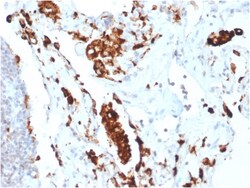
Biotium Napsin A (Lung Adenocarcinoma Marker)(NAPSA/1865R), 0.2mg/mL
Napsin is a pepsin-like aspartic proteinase connected with maturation of surfactant protein B. There are two closely related napsins, napsin A and napsin B. Napsin A is expressed as a single chain protein. Immunohistochemical studies revealed high expression levels of napsin A in human lung and kidney but low expression in spleen. Napsin A is expressed in type II pneumocytes and in adenocarcinomas of lung. The high specificity expression of napsin A in adenocarcinomas of lung is useful to distinguish primary lung adenocarcinomas from adenocarcinomas of other organs. Primary antibodies are available purified, or with a selection of fluorescent CF Dyes and other labels. CF Dyes offer exceptional brightness and photostability. Note: Conjugates of blue fluorescent dyes like CF405S and CF405M are not recommended for detecting low abundance targets, because blue dyes have lower fluorescence and can give higher non-specific background than other dye colors.
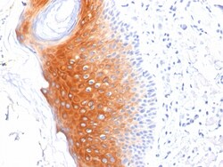
Biotium Cytokeratin 1 (Suprabasal Keratinocyte Marker)(LHK1), Biotin conjugate, 0.1mg/mL
Keratins are a family of intermediate filament proteins that assemble into filaments through forming heterodimers of one type I keratin (keratins 9 to 23) and one type II keratin (keratins 1 to 8). Keratins demonstrate tissue and differentiation specific expression profiles. Cytokeratin 1 is a hetero-tetramer of two type I as well as two type II keratins, keratin-10 (KRT10). Keratin 1 is differentiation specific keratin that is one of the predominant keratins in suprabasal keratinocytes in stratified epithelia. Mutations in keratin 1 cause epidermolytic hyperkeratosis. Primary antibodies are available purified, or with a selection of fluorescent CF Dyes and other labels. CF Dyes offer exceptional brightness and photostability. Note: Conjugates of blue fluorescent dyes like CF405S and CF405M are not recommended for detecting low abundance targets, because blue dyes have lower fluorescence and can give higher non-specific background than other dye colors.
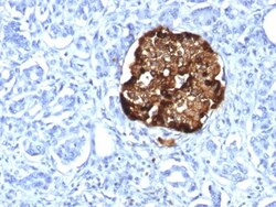
Biotium Chromogranin A / CHGA (Neuroendocrine Marker) (CHGA/1815R), CF488A conjugate, 0.1mg/mL
Chromogranin A is present in neuroendocrine cells throughout the body, including the neuroendocrine cells of the large and small intestine, adrenal medulla and pancreatic islets. It is an excellent marker for carcinoid tumors, pheochromocytomas, paragangliomas, and other neuroendocrine tumors. Co-expression of chromogranin A and neuron specific enolase (NSE) is common in neuroendocrine neoplasms. Reportedly, co-expression of certain keratins and chromogranin indicates neuroendocrine lineage. The presence of strong anti-chromogranin staining and absence of anti-keratin staining should raise the possibility of paraganglioma. The co-expression of chromogranin and NSE is typical of neuroendocrine neoplasms. Most pituitary adenomas and prolactinomas readily express chromogranin.Primary antibodies are available purified, or with a selection of fluorescent CF Dyes and other labels. CF Dyes offer exceptional brightness and photostability. Note: Conjugates of blue fluorescent dyes
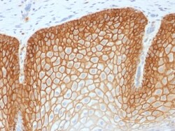
Biotium CD44v4 (Marker of Tumor Metastasis) (CD44v4/1700R), CF740 conjugate, 0.1mg/mL
This antibody recognizes an epitope encoded by exon v4 on the variant portion of human CD44. The CD44 molecule belongs to a family of cellular adhesion molecules found on a wide range of normal and malignant cells in epithelial, mesothelial and hemopoiesis tissues. CD44 is a single gene with 20 exons, of which 10 are normally expressed to encode the basic CD44 (H-CAM) molecule. The additional 10 exons (v1 to v10) are only expressed by alternative splicing of the nuclear RNA. The expression of specific cell adhesion molecule CD44 splice variants has been reported to be associated with metastasis in certain human malignancies.Primary antibodies are available purified, or with a selection of fluorescent CF Dyes and other labels. CF Dyes offer exceptional brightness and photostability. Note: Conjugates of blue fluorescent dyes like CF405S and CF405M are not recommended for detecting low abundance targets, because blue dyes have lower fluorescence and can give higher non-spec
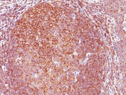
Biotium BCL10 (MALT-Lymphoma Marker)(151), CF568 conjugate, 0.1mg/mL
B-cell lymphoma/leukemia 10 (BCL10) is a signaling protein that promotes apoptosis, pro-caspase-9 maturation, and activation of NF-kappa-B via NIK and IKK. BCL10 contains an N-terminal caspase recruitment domain (CARD), which is found in a number of apoptotic regulatory molecules. BCL10 is involved in the adaptive immune response, and may be an adapter protein between upstream TNFR1-TRADD-RIP complex and the downstream NIK-IKK-IKAP complex. A BCL10 translocation is recurrent in low-grade mucosa-associated lymphoid tissue (MALT lymphoma). Primary antibodies are available purified, or with a selection of fluorescent CF Dyes and other labels. CF Dyes offer exceptional brightness and photostability. Note: Conjugates of blue fluorescent dyes like CF405S and CF405M are not recommended for detecting low abundance targets, because blue dyes have lower fluorescence and can give higher non-specific background than other dye colors.
Biotium Chromogranin A / CHGA (Neuroendocrine Marker), CF647 conjugate, 0.1mg/mL
Chromogranin A is present in neuroendocrine cells throughout the body, including the neuroendocrine cells of the large and small intestine, adrenal medulla and pancreatic islets. It is an excellent marker for carcinoid tumors, pheochromocytomas, paragangliomas, and other neuroendocrine tumors. Co-expression of chromogranin A and neuron specific enolase (NSE) is common in neuroendocrine neoplasms. Reportedly, co-expression of certain keratins and chromogranin indicates neuroendocrine lineage. The presence of strong anti-chromogranin staining and absence of anti-keratin staining should raise the possibility of paraganglioma. The co-expression of chromogranin and NSE is typical of neuroendocrine neoplasms. Most pituitary adenomas and prolactinomas readily express chromogranin. Primary antibodies are available purified, or with a selection of fluorescent CF Dyes and other labels. CF Dyes offer exceptional brightness and photostability. Note: Conjugates of blue fluorescent dyes
Biotium AIF1 / Iba1 (Microglia Marker) (AIF1/1909), Biotin conjugate, 0.1mg/mL
Allograft Inflammatory Factor 1 (also known as AIF1 or IBA1) is a cytoplasmic, calcium-binding protein that is thought to play a role in macrophage activation and function. AIF1, containing two EF domains, is induced by cytokines and Interferons. In an unstimulated state, AIF1 colocalizes with actin, and upon stimulation, translocates to lamellipodia. It is also a marker of human microglia and is expressed by macrophages in injured skeletal muscle. The gene encoding AIF1 resides in the tumor necrosis factor (TNF) cluster of genes, located in the region represented by the human major histocompatibility complex (MHC).Primary antibodies are available purified, or with a selection of fluorescent CF Dyes and other labels. CF Dyes offer exceptional brightness and photostability. Note: Conjugates of blue fluorescent dyes like CF405S and CF405M are not recommended for detecting low abundance targets, because blue dyes have lower fluorescence and can give higher non-specific
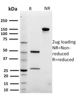
Biotium ARF1 (Golgi Apparatus Marker)(3F1), CF568 conjugate, 0.1mg/mL
The ADP-ribosylation factor (ARF) family comprises a group of structurally and functionally conserved proteins, which are members of the Ras superfamily of regulatory GTP-binding proteins. The ARF family is divided functionally into the ARF and the ARF-like proteins. ARF's share more than 60% sequence identity, appear to be ubiquitous in eukaryotes, and are highly conserved evolutionarily. ARF is involved in intracellular protein traffic to and within the Golgi complex. ARF has a number of disparate activities including maintenance of organelle integrity, assembly of coat proteins, as a co-factor for cholera toxin and as an activator of phospholipase D. Primary antibodies are available purified, or with a selection of fluorescent CF Dyes and other labels. CF Dyes offer exceptional brightness and photostability. Note: Conjugates of blue fluorescent dyes like CF405S and CF405M are not recommended for detecting low abundance targets, because blue dyes have lower fluorescenc
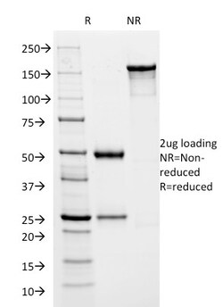
Biotium HSV1 (Herpes Simplex Virus Type I) ICP8(HSVI/2045), CF568 conjugate, 0.1mg/mL
The antibody reacts with HSV type 1 specific antigen. It is suitable for detection of HSV in human cellular material obtained from superficial lesions or biopsies and for the early identification of HSV in infected tissue cultures. The herpes simplex virus (HSV) (also known as cold sore, night fever or fever blister) is a virus that causes a contagious disease. There are two main types of Herpes Simplex Virus (HSV), 1 and 2. The HSV-1 strain generally appears in the orafacial organs. HSV2 usually resides in the sacral ganglion at the base of the spine. All herpes viruses are morphologically identical: they have a large double-stranded DNA genome and the virion consists of an icosahedral nucleo-capsid, which is surrounded by a lipid bilayer envelope. ICP8, the HSV1 encoded single-strand DNA (ssDNA)-binding protein, is the major DNA binding protein of HSV1. ICP8 promotes single-stranded DNA to assemble into a homologous duplex plasmid producing a displacement loop. At higher concent