
Mouse Secondary Antibodies

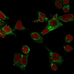
Biotium GPX4 / MCSP(LHM 2), Biotin conjugate, 0.1mg/mL
The antioxidant enzyme glutathione peroxidase 4 (GPx4) catalyzes the reduction of peroxides through oxidation of cellular glutathione.Primary antibodies are available purified, or with a selection of fluorescent CF Dyes and other labels. CF Dyes offer exceptional brightness and photostability. Note: Conjugates of blue fluorescent dyes like CF405S and CF405M are not recommended for detecting low abundance targets, because blue dyes have lower fluorescence and can give higher non-specific background than other dye colors.

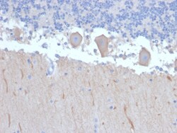
Biotium ATG5 (Autophagy Marker)(ATG5/2492), CF740 conjugate, 0.1mg/mL
Autophagy protein 5 (ATG5), in combination with autophagy protein 12, functions as an E1-like activating enzyme in a ubiquitin-like conjugating system. ATG5 is involved in several cellular processes, including autophagic vesicle formation, mitochondrial quality control after oxidative damage, negative regulation of the innate antiviral immune response, lymphocyte development and proliferation, MHC II antigen presentation, adipocyte differentiation, and apoptosis. The ATG5 protein is essential for autophagy; a process that is usually beneficial for cells to self-degrade their own components when they are no longer useful. Primary antibodies are available purified, or with a selection of fluorescent CF Dyes and other labels. CF Dyes offer exceptional brightness and photostability. Note: Conjugates of blue fluorescent dyes like CF405S and CF405M are not recommended for detecting low abundance targets, because blue dyes have lower fluorescence and can give higher non-specifi
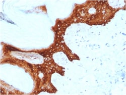
Biotium 14-3-3 Sigma / Stratifin(CPTC-SFN-2), CF740 conjugate, 0.1mg/mL
Stratifin is a p53-induced tumor suppressor gene which is activated in response to DNA damage, causing cell cycle arrest at G2 phase by blocking cdc2-cyclin B1 complex from entering the nucleus. It is inactivated in breast, lung, prostate, liver and gastric cancer. It is associated with poor prognosis when its down-regulation is observed in epithelial ovarian cancer. SFN expression could contribute to cancer cell proliferation and the development and/or progression of human gastrointestinal cancer. Primary antibodies are available purified, or with a selection of fluorescent CF Dyes and other labels. CF Dyes offer exceptional brightness and photostability. Note: Conjugates of blue fluorescent dyes like CF405S and CF405M are not recommended for detecting low abundance targets, because blue dyes have lower fluorescence and can give higher non-specific background than other dye colors.
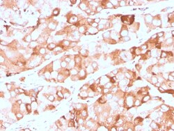
Biotium Prolactin Receptor (hPRL Receptor)(rPRLR/742), CF568 conjugate, 0.1mg/mL
Recognizes a protein of 70 kDa, identified as prolactin receptor. Prolactin is a pituitary hormone involved in the stimulation of milk production, salt and water regulation, growth, development and reproduction. The initial step in its action is the binding to a specific membrane receptor (prolactin receptor), which belongs to the superfamily of class 1 cytokine receptors. The function of the prolactin receptor is mediated, at least in part, by two families of signaling molecules: Janus kinases and signal transducers and activators of transcription. Primary antibodies are available purified, or with a selection of fluorescent CF Dyes and other labels. CF Dyes offer exceptional brightness and photostability. Note: Conjugates of blue fluorescent dyes like CF405S and CF405M are not recommended for detecting low abundance targets, because blue dyes have lower fluorescence and can give higher non-specific background than other dye colors.
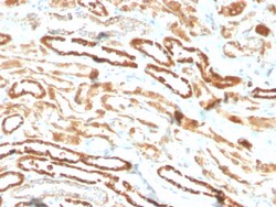
Biotium Erythropoietin (EPO) (Marker of Placentation Disorders)(EPO/3793R), CF488A conjugate, 0.1mg/mL
Recognizes a protein of about 37 kDa, which is identified as Erythropoietin (EPO). Erythropoietin is a secreted, glycosylated cytokine hormone composed of four alpha helical bundles. It is the primary factor responsible for regulating erythropoiesis during steady-state conditions and in response to blood loss and hemorrhage in the adult organism. Erythropoietin is synthesized by the kidney and stimulates the proliferation and maturation of bone marrow erythroid precursor cells. The protein is found in the plasma and regulates red cell production by promoting erythroid differentiation and initiating hemoglobin synthesis. Primary antibodies are available purified, or with a selection of fluorescent CF Dyes and other labels. CF Dyes offer exceptional brightness and photostability. Note: Conjugates of blue fluorescent dyes like CF405S and CF405M are not recommended for detecting low abundance targets, because blue dyes have lower fluorescence and can give higher non-specific
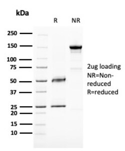
Biotium BAP1 (BRCA1 Associated Protein 1)(BAP1/2667), CF568 conjugate, 0.1mg/mL
The BRCA 1 Associated Protein 1 (BAP1) protein belongs to the ubiquitin C-terminal hydrolase subfamily of de-ubiquitination enzymes that are involved in the removal of ubiquitin from proteins. The encoded enzyme binds to the breast cancer type 1 susceptibility protein (BRCA1) via the RING finger domain of the latter and acts as a tumor suppressor. In addition, the enzyme may be involved in regulation of transcription, regulation of cell cycle and growth, response to DNA damage and chromatin dynamics. Germ line mutations in this gene may be associated with tumor predisposition syndrome (TPDS), which involves increased risk of cancers including malignant mesothelioma, Uveal melanoma and cutaneous melanoma. Primary antibodies are available purified, or with a selection of fluorescent CF Dyes and other labels. CF Dyes offer exceptional brightness and photostability. Note: Conjugates of blue fluorescent dyes like CF405S and CF405M are not recommended for detecting low abundan

Biotium GATA-3 (Breast and Urothelial Marker)(GATA3/2441), CF740 conjugate, 0.1mg/mL
GATA-3 is a zinc finger transcription factor and plays an important role in promoting and directing cell proliferation, development, and differentiation in many tissues and cell types. GATA-3 expression is primarily seen in breast and urothelial carcinomas. 'Therefore, GATA3 antibody can be useful in the identification of unknown primary carcinoma when carcinomas of the breast or bladder are a possibility. Primary antibodies are available purified, or with a selection of fluorescent CF Dyes and other labels. CF Dyes offer exceptional brightness and photostability. Note: Conjugates of blue fluorescent dyes like CF405S and CF405M are not recommended for detecting low abundance targets, because blue dyes have lower fluorescence and can give higher non-specific background than other dye colors.
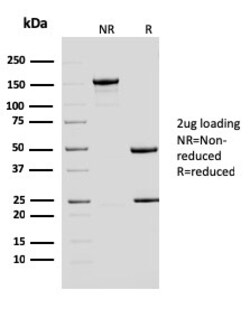
Biotium ERCC1 / RAD10 (Tumor Progression Marker)(ERCC1/2683), CF568 conjugate, 0.1mg/mL
Recognizes a protein of 110 kDa, identified as Excision Repair Cross Complementing 1 (ERCC1). It is a mammalian nucleotide excision repair (NER) enzyme involved in repair of damaged DNA. ERCC1 is a homologous to RAD10 in Saccharomyces cerevisiae, which is required in mitotic intrachromosomal recombination and repair. ERCC1 is required in repair of cisplatin-induced DNA adducts and ultraviolet (UV)-induced DNA damage. High expression of ERCC1 has been linked to tumor progression in a variety of cancers including non-small cell lung cancer (NSCLC), squamous cell carcinoma of the head, ovarian cancer and esophageal cancer. Primary antibodies are available purified, or with a selection of fluorescent CF Dyes and other labels. CF Dyes offer exceptional brightness and photostability. Note: Conjugates of blue fluorescent dyes like CF405S and CF405M are not recommended for detecting low abundance targets, because blue dyes have lower fluorescence and can give higher non-specific

Biotium Gastrin(GAST/2635), CF568 conjugate, 0.1mg/mL
Gastrin, is a hormone that normally formed by mucosal cells in the gastric antrum and by the D cells of the pancreatic islets. Its primary function is to stimulate secretion of HCl by the gastric mucosa. HCl, in turn, inhibits gastrin formation. It also responsible for stimulating smooth muscle contraction and increasing blood circulation and water secretion in the stomach and intestine. Gastrin is regulated by epidermal growth factor in both mice and humans. Gastrin is excreted in excess by pancreatic tumors in the Zollinger-Ellison syndrome. Gastrin-Releasing Peptide (GRP) stimulates the release of gastrin as well as other gastrointestinal hormones and also acts as an autocrine growth factor for certain cell types. High levels of GRP are found in the human lung just after birth and levels decrease thereafter in parallel with the observed disease in a number of pulmonary neuroendocrine cells. GRP is known to promote lung tumorigenesis in model systems. Primary antibodies are
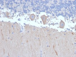
Biotium ATG5 (Autophagy Marker)(ATG5/2492), Biotin conjugate, 0.1mg/mL
Autophagy protein 5 (ATG5), in combination with autophagy protein 12, functions as an E1-like activating enzyme in a ubiquitin-like conjugating system. ATG5 is involved in several cellular processes, including autophagic vesicle formation, mitochondrial quality control after oxidative damage, negative regulation of the innate antiviral immune response, lymphocyte development and proliferation, MHC II antigen presentation, adipocyte differentiation, and apoptosis. The ATG5 protein is essential for autophagy; a process that is usually beneficial for cells to self-degrade their own components when they are no longer useful. Primary antibodies are available purified, or with a selection of fluorescent CF Dyes and other labels. CF Dyes offer exceptional brightness and photostability. Note: Conjugates of blue fluorescent dyes like CF405S and CF405M are not recommended for detecting low abundance targets, because blue dyes have lower fluorescence and can give higher non-specifi
Biotium BOB.1 (B-Cell Marker) (BOB1/2423), CF568 conjugate, 0.1mg/mL
BOB-1 is a transcriptional coactivator that specifically associates with either POU2F1/OCT1 or POU2F2/OCT2, boosting the promoter activity of those complexes. It is essential for the response of B-cells to antigens, required for the formation of germinal centers, and regilates IL-6 expression. BOB-1 expression in a variety of established B-cell lines, representing different stages of B-cell development, has suggested a constitutive, B-cell-specific expression pattern. LP cells in nodular lymphocyte predominant Hodgkin lymphoma, because they are germinal center-derived, are consistently immuno-positive for BOB-1. Conversely, only some cases of classical Hodgkin lymphoma show BOB-1 immuno-reactivity within the Hodgkin and Reed-Sternberg cells. Expression of BOB-1 has been reported in follicular center cell lymphoma, diffuse large B-cell lymphoma and some cases of acute myeloid leukemia. B-CLL, marginal zone lymphoma, and mantle cell lymphoma may show weak to moderate immunoreactivit
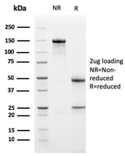
Biotium Bcl-X (Apoptosis Marker)(BCL2L1/1762), CF568 conjugate, 0.1mg/mL
Bcl-x belongs to the Bcl-2 family of proteins and plays a crucial role in apoptosis. Bcl-X has two isoforms: Bcl-XL (long), a 26 kDa protein which suppresses cell death, and Bcl-XS (short), a 178 amino acid protein lacking a 63 amino acid domain which functions as a dominant inhibitor of Bcl-2 and promotes apoptosis. This MAb reacts with both Bcl-XS and Bcl-XL proteins, and shows no cross-reaction with Bcl-2 or Bax proteins.Primary antibodies are available purified, or with a selection of fluorescent CF Dyes and other labels. CF Dyes offer exceptional brightness and photostability. Note: Conjugates of blue fluorescent dyes like CF405S and CF405M are not recommended for detecting low abundance targets, because blue dyes have lower fluorescence and can give higher non-specific background than other dye colors.
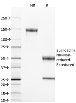
Biotium CD40 / TNFRSF5 / CD40L-Receptor(T8P2G4*A6), CF568 conjugate, 0.1mg/mL
This antibody is specific for the soluble form of CD40. It is a receptor on antigen-presenting cells of the immune system and is essential for mediating a broad variety of immune and inflammatory responses including T cell-dependent immunoglobulin class switching, memory B cell development, and germinal center formation. AT-hook transcription factor AKNA is reported to coordinately regulate the expression of this receptor and its ligand, which may be important for homotypic cell interactions. Adaptor protein TNFR2 interacts with this receptor and serves as a mediator of the signal transduction. The interaction of this receptor and its ligand is found to be necessary for amyloid-beta-induced microglial activation, and thus is thought to be an early event in Alzheimer disease pathogenesis. CD40 is expressed on B-lymphocytes, follicular dendritic cells, bone marrow-derived dendritic cells, thymic epithelium, and interdigitating cells in the T-cell zones of secondary lymphoid org
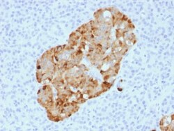
Biotium Chromogranin A / CHGA (Neuroendocrine Marker) (CHGA/1731R), CF568 conjugate, 0.1mg/mL
Chromogranin A is present in neuroendocrine cells throughout the body, including the neuroendocrine cells of the large and small intestine, adrenal medulla and pancreatic islets. It is an excellent marker for carcinoid tumors, pheochromocytomas, paragangliomas, and other neuroendocrine tumors. Co-expression of chromogranin A and neuron specific enolase (NSE) is common in neuroendocrine neoplasms. Reportedly, co-expression of certain keratins and chromogranin indicates neuroendocrine lineage. The presence of strong anti-chromogranin staining and absence of anti-keratin staining should raise the possibility of paraganglioma. The co-expression of chromogranin and NSE is typical of neuroendocrine neoplasms. Most pituitary adenomas and prolactinomas readily express chromogranin.Primary antibodies are available purified, or with a selection of fluorescent CF Dyes and other labels. CF Dyes offer exceptional brightness and photostability. Note: Conjugates of blue fluorescent dyes
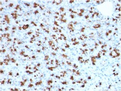
Biotium BARX1 (Prognostic Biomarker in Hepatocellular Carcinoma)(BARX1/2760), CF568 conjugate, 0.1mg/mL
BarX1 is a member of the Bar subclass of homeobox transcription factors. Studies of the Mouse and chick homolog suggest the encoded protein may play a role in developing teeth and craniofacial mesenchyme of neural crest origin. The protein may also be associated with differentiation of stomach epithelia. Down-regulation of Barx1 promotes hepatocellular carcinoma cell (HCC) migration, invasion and metastasis, whereas up-regulation of Barx1 inhibits HCC migration, invasion and metastasis. The loss of Barx1 expression represents a prognostic biomarker in human HCC. Primary antibodies are available purified, or with a selection of fluorescent CF Dyes and other labels. CF Dyes offer exceptional brightness and photostability. Note: Conjugates of blue fluorescent dyes like CF405S and CF405M are not recommended for detecting low abundance targets, because blue dyes have lower fluorescence and can give higher non-specific background than other dye colors.