
Mouse Secondary Antibodies


Biotium Lewis B(2-25LE), CF740 conjugate, 0.1mg/mL
The Lewis histo-blood group system comprises a set of fucosylated glycosphingolipids that are synthesized by exocrine epithelial cells and circulate in body fluids. The glycosphingolipids function in embryogenesis, tissue differentiation, tumor metastasis, inflammation, and bacterial adhesion. They are secondarily absorbed to red blood cells giving rise to their Lewis phenotype. This gene is a member of the fucosyltransferase family, which catalyzes the addition of fucose to precursor polysaccharides in the last step of Lewis antigen biosynthesis. It encodes an enzyme with alpha(1,3)-fucosyltransferase and alpha(1,4)-fucosyltransferase activities. Lewis blood group antigens are carbohydrate moieties structurally integrated in mucous secretions. Lewis antigen system alterations have been described in gastric carcinoma and associated lesions. Anomalous expression of Lewis B antigen has been found in some non-secretory gastric carcinomas and colorectal cancers.Primary antibodies

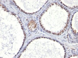
Biotium CD41a(ITGA2B/1036), CF740 conjugate, 0.1mg/mL
Reacts with a calcium-dependent complex of CD41/CD61, a dimer of 90 kDa and 140 kDa present on the membrane of normal platelets and megakaryocytes. CD41/CD61 is also known as platelet glycoprotein GPIIb/GPIIIa or integrin IIa/3. This complex is the receptor of fibrinogen, fibronectin and von Willebrand factor, and mediates platelet adhesion and aggregation.Primary antibodies are available purified, or with a selection of fluorescent CF Dyes and other labels. CF Dyes offer exceptional brightness and photostability. Note: Conjugates of blue fluorescent dyes like CF405S and CF405M are not recommended for detecting low abundance targets, because blue dyes have lower fluorescence and can give higher non-specific background than other dye colors.
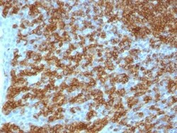
Biotium CD45RA(PTPRC/1148), 1mg/mL
Recognizes a protein of 205 kDa-220 kDa, identified as CD45RA (Workshop III). CD45RA is isoforms of the human leukocyte common antigen (CD45). Human CD45 contains three exons which encode peptide segments designated A, B and C, respectively. The differential splicing of the exons generates at least five isoforms, ABC, AB, BC, B and O. This antibody reacts with ABC and BC isoforms. CD45RA is expressed on 40-50% of peripheral CD4 T-cells, 50% of peripheral CD8 T-cells, B-cells, and leukemic B-cell lines. T-cells expressing CD45RA are naive or virgin T-cells. T-cells expressing CD45RO are memory T-cells. CD45RA and CD45RO define complementary, predominantly non-overlapping populations of resting peripheral T-cells. This MAb is useful in study on the subpopulation of CD4 or CD8 T-cells. It can especially be used to differentiate T-cell lymphomas (CD45RO positive) from B cell lymphomas (CD45RA positive).Primary antibodies are available purified, or with a selection of fluoresc
Biotium Helicobacter pylori(B-53.4), 0.2mg/mL
The spiral shaped bacteriumHelicobacter pyloriis strongly associated with inflammation of the stomach and is also implicated in the development of gastric malignancy.H. pyloriis known to cause peptic ulcers and chronic gastritis in human. It is associated with duodenal ulcers and may be involved in development of adenocarcinoma and low-grade lymphoma of mucosa associated lymphoid tissue in the stomach.This antibody stains the individual H. pylori bacterium when it presents on the surface of the epithelium or in the cytoplasm of the epithelial cells in biopsy tissue sections from the antrum and body of the stomach.
Biotium CD7(124.1D1), Biotin conjugate, 0.1mg/mL
Recognizes a protein of 40 kDa, identified as CD7 (Workshop IV; Code T155). CD7 is a member of the immunoglobulin gene superfamily. Its N-terminal amino acids 1-107 are highly homologous to Ig kappa-L chains whereas the carboxyl-terminal region of the extracellular domain is proline-rich and has been postulated to form a stalk from which the Ig domain projects. CD7 is expressed on the majority of immature and mature T-lymphocytes, and T cell leukemia. It is also found on natural killer cells, a small subpopulation of normal B cells and on malignant B cells. Cross-linking surface CD7 positively modulates T cell and NK cell activity as measured by calcium fluxes, expression of adhesion molecules, cytokine secretion and proliferation. CD7 associates directly with phosphoinositol 3'-kinase. CD7 ligation induces production of D-3 phosphoinositides and tyrosine phosphorylation.Primary antibodies are available purified, or with a selection of fluorescent CF Dyes and other labels. C
Biotium NOX4 / NADPH Oxidase 4 (NOX4/1245), CF488A conjugate, 0.1mg/mL
The superoxide-generating NADPH oxidase includes a membrane-bound flavocytochrome containing two subunits, gp91-phox and p22-phox, and the cytosolic proteins p47-phox and p67-phox. During activation of the NADPH oxidase, p47-phox and p67-phox migrate to the plasma membrane where they associate with the flavocytochrome, cytochrome b558, to form the active enzyme complex. The p22 and gp91-phox subunits also function as surface O2 sensors that initiate cellular signaling in response to hypoxic conditions. NOX4 is a renal gp91-phox homolog highly expressed at the site of erythropoietin production in the proximal convoluted tubule epithelial cells of the renal cortex. It is also expressed in fetal tissues, placenta, glioblastoma and vascular cells.Primary antibodies are available purified, or with a selection of fluorescent CF Dyes and other labels. CF Dyes offer exceptional brightness and photostability. Note: Conjugates of blue fluorescent dyes like CF405S and CF405M are no
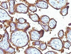
Biotium EGFR(31G7), CF568 conjugate, 0.1mg/mL
This MAb recognizes a protein of 170 kDa, identified as EGFR. EGFR is type I receptor tyrosine kinase with sequence homology to erbB-1, -2, -3 -4 or HER-1, -2, -3 -4. It binds to Epidermal Growth Factor (EGF), Transforming Growth Factor-a (TGF-a), Heparin-binding EGF (HB-EGF), amphiregulin, betacellulin and epiregulin. EGFR is overexpressed in tumors of breast, brain, bladder, lung, gastric, head & neck, esophagus, cervix, vulva, ovary, and endometrium. It is predominantly present in squamous cell carcinomas.Primary antibodies are available purified, or with a selection of fluorescent CF Dyes and other labels. CF Dyes offer exceptional brightness and photostability. Note: Conjugates of blue fluorescent dyes like CF405S and CF405M are not recommended for detecting low abundance targets, because blue dyes have lower fluorescence and can give higher non-specific background than other dye colors.
Biotium CD63 (Late Endosomes Marker) (LAMP3/2990R), 0.2mg/mL
This MAb recognizes protein of 26 kDa-60 kDa, which is identified as CD63. Its epitope is different from that of MAb LAMP3/529. The tetraspanins are integral membrane proteins expressed on cell surface and granular membranes of hematopoietic cells and are components of multi-molecular complexes with specific integrins. The tetraspanin CD63 is a lysosomal membrane glycoprotein that translocates to the plasma membrane after platelet activation. CD63 is expressed on activated platelets, monocytes and macrophages, and is weakly expressed on granulocytes, T cell and B cells. It is located on the basophilic granule membranes and on the plasma membranes of lymphocytes and granulocytes. CD63 is a member of the TM4 superfamily of leukocyte glycoproteins that includes CD9, CD37 and CD53, which contain four transmembrane regions. CD63 may play a role in phagocytic and intracellular lysosome-phagosome fusion events. CD63 deficiency is associated with Hermansky-Pudlak syndrome and is strongly
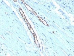
Biotium Blood Group Antigen A(HE-193), 1mg/mL
This antibody is applicable for staining ABO blood group A (monofucosyl and difucosyl A antigens with chain types 1, 2, 3, 4, 5, 6). It also recognizes Forssman antigen. The histo-blood group ABO involves three carbohydrate antigens: A, B, and H. Blood group antigens are generally defined as molecules formed by sequential addition of saccharides to the carbohydrate side chains of lipids and proteins detected on erythrocytes and certain epithelial cells. Blood group related antigens represent a group of carbohydrate determinants carried on both glycolipids and glycoproteins. They are usually mucin-type, and are detected on erythrocytes, certain epithelial cells, and in secretions of certain individuals. Sixteen genetically and biosynthetically distinct but inter-related specificities belong to this group of antigens, including A, B, H, Lewis A, Lewis B, Lewis X, Lewis Y, and precursor type 1 chain antigen.This antibody is available purified with BSA/azide at 200 ug/mL, or
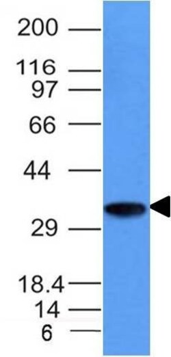
Biotium CD74(CLIP/1133), CF568 conjugate, 0.1mg/mL
Recognizes proteins of 33, 35 and 41 kDa, which are identified as various isoforms of CD74. Its epitope is localized in the extracellular domain of CD74. CD74 is a type II transmembrane protein which binds to the peptide binding groove of newly synthesized MHC class II alpha/beta heterodimers and prevents their premature association with endogenous polypeptides. The CD74 molecule plays a critical role in the presentation of peptides, by the MHC class II antigens, to CD4 positive lymphocytes. CD74 is expressed on MHC class II positive cells including B cells, a subset of activated T cells, monocytes, and dendritic cells and by various types of carcinomas. CD74 is expressed primarily by antigen presenting cells, such as B-lymphocytes (from before the pre-B cell stage to before the plasma cell stage), macrophages, and monocytes, and many epithelial cells. Anti-CD74 stains predominantly germinal center lymphocytes and B-cell lymphomas, but rarely T-cell lymphomas. Anti-CD74 has b
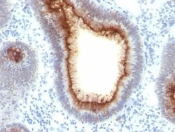
Biotium CD66 (CEA)(C66/1009), CF488A conjugate, 0.1mg/mL
This antibody recognizes proteins of 80-200 kDa, identified as different members of CEA family. CEA is synthesized during development in the fetal gut and is re-expressed in increased amounts in intestinal carcinomas and several other tumors. This MAb does not react with nonspecific cross-reacting antigen (NCA) and with human polymorphonuclear leucocytes. It shows no reaction with a variety of normal tissues and is suitable for staining of formalin/paraffin tissues. CEA is not found in benign glands, stroma, or malignant prostatic cells. Antibody to CEA is useful in detecting early foci of gastric carcinoma and in distinguishing pulmonary adenocarcinomas (60-70% are CEA positive) from pleural mesotheliomas (rarely or weakly CEA positive). Anti-CEA positivity is seen in adenocarcinomas from the lung, colon, stomach, esophagus, pancreas, gallbadder, urachus, salivary gland, ovary, and endocervix.Primary antibodies are available purified, or with a selection of fluorescent CF
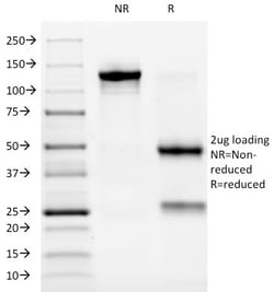
Biotium ALK (Anaplastic Lymphoma Kinase) / CD246(ALK/1032), 0.2mg/mL
The wild-type anaplastic lymphoma kinase (ALK) protein is a 200 kDa transmembrane receptor tyrosine kinase. Its expression is restricted to a few scattered cells in the nervous system (some glial cells and neurons, and a few endothelial cells and pericytes). ALK has been found to be rearranged, mutated, or amplified in a series of tumors. The hybrid gene, NPM-ALK, created by the t(2;5)(p23;q35) chromosomal translocation, encodes part of the nucleolar phosphoprotein nucleophosmin (NPM), joined to the entire cytoplasmic portion of ALK. As a consequence, the ALK gene comes under the control of the NPM promoter, which induces a permanent and ubiquitous transcription of the NPM-ALK hybrid gene, resulting in the production of a 80 kDa NPM-ALK chimeric protein. This translocation is found in anaplastic large cell lymphomas (ALCL). Reportedly, expression of ALK indicates a better prognosis. Approximately 5%-10% of non-small cell lung carcinomas also express ALK protein, producing a c
Biotium CD7(124.1D1), CF488A conjugate, 0.1mg/mL
Recognizes a protein of 40 kDa, identified as CD7 (Workshop IV; Code T155). CD7 is a member of the immunoglobulin gene superfamily. Its N-terminal amino acids 1-107 are highly homologous to Ig kappa-L chains whereas the carboxyl-terminal region of the extracellular domain is proline-rich and has been postulated to form a stalk from which the Ig domain projects. CD7 is expressed on the majority of immature and mature T-lymphocytes, and T cell leukemia. It is also found on natural killer cells, a small subpopulation of normal B cells and on malignant B cells. Cross-linking surface CD7 positively modulates T cell and NK cell activity as measured by calcium fluxes, expression of adhesion molecules, cytokine secretion and proliferation. CD7 associates directly with phosphoinositol 3'-kinase. CD7 ligation induces production of D-3 phosphoinositides and tyrosine phosphorylation.Primary antibodies are available purified, or with a selection of fluorescent CF Dyes and other labels. C
Biotium Galectin-13 (GAL13) / Placental Protein 13 (PP13) (PP13/1162), Biotin conjugate, 0.1mg/mL
This antibody recognizes a 32 kDa protein, which is identified as homodimer of galectin-13 (also known as PP13). Galectins are a family of soluble beta-galactoside-binding lectins that modulate cell-to-cell adhesion and cell-to-extracellular matrix (ECM) interactions and play a role in tumor progression, pre-mRNA splicing and apoptosis. One such member, galectin-13, is a 139 amino acid protein that contains one galectin domain. Possessing lysophospholipase activity, galectin-13 exists as a disulfide-linked homodimer. Galectin-13 is suggested to have a developmental role in the placenta and may display immunobiological activity in feto-maternal blood-spaces. Screening for galectin-13 in maternal serum during the first trimester of pregnancy may serve as a diagnostic marker to predict preterm pre-eclampsia.Primary antibodies are available purified, or with a selection of fluorescent CF Dyes and other labels. CF Dyes offer exceptional brightness and photostability. Note: Co
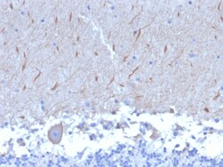
Biotium ATG5 (Autophagy Marker)(ATG5/2553), 0.2mg/mL
Autophagy protein 5 (ATG5), in combination with autophagy protein 12, functions as an E1-like activating enzyme in a ubiquitin-like conjugating system. ATG5 is involved in several cellular processes, including autophagic vesicle formation, mitochondrial quality control after oxidative damage, negative regulation of the innate antiviral immune response, lymphocyte development and proliferation, MHC II antigen presentation, adipocyte differentiation, and apoptosis. The ATG5 protein is essential for autophagy; a process that is usually beneficial for cells to self-degrade their own components when they are no longer useful. Primary antibodies are available purified, or with a selection of fluorescent CF Dyes and other labels. CF Dyes offer exceptional brightness and photostability. Note: Conjugates of blue fluorescent dyes like CF405S and CF405M are not recommended for detecting low abundance targets, because blue dyes have lower fluorescence and can give higher non-specifi