
Mouse Secondary Antibodies


Biotium Cytokeratin 15 (KRT15) (Basal Cell Marker) (LHK15), CF740 conjugate, 0.1mg/mL
Keratins are a family of intermediate filament proteins that assemble into filaments through forming heterodimers of one type I keratin (keratins 9 to 23) and one type II keratin (keratins 1 to 8). Keratins demonstrate tissue and differentiation specific expression profiles. Keratin 15 is a type I keratin which is expressed only in basal keratinocytes in stratified epithelia and does not appear to have a natural type II expression partner. Keratin 15 is down regulated in activated keratinocytes. Cytokeratin 15 is a specific marker of stem cells of the hair-follicle bulge and may be a useful marker for diagnosis between basal cell carcinoma (BCC) and trichoepithelioma. Trichoblastoma are benign neoplasms of follicular differentiation frequently found in nevus sebaceous. Many morphologic features are shared with nodular basal cell carcinoma, sometimes rendering a diagnosis difficult. Trichoblastoma and BCC show variable expression of Cytokeratin 15 and Cytokeratin 19, and absen

Biotium Villin (GI-Mucosal & Urogenital Brush Border Marker) (VIL1/1314), CF488A conjugate, 0.1mg/mL
This antibody recognizes a protein of 95 kDa, which is identified as villin. It is a major constituent in the microvilli, which compose the brush border of epithelial cells forming absorptive surfaces of the intestinal and renal proximal tubular epithelia. Anti-Villin labels the brush border area in the gastrointestinal mucosal epithelium and urogenital tract. Among neoplasms, villin is predominantly expressed in tumors of colorectal origin. Antibody to villin is useful in identifying malignant cells from primary and metastatic colorectal carcinomas. This antibody also labels Merkel cells of the skin.Primary antibodies are available purified, or with a selection of fluorescent CF Dyes and other labels. CF Dyes offer exceptional brightness and photostability. Note: Conjugates of blue fluorescent dyes like CF405S and CF405M are not recommended for detecting low abundance targets, because blue dyes have lower fluorescence and can give higher non-specific background than oth
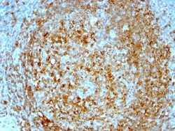
Biotium CD74(BU45), CF740 conjugate, 0.1mg/mL
Recognizes proteins of 33, 35 and 41 kDa, which are identified as various isoforms of CD74. Its epitope is localized in the extracellular domain of CD74. CD74 is a type II transmembrane protein which binds to the peptide binding groove of newly synthesized MHC class II alpha/beta heterodimers and prevents their premature association with endogenous polypeptides. The CD74 molecule plays a critical role in the presentation of peptides, by the MHC class II antigens, to CD4 positive lymphocytes. CD74 is expressed on MHC class II positive cells including B cells, a subset of activated T cells, monocytes, and dendritic cells and by various types of carcinomas. CD74 is expressed primarily by antigen presenting cells, such as B-lymphocytes (from before the pre-B cell stage to before the plasma cell stage), macrophages, and monocytes, and many epithelial cells. Anti-CD74 stains predominantly germinal center lymphocytes and B-cell lymphomas, but rarely T-cell lymphomas. Anti-CD74 has b


Biotium Factor XIIIa (Coagulation Factor XIIIA Chain) (F13A1/1447), CF488A conjugate, 0.1mg/mL
This antibody recognizes a protein of 83 kDa, which is identified as Factor XIIIa. It has been identified in platelets, megakaryocytes, and fibroblast-like mesenchymal or histiocytic cells in the placenta, uterus, and prostate, monocytes and macrophages and dermal dendritic cells. Anti-factor XIIIa has been found to be useful in differentiating between dermatofibroma (almost all cases are positive), dermatofibrosarcoma protuberans (-/ ) and desmoplastic malignant melanoma (-). Anti-factor XIIIa positivity is also seen in capillary hemagioblastoma, hemangioendothelioma, hemangiopericytoma, xanthogranuloma, xanthoma, hepatocellular carcinoma, glomus tumor, and meningioma.Primary antibodies are available purified, or with a selection of fluorescent CF Dyes and other labels. CF Dyes offer exceptional brightness and photostability. Note: Conjugates of blue fluorescent dyes like CF405S and CF405M are not recommended for detecting low abundance targets, because blue dyes have
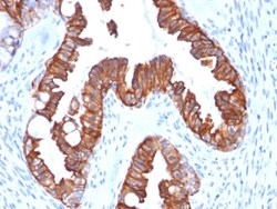
Biotium Cytokeratin 7 (Glandular and Transitional Epithelial Marker) (rOV-TL12/30), Biotin conjugate, 0.1mg/mL
This antibody recognizes an intermediate filament protein (IFP) of 55 kDa, which is identified as cytokeratin 7. This MAb is highly specific to cytokeratin 7 and shows no cross-reaction with other IFPs. Cytokeratin 7 is a basic cytokeratin, which is found in most glandular and transitional epithelia but not in the stratified squamous epithelia. Keratin 7 is expressed in the epithelial cells of ovary, lung, and breast but not of colon, prostate, or gastrointestinal tract. This MAb is highly useful in distinguishing ovarian carcinomas (keratin 7 ) from colon carcinomas (keratin 7-).Primary antibodies are available purified, or with a selection of fluorescent CF Dyes and other labels. CF Dyes offer exceptional brightness and photostability. Note: Conjugates of blue fluorescent dyes like CF405S and CF405M are not recommended for detecting low abundance targets, because blue dyes have lower fluorescence and can give higher non-specific background than other dye colors.
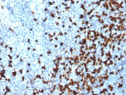
Biotium TCL1 (T-Cell Marker)(TCL1/2079), CF740 conjugate, 0.1mg/mL
T-cell leukemia/Lymphoma Protein 1A (TCL-1A), also known as p14TCL1 is a product of the TCL1 gene that is involved in T-cell prolymphocytic leukemia (TPLL). T-PLL is a rare form of mature T-cell leukemia, which is consistently associated with chromosomal rearrangements characterized by the juxtaposition of the TCRA locus on chromosome 14q11 and the TCL1A gene on 14q32. 13. TCL1 is overexpressed in Burkitt lymphoma, the majority of AIDS-related non-Hodgkin lymphoma-designated immunoblastic plasmacytoid lymphoma, lymphoblastic lymphoma, chronic lymphocytic leukemia, mantle cell lymphoma, follicular lymphoma, diffuse large B-cell lymphoma, and primary cutaneous B-cell lymphoma. Primary antibodies are available purified, or with a selection of fluorescent CF Dyes and other labels. CF Dyes offer exceptional brightness and photostability. Note: Conjugates of blue fluorescent dyes like CF405S and CF405M are not recommended for detecting low abundance targets, because blue dyes have
Biotium Histone H1(1415-1), CF740 conjugate, 0.1mg/mL
Eukaryotic histones are basic, water-soluble nuclear proteins that form hetero-octameric nucleosome particles. They wrap 146 base pairs of DNA in a left-handed supealpha-helicalturn sequentially to form chromosomal fiber. Two molecules of each of the four core histones (H2A, H2B, H3, and H4) form the octamer; formed of two H2A-H2B dimers and two H3-H4 dimers, forming two nearly symmetrical halves by tertiary structure. Over 80% of nucleosomes contain the linker Histone H1, derived from an intronless gene that interacts with linker DNA between nucleosomes and mediates compaction into higher order chromatin. Histones are subject to posttranslational modification by enzymes primarily on their N-terminal tails, but also in their globular domains. Such modifications include methylation, citrullination, acetylation, phosphorylation, sumoylation, ubiquitination and ADP-ribosylation.Primary antibodies are available purified, or with a selection of fluorescent CF Dyes and other lab
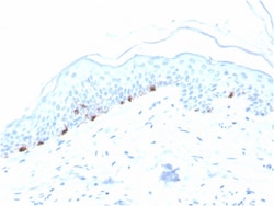
Biotium Tyrosinase-Related Protein-1 (TYRP-1) (Melanoma Marker)(TYRP1/3280), CF488A conjugate, 0.1mg/mL
Reacts with a 75 kDa melanocyte-specific gene product, identified as Tyrosinase-related protein-1 (TRP-1). It is involved in melanin synthesis. TRP1 is present on the melanosomal membranes of melanoma, normal melanocytes and nevi. Recent evidence suggests that TRP-1 is involved in maintaining stability of tyrosinase protein and modulating its catalytic activity. TRP-1 is also involved in maintenance of melanosome ultrastructure and affects melanocyte proliferation and cell death. Primary antibodies are available purified, or with a selection of fluorescent CF Dyes and other labels. CF Dyes offer exceptional brightness and photostability. Note: Conjugates of blue fluorescent dyes like CF405S and CF405M are not recommended for detecting low abundance targets, because blue dyes have lower fluorescence and can give higher non-specific background than other dye colors.
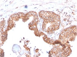
Biotium CD47 / IAP (Integrin Associated Protein)(CD47/2937), Biotin conjugate, 0.1mg/mL
This antibody reacts with Ig domain of CD47 protein. CD47, originally named integrin-associated protein (IAP), is a 50 kDa protein containing five membrane-spanning sequences and a short cytoplasmic tail. CD47 plays a role in both cell adhesion by acting as an adhesion receptor for THBS1 on platelets, and in the modulation of integrins. It is important in memory formation and synaptic plasticity in the hippocampus. CD47 may play a role in membrane transport and/or integrin dependent signal transduction. Primary antibodies are available purified, or with a selection of fluorescent CF Dyes and other labels. CF Dyes offer exceptional brightness and photostability. Note: Conjugates of blue fluorescent dyes like CF405S and CF405M are not recommended for detecting low abundance targets, because blue dyes have lower fluorescence and can give higher non-specific background than other dye colors.
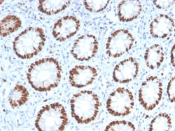
Biotium MCM6 (Proliferation Marker)(MCM6/2999), 0.2mg/mL
The mini-chromosome maintenance (MCM) family of proteins, including MCM2, MCM3, MCM6 (Cdc21), MCM5 (Cdc46), MCM6 (Mis5) and MCM7 (Cdc47), are regulators of DNA replication that act to ensure replication occurs only once in the cell cycle. Expression of MCM proteins increases during cell growth, peaking at G1/S phase. The MCM proteins each contain an ATP-binding motif, which is predicted to mediate ATP-dependent opening of double-stranded DNA. MCM proteins are regulated by E2F transcription factors, which induce MCM expression, and by protein kinases, which interact with MCM proteins to maintain the post-replicative state of the cell. MCM2/MCM6 complexes function as substrates for Cdc2/cyclin B in vitro. Primary antibodies are available purified, or with a selection of fluorescent CF Dyes and other labels. CF Dyes offer exceptional brightness and photostability. Note: Conjugates of blue fluorescent dyes like CF405S and CF405M are not recommended for detecting low abundanc
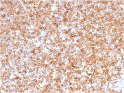
Biotium HLA-DP/-DQ/-DR (MHC II)(CR3/43), 1mg/mL
Reacts with a common epitope of human major histocompatibility (MHC) class II antigens, HLA-DP, -DQ and -DR. Human MHC class II antigens are transmembrane glycoproteins composed of an chain (36 kDa) and a chain (27 kDa). They are expressed primarily on antigen presenting cells such as B lymphocytes, monocytes, macrophages, and thymic epithelial cells and are also present on activated T lymphocytes. Human MHC class II genes are located in the HLA-D region that encodes at least six and ten chain genes. Three loci, DR, DQ and DP, encode the major expressed products of the human class II region. The human MHC class II molecules bind intracellularly processed peptides and present them to T-helper cells. They, therefore, have a critical role in the initiation of the immune response. It has been shown that some autoimmune diseases are associated with certain class II alleles.Primary antibodies are available purified, or with a selection of fluorescent CF Dyes and other labels.
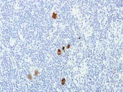
Biotium Epstein-Barr Virus (LMP-1)(CS1-4), CF740 conjugate, 0.1mg/mL
This antibody is a mixture of four different monoclonal antibodies. This antibody is specific to 60 kDa latent membrane protein (LMP-1) encoded by the BNLF1 gene of the EBV. Each clone reacts with different epitopes on the hydrophilic C-terminus of the cytoplasmic domain of LMP-1. This antibody stains strongly with EBV-positive lymphoblastoid cell lines and EBV infected B cell immunoblasts in infectious mononucleosis. EBV, also designated human herpesvirus 4 (HHV-4), is a member of the herpesvirus family and is one of the most common human viruses. EBV infects B cells and, though often asymptomatic, it can cause infectious mononucleosis, a disease characterized by fatigue, fever, sore throat and muscle soreness. Primary antibodies are available purified, or with a selection of fluorescent CF Dyes and other labels. CF Dyes offer exceptional brightness and photostability. Note: Conjugates of blue fluorescent dyes like CF405S and CF405M are not recommended for detecting low
Biotium Muscle Specific Actin(HHF35 + MSA/953), CF488A conjugate, 0.1mg/mL
This antibody recognizes actin of skeletal, cardiac, and smooth muscle cells. It is not reactive with other mesenchymal cells except for myoepithelium. Actin can be resolved on the basis of its isoelectric points into three distinctive components: alpha, beta, and gamma in order of increasing isoelectric point. Anti-muscle specific actin recognizes alpha and gamma isotypes of all muscle groups. Non-muscle cells such as vascular endothelial cells and connective tissues are non-reactive. Also, neoplastic cells of non-muscle-derived tissue such as carcinomas, melanomas, and lymphomas are negative.It stains tumors of smooth muscle (leiomyomas and leiomyosarcomas) as well as skeletal muscle (rhabdomyomas and rhabdomyosarcomas).Primary antibodies are available purified, or with a selection of fluorescent CF Dyes and other labels. CF Dyes offer exceptional brightness and photostability. Note: Conjugates of blue fluorescent dyes like CF405S and CF405M are not recommended for detectin