
Mouse Secondary Antibodies

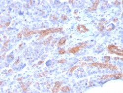
Biotium MRP3 (Multidrug Resistance-Associated Protein 3)(ABCC3/2971), Biotin conjugate, 0.1mg/mL
The two members of the large family of ABC transporters known to confer multidrug resistance in human cancer cells are the MDR1 P-glycoprotein and the multidrug-resistance protein MRP1. MRP1 is an integral membrane protein that contains an MDR-like core, an N-terminal membrane-bound region and a cytoplasmic linker, and it is expressed in various cerebral cells, as well as in lung, testis and peripheral blood. The MRP gene family also includes MRP2, which is alternatively designated cMOAT (for canalicular multispecific organic anion transporter) and MRP3, which are both conjugate export pumps exp- ressed predominantly in hepatocytes. MRP2 localizes exclusively to the apical membrane and is constitutively expressed at a high level in normal liver cells. Conversely, MRP3 localizes to the basolateral membrane where it also medi- ates the transport of the organic anion S-(2, 4-dinitrophenyl-) glutathione toward the basolateral side of the membrane. MRP3 is normally expressed at co


Biotium VEGI (Vascular Endothelial Growth Inhibitor) (VEGI/1283), 0.2mg/mL
VEGI is an anti-angiogenic cytokine that belongs to tumor necrosis factor superfamily, member 15 (TNFSF15). This protein is abundantly expressed in endothelial cells, but is not expressed in either B or T cells. The expression of this protein is inducible by TNF and IL-1 alpha. This cytokine is a ligand for receptor TNFRSF25 and decoy receptor TNFRSF21/DR6. It can activate NF-kappaB and MAP kinases, and acts as an autocrine factor to induce apoptosis in endothelial cells. This cytokine is also found to inhibit endothelial cell proliferation, and thus may function as an angiogenesis inhibitor. Reduced expression of VEGI has been reported as a marker of poor prognosis in breast cancer.Primary antibodies are available purified, or with a selection of fluorescent CF Dyes and other labels. CF Dyes offer exceptional brightness and photostability. Note: Conjugates of blue fluorescent dyes like CF405S and CF405M are not recommended for detecting low abundance targets, because bl

Biotium c-Myc Oncoprotein(MYC275 + MYC909), CF568 conjugate, 0.1mg/mL
This antibody recognizes a transcription factor of 64-67 kDa, identified as c-myc. This MAb shows no cross-reaction with v-myc. c-myc is involved in the control of cell proliferation and differentiation and is amplified and/or over-expressed in a variety of tumors. Over-expression of c-myc protein occurs frequently in luminal cells of prostate intraepithelial neoplasia as well as in most primary carcinomas and metastatic disease. Rearrangement of the MYC gene is found in 3% to 16% of diffuse large B-cell lymphoma (DLBCL's) and in nearly 100% of Burkitt lymphomas (BL). Identifying MYC status is important in establishing final diagnosis of DLBCL, BL, or B-cell lymphoma, with features intermediate between DLBCL and BL as well as in differential diagnoses of the lymphomas.

Biotium Cytokeratin 7 (Glandular and Transitional Epithelial Marker) (KRT7/1499R), Biotin conjugate, 0.1mg/mL
This antibody recognizes an intermediate filament protein (IFP) of 55 kDa, which is identified as cytokeratin 7. This MAb is highly specific to cytokeratin 7 and shows no cross-reaction with other IFPs. Cytokeratin 7 is a basic cytokeratin, which is found in most glandular and transitional epithelia but not in the stratified squamous epithelia. Keratin 7 is expressed in the epithelial cells of ovary, lung, and breast but not of colon, prostate, or gastrointestinal tract. This MAb is highly useful in distinguishing ovarian carcinomas (keratin 7 ) from colon carcinomas (keratin 7-).Primary antibodies are available purified, or with a selection of fluorescent CF Dyes and other labels. CF Dyes offer exceptional brightness and photostability. Note: Conjugates of blue fluorescent dyes like CF405S and CF405M are not recommended for detecting low abundance targets, because blue dyes have lower fluorescence and can give higher non-specific background than other dye colors.
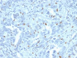
Biotium Cyclin A2 (S- & G2-phase Cyclin)(CCNA2/2333), CF488A conjugate, 0.1mg/mL
Recognizes a protein of 54 kDa, which is identified as cyclin A. Its epitope is located amino acids 144-148 of human Cyclin A2. Cyclins are regulatory subunits of the cyclin-dependent kinases (cdk's) and they control transition at different specific phases of the cell cycle. The temporal expression of cyclins is tightly regulated and subsequently plays a critical role in controlling the enzymatic activity of cdk's. These cyclin/cdk complexes are essential for passage through specific stages in the cell cycle. In mammalian somatic cells, cyclin A is required for S-phase and passage through G2-phase. The D and E type cyclins regulate the passage of G1, while cyclin B is a critical regulator of mitosis. Mutation or disruption of normal cyclin A expression causes cells to arrest in G2-phase. Primary antibodies are available purified, or with a selection of fluorescent CF Dyes and other labels. CF Dyes offer exceptional brightness and photostability. Note: Conjugates of blue fluores
Biotium CD99 / MIC2 (Ewing's Sarcoma Marker), CF568 conjugate, 0.1mg/mL
Recognizes a sialoglycoprotein of 27-32 kDa, identified as CD99, or MIC2 gene product, or E2 antigen. MIC2 gene is located in the pseudo-autosomal region of the human X and Y chromosome. MIC2 gene encodes two distinct proteins, which are produced by alternative splicing of the CD99 gene transcript and are identified as bands of 30 and 32 kDa (p30/32). Although its function is not fully understood, CD99 is implicated in various cellular processes including homotypic aggregation of T cells, upregulation of T cell receptor and MHS molecules, apoptosis of immature thymocytes and leukocyte diapedesis. CD99 is expressed on the cell membrane of some lymphocytes, cortical thymocytes, and granulosa cells of the ovary. Most pancreatic islet cells, Sertoli cells of the testis, and some endothelial cells express this antigen. Mature granulocytes express very little or no CD99. MIC2 is strongly expressed on Ewing's sarcoma cells and primitive peripheral neuroectodermal tumors. Primary ant

Biotium Alpha-1-Antichymotrypsin (SERPINA3) (Histiocytoma Marker) (AACT/1451 + AACT/1452), CF740 conjugate, 0.1mg/mL
Alpha-1 Antichymotrypsin (AACT) is a plasma protease inhibitor synthesized in the liver as a single glycopeptide chain. In human, the normal serum level of AACT is about one-tenth that of alpha-1-antitrypsin (AAT), with which it shares nucleic acid and protein sequence homology. Both are major acute phase reactants; their concentrations in plasma increase in response to trauma, surgery and infection. Elevated levels of AACT are widely, but not universally, reported in the cerebrospinal fluid and plasma of AD patients. Prostate-specific antigen (PSA) and its SDS-stable complex with AACT are in widespread use as markers for the diagnosis of prostate cancer. AACT deficiency may also be a possible cause of chronic liver disease. AACT antibody reacts with histiocytes and histiocytic neoplasms. It is widely used to identify histiocytes and tumors derived from them. Acinar tumors of the pancreas and salivary gland may also exhibit AACT positivity.Primary antibodies are available pur
Biotium Mucin 5AC (MUC5AC)(MUC5AC/917 + 45M1), 1mg/mL
Mucin 5AC glycoprotein (MUC5AC) is a 641 kDa glycoprotein belonging to the superfamily of mucins. Mucins are high molecular weight glycoproteins produced by epithelial cells and can be divided into two families; secretory mucins and membrane bound mucins. MUC5AC is a mucus-forming secreted mucin that is found in normal gastric and tracheo-bronchial mucosa, but absent from normal colon. MUC5AC expression is present in primary ovarian mucinous cancer but usually absent in colorectal adenocarcinoma, thus showing an expression pattern opposite to MUC2. Together with a panel of antibodies, Anti-MUC5AC may be useful for differential identification of primary mucinous ovarian tumors from colon adenocarcinoma metastatic to the ovary. MUC5AC antibodies may also be useful for identification of intestinal metaplasia as well as in the identification of pancreatic carcinoma and pre-cancerous changes vs. normal pancreas.Primary antibodies are available purified, or with a selection of fluoresce
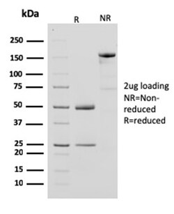
Biotium Creatine Phosphokinase-BB (CK-BB)(CPTC-CKB-2), CF488A conjugate, 0.1mg/mL
Creatine kinases (CK) are a large family of isoenzymes that regulate levels of ATP in subcellular compartments, where they provide ATP at sites of fluctuating energy demand by the transfer of phosphates between creatine and adenine nucleotides. CKs provide the energy of phosphate hydrolysis necessary to drive the normal function of many cellular systems. In cells, the cytosolic CK enzymes consist of two subunits, which can be either B (brain type) or M (muscle type). There are three different isoenzymes: CKMM, CKBB and CKMB. This MAb recognizes the CKBB isoenzyme and does not react with the B subunit in CKMB. Primary antibodies are available purified, or with a selection of fluorescent CF Dyes and other labels. CF Dyes offer exceptional brightness and photostability. Note: Conjugates of blue fluorescent dyes like CF405S and CF405M are not recommended for detecting low abundance targets, because blue dyes have lower fluorescence and can give higher non-specific background than
Biotium AKT1 (Prognostic Marker for Neuroendocrine Tumors) (AKT1/2552), CF568 conjugate, 0.1mg/mL
This antibody recognizes a protein of 62 kDa, which is identified as AKT1. The serine/threonine kinase Akt family contains several members, including Akt1 (also designated PKB or RacPK), Akt2 (also designated PKBbeta or RacPK-beta) and Akt3 (also designated PKB-gamma or thymoma viral proto-oncogene 3), which exhibit sequence homology with the protein kinase A and C families and are encoded by the c-Akt proto-oncogene. All members of the Akt family have a Pleckstrin homology domain. Akt1 and Akt2 are activated by PDGF stimulation. This activation is dependent on PDGFR-beta tyrosine residues 740 and 751, which bind the subunit of the phosphatidylinositol 3-kinase (PI 3-kinase) complex. Activation of Akt1 by insulin or insulin-growth factor-1 (IGF-1) results in phosphorylation of both Thr 308 and Ser 473. Akt proteins become phosphorylated and activated in insulin/IGF-1-stimulated cells by an upstream kinase(s), and the activation of Akt1 and Akt2 is inhibited by the PI kinase inhibi

Biotium Desmoglein-1 (DSG1) (18D4), 0.2mg/mL
This antibody recognizes a protein of ~150 kDa, identified as Desmoglein-1 (DSG1). Desmoglein-1 is a member of the desmosomal cadherin family. Desmosomes are intercellular adhering junctions that represent cell surface attachment sites for intermediate filament. Desmocollins and desmogleins are the main desmosomal transmembrane proteins. Desmogleins consist of Dsg1, Dsg2, Dsg3, and Dsg4 isoforms. Within the desmosome, the extracellular domain of desmoglein is essential for calcium dependent heterophilic binding to the desmocollins, whereas the intracellular domain is essential for binding to the desmosomal plaque protein, plakoglobin. Desmoglein 1 is synthesized exclusively in the suprabasal layers. Intact and functionally active desmoglein 1 is essential to epidermal integrity.Primary antibodies are available purified, or with a selection of fluorescent CF Dyes and other labels. CF Dyes offer exceptional brightness and photostability. Note: Conjugates of blue fluorescent
Biotium Cytochrome C(CYCS/1010), CF488A conjugate, 0.1mg/mL
Cytochrome c is a well-characterized mobile electron transport protein that is essential to energy conversion in all aerobic organisms. In mammalian cells, this highly conserved protein is normally localized to the mitochondrial inter-membrane space. More recent studies have identified cytosolic cytochrome c as a factor necessary for activation of apoptosis. During apoptosis, cytochrome c is trans-located from the mitochondrial membrane to the cytosol, where it is required for activation of caspase-3 (CPP32). Overexpression of Bcl-2 has been shown to prevent the translocation of cytochrome c, thereby blocking the apoptotic process. Overexpression of Bax has been shown to induce the release of cytochrome c and to induce cell death. The release of cytochrome c from the mitochondria is thought to trigger an apoptotic cascade, whereby Apaf-1 binds to Apaf-3 (caspase-9) in a cytochrome c-dependent manner, leading to caspase-9 cleavage of caspase-3. This MAb recognizes total cytochrome
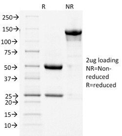
Biotium Cycloxygenase-2 (COX-2)(COX2/1941), CF568 conjugate, 0.1mg/mL
Prostaglandins are a diverse group of autocrine and paracrine hormones that mediate many cellular and physiologic processes. Prostaglandin H2 (PGH2) is an intermediate molecule in formation of the prostaglandins. Cyclooxygenase-1 (Cox-1) and cyclooxygenase-2 (Cox-2) are prostaglandin synthases that catalyze the formation of PGH2 from arachidonic acid (AA). Cox-1 and Cox-2 are isozymes of prostaglandin-endoperoxidase synthase (PTGS). Cox-1 is constitutively expressed in most tissues and is thought to serve in general housekeeping functions. Cox-2 is efficiently induced in migratory cells responding to pro-inflammatory stimuli and is considered to be an important mediator of inflammation. Both enzymes are targets for the nonsteroidal therapeutic anti-inflammatory drugs, NSAIDs. COX2 expression is significantly increased in 85-90% of human colorectal adenocarcinomas whereas levels of COX-1 are not changed. Primary antibodies are available purified, or with a selection of fluorescent

Biotium P-Cadherin (CDH3) (6A9), Biotin conjugate, 0.1mg/mL
This antibody recognizes a protein of 116 kDa, identified as P-Cadherin-1 (CDH3). It is a calcium-dependent cell-cell adhesion glycoprotein comprised of five extracellular cadherin repeats, a transmembrane region and a highly conserved cytoplasmic tail. This gene is located in a six-cadherin cluster in a region on the long arm of chromosome 16 that is involved in loss of heterozygosity events in breast and prostate cancer. In addition, aberrant expression of this protein is observed in cervical adenocarcinomas. Mutations in this gene have been associated with congenital hypotrichosis with juvenile macular dystrophy.Primary antibodies are available purified, or with a selection of fluorescent CF Dyes and other labels. CF Dyes offer exceptional brightness and photostability. Note: Conjugates of blue fluorescent dyes like CF405S and CF405M are not recommended for detecting low abundance targets, because blue dyes have lower fluorescence and can give higher non-specific back
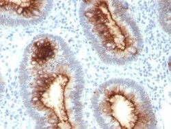
Biotium CD66 (CEA)(C66/1030), CF488A conjugate, 0.1mg/mL
This antibody recognizes proteins of 80-200 kDa, identified as different members of CEA family. CEA is synthesized during development in the fetal gut and is re-expressed in increased amounts in intestinal carcinomas and several other tumors. This MAb does not react with nonspecific cross-reacting antigen (NCA) and with human polymorphonuclear leucocytes. It shows no reaction with a variety of normal tissues and is suitable for staining of formalin/paraffin tissues. CEA is not found in benign glands, stroma, or malignant prostatic cells. Antibody to CEA is useful in detecting early foci of gastric carcinoma and in distinguishing pulmonary adenocarcinomas (60-70% are CEA positive) from pleural mesotheliomas (rarely or weakly CEA positive). Anti-CEA positivity is seen in adenocarcinomas from the lung, colon, stomach, esophagus, pancreas, gallbadder, urachus, salivary gland, ovary, and endocervix.Primary antibodies are available purified, or with a selection of fluorescent CF