
Mouse Secondary Antibodies


Biotium Cytokeratin 10 (Suprabasal Epithelial Marker) (KRT10/844 + KRT10/1275), 0.2mg/mL
This MAb recognizes a protein of 56.5 kDa, identified as cytokeratin 10 (CK10). CK10 is expressed in all suprabasal layers of the epidermis. In the epidermis, expression of CK10 strictly parallels the extent of differentiation; it is absent in the basal layer, appears in the first suprabasal layers and increases in concentration towards the granular layer. However, CK10 is rarely detected in early stages of vulvar squamous carcinomas (tumors less than 2 cm, clinical stage I) regardless of the tumor grade. In larger and more advanced tumors (greater than 2 cm, clinical stages II and III), CK10 is detected very frequently. Expression of CK10 is related to maturation of malignant keratinocytes, being preferentially detected in more-differentiated parts.Primary antibodies are available purified, or with a selection of fluorescent CF Dyes and other labels. CF Dyes offer exceptional brightness and photostability. Note: Conjugates of blue fluorescent dyes like CF405S and CF405M

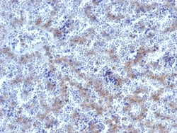
Biotium Glypican-3 (GPC3) (Hepatocellular Carcinoma Marker), CF568 conjugate, 0.1mg/mL
Glypican-3 (GPC3) is a glycosylphospatidyl inositol-anchored membrane protein, which may also be found in a secreted form. Anti-GPC3 has been identified as a useful tumor marker for the diagnosis of hepatocellular carcinoma (HCC), hepatoblastoma, melanoma, testicular germ cell tumors, and Wilm's tumor. In patients with HCC, GPC3 is overexpressed in neoplastic liver tissue and elevated in serum, but is undetectable in normal liver, benign liver, and the serum of healthy donors. GPC3 expression is also found to be higher in HCC liver tissue than in cirrhotic liver or liver with focal lesions such as dysplastic nodules and areas of hepatic adenoma (HA) with malignant transformation. In the context of testicular germ cell tumors, GPC3 expression is up regulated in certain histologic subtypes, specifically yolk sac tumors and choriocarcinoma. A high level of GPC3 expression is also found in some types of embryonal tumors, such as Wilm's tumor and hepatoblastoma, with a low or unde
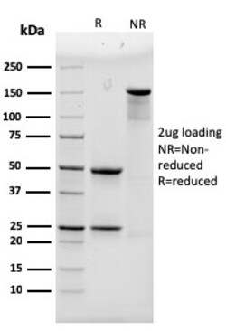
Biotium Alpha Actinin 4 / ACTN4(93), Biotin conjugate, 0.1mg/mL
Alpha actinins belong to the spectrin gene superfamily which represents a diverse group of cytoskeletal proteins, including the alpha and beta spectrins and dystrophins. There are four tissue-specific -actinins, namely -actinin-1, -actinin-2, -actinin-3 and -actinin-4, which are localized to muscle and non-muscle cells, including skeletal, cardiac and smooth muscle cells, as well as within the cytoskeleton. Alpha actinin is an actin-binding protein with multiple roles in different cell types. In nonmuscle cells, the cytoskeletal isoform is found along microfilament bundles and adherens-type junctions, where it is involved in binding actin to the membrane. In contrast, skeletal, cardiac, and smooth muscle isoforms are localized to the Z-disc and analogous dense bodies, where they help anchor the myofibrillar actin filaments. This gene encodes a nonmuscle, alpha actinin isoform which is concentrated in the cytoplasm, and thought to be involved in metastatic processes. Each -act
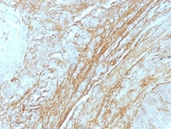
Biotium Tenascin(T2H5), CF740 conjugate, 0.1mg/mL
In Western blotting, this antibody reacts with two bands of ~MW of 210 kDa and 300 kDa, identified as two isoforms of Tenascin C. Specificity of this MAb is validated by sequential immunoprecipitation with a PAb against Tenascin C. Tenascin C is a multifunctional, disulfide-linkedhexameric extracellular matrix glycoprotein expressed in association with mesenchymal epithelial interactions during development and in the neo-vasculature and stroma of undifferentiated tumors. In adults, it is restricted to certain epithelial-stromal interfaces and increases markedly in hyper-proliferative diseases and in stroma of many neoplasms, including gliomas, breast, squamous and lung carcinomas.Primary antibodies are available purified, or with a selection of fluorescent CF Dyes and other labels. CF Dyes offer exceptional brightness and photostability. Note: Conjugates of blue fluorescent dyes like CF405S and CF405M are not recommended for detecting low abundance targets, because blue
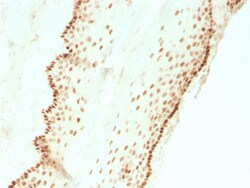
Biotium SOX2 (Embryonic Stem Cell Marker)(SOX2/3811R), 0.2mg/mL
SOX2 is required for stem cell maintenance in the central nervous system, and it also regulates gene expression in the stomach. SOX2 is necessary for regulating multiple transcription factors that affect Oct 3/4 expression. An essential function of SOX2 is to stabilize embryonic stem cells in a pluripotent state by maintaining the requisite level of Oct 3/4 expression. Reportedly, SOX2 is associated with aggressive phenotypes of breast, head and neck, gastric, colorectal, bladder, and small cell lung cancers. However, SOX2 is expressed in a high percentage of lung squamous cell carcinomas and has been shown to be an independent favorable prognostic marker. Primary antibodies are available purified, or with a selection of fluorescent CF Dyes and other labels. CF Dyes offer exceptional brightness and photostability. Note: Conjugates of blue fluorescent dyes like CF405S and CF405M are not recommended for detecting low abundance targets, because blue dyes have lower fluoresc

Biotium Podoplanin (PDPN) (Lymphatic Endothelial & Mesothelial Marker) (PDPN/1433), Biotin conjugate, 0.1mg/mL
This antibody recognizes a muco-protein of 38-43 kDa, which is identified Podoplanin (PDPN). It localizes in stromal cells of peripheral lymphoid tissue and thymic epithelial cells. As a regulator of the lymphatic endothelium, podoplanin probably plays a role in maintaining the unique shape of podocytes. It is selectively expressed in lymphatic endothelium as well as lymphoangiomas, Kaposi'sarcomas, and in a subset of angiosarcomas with probable lymphatic differentiation. Recent studies have also shown podoplanin to be a highly sensitive and relatively specific marker for epithelioid mesothelioma. Therefore, it can be used in a panel to distinguish mesotheliomas or mesothelial cells from pulmonary carcinomas.Primary antibodies are available purified, or with a selection of fluorescent CF Dyes and other labels. CF Dyes offer exceptional brightness and photostability. Note: Conjugates of blue fluorescent dyes like CF405S and CF405M are not recommended for detecting low abu
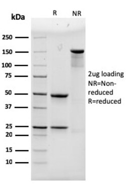
Biotium Alpha Actinin 4 / ACTN4(93), CF488A conjugate, 0.1mg/mL
Alpha actinins belong to the spectrin gene superfamily which represents a diverse group of cytoskeletal proteins, including the alpha and beta spectrins and dystrophins. There are four tissue-specific -actinins, namely -actinin-1, -actinin-2, -actinin-3 and -actinin-4, which are localized to muscle and non-muscle cells, including skeletal, cardiac and smooth muscle cells, as well as within the cytoskeleton. Alpha actinin is an actin-binding protein with multiple roles in different cell types. In nonmuscle cells, the cytoskeletal isoform is found along microfilament bundles and adherens-type junctions, where it is involved in binding actin to the membrane. In contrast, skeletal, cardiac, and smooth muscle isoforms are localized to the Z-disc and analogous dense bodies, where they help anchor the myofibrillar actin filaments. This gene encodes a nonmuscle, alpha actinin isoform which is concentrated in the cytoplasm, and thought to be involved in metastatic processes. Each -act
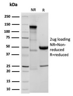
Biotium Ornithine Decarboxylase-1 (ODC-1)(ODC1/3636R), CF568 conjugate, 0.1mg/mL
Recognizes a 53 kDa protein, identified as the Ornithine Decarboxylase (ODC-1). ODC is the initial and rate-limiting enzyme in the biosynthetic pathway of polyamines and is involved in the conversion of ornithine to putrescine. The biological activity of ODC-1 is rapidly induced in response to virtually all agents known to promote cell proliferation including hormones, drugs, growth factors, mitogens, and tumor promoters. Reportedly, ODC mRNA levels are elevated in lung carcinomas as well as in colon adenomas and carcinomas. ODC activity in colorectal carcinomas is greater than those in adenomas and normal mucosa. Primary antibodies are available purified, or with a selection of fluorescent CF Dyes and other labels. CF Dyes offer exceptional brightness and photostability. Note: Conjugates of blue fluorescent dyes like CF405S and CF405M are not recommended for detecting low abundance targets, because blue dyes have lower fluorescence and can give higher non-specific backg

Biotium Clathrin, Light Chain (CLC/1421), 0.2mg/mL
This antibody recognizes proteins of 31-44 kDa, which are identified as Clathrin Light Chains (both A & B). Clathrin is composed of three heavy chains and three light chains, which associate non-covalently to form a triskelion structure. Clathrin light chain regulates the self-assembly of triskelions onto intracellular membranes. Clathrin light chain subunits (LCA and LCB) contribute to regulation of coated vesicle formation to sort proteins during receptor-mediated endocytosis and organelle biogenesis. LCA and LCB are encoded by two discrete genes. They share only 60% homology, and have certain features in common. Both LCA and LCB undergo alternative mRNA splicing, which results in the generation of tissue-specific isoforms.Primary antibodies are available purified, or with a selection of fluorescent CF Dyes and other labels. CF Dyes offer exceptional brightness and photostability. Note: Conjugates of blue fluorescent dyes like CF405S and CF405M are not recommended for
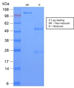
Biotium TAG-72 / CA72.4 (Tumor-Associated Glycoprotein)(CA72/2869R), CF488A conjugate, 0.1mg/mL
Recognizes an oncofetal antigen of 220 kDa, identified as a tumor-associated glycoprotein (TAG-72) with properties of a mucin. This MAb defines the mucin-carried sialylated-Tn epitope. TAG-72 is usually expressed by adenocarcinomas, but is negative in mesotheliomas. Studies have reported that this antibody has 80% sensitivity and 93% specificity for pulmonary adenocarcinoma. Therefore, TAG-72 is a useful marker to distinguish between mesothelioma and adenocarcinoma. However, false positive reactions can occur so results must be interpreted with the utmost caution. This antibody may be useful in the differentiation of non-small cell carcinomas from small cell carcinomas of the lung. The combined use of anti-TAG-72 and anti-GCDFP-15 is valuable in the diagnosis of apocrine carcinoma. Primary antibodies are available purified, or with a selection of fluorescent CF Dyes and other labels. CF Dyes offer exceptional brightness and photostability. Note: Conjugates of blue fluoresc
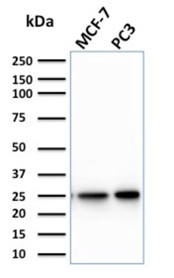
Biotium Calcineurin B homologous protein 2 / HCC Antigen 520(CPTC-CHP2-1), Biotin conjugate, 0.1mg/mL
Calcineurin B homologous protein 2, also known as hepatocellular carcinoma-associated antigen 520, is a 196 amino acid protein that contains 4 EF-hand domains and plays a potential role in transmembrane Na/H exchange. By binding to and activating NHE-1, CHP2 increases the pH and works to protect cells from serum deprivation-induced death. Though not typically detected in normal tissues, CHP2 is highly expressed in malignantly transformed cells and is therefore considered to be a tumor-associated antigen. Ectopic expression of CHP2 promotes proliferation of HEK293 cells and knockdown of CHP2 mRNA in HepG2 cells inhibits cell proliferation. Like calcineurin B, CHP2 can bind to and stimulate phosphatase activity of calcineurin A and activate the calcineurin/NFAT signaling pathway. Primary antibodies are available purified, or with a selection of fluorescent CF Dyes and other labels. CF Dyes offer exceptional brightness and photostability. Note: Conjugates of blue fluorescent dyes
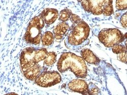
Biotium Mucin 6 (MUC6)(CLH5), CF740 conjugate, 0.1mg/mL
The MUC6 gastric mucin is a secreted glycoprotein that plays an essential role in epithelial cyto-protection from acid, proteases, pathogenic microorganisms, and mechanical trauma in the gastrointestinal tract. Mucin 6 expression is highest in the stomach and gall bladder, with lower expression in the terminal ileum and right colon. In gastric cancer, Mucin 6 has an altered expression. In normal stomach, Mucin 6 is associated with Lewis type 2; Mucin 6 is also expressed in gastric metaplasia, duodenum and pancreas. Mucin 6 is a secretory mucin, located in the deeper mucosal folds of human gall bladder, and its expression is altered with increasing degrees of inflammation.Primary antibodies are available purified, or with a selection of fluorescent CF Dyes and other labels. CF Dyes offer exceptional brightness and photostability. Note: Conjugates of blue fluorescent dyes like CF405S and CF405M are not recommended for detecting low abundance targets, because blue dyes have
Biotium Galectin-1 / Human Placental Lactogen (hPL) (GAL1/2499R), CF568 conjugate, 0.1mg/mL
Galectin-1 is a member of the beta-galactoside-binding family and is a dimeric protein of 14 kD participating in a variety of normal and pathological processes, including cancer progression. Galectin-1 can affect the proliferation of normal and malignant cells. Inhibition of cell growth is observed in a lactose-dependent manner as lower concentrations of the lectin stimulate cell proliferation. Galectin-1 may also be implicated in the induction of apoptosis of activated T cells through the binding of exogenous galectin-1 to CD45 molecules present on the surface of lymphocytes. Galectin-1, reported to be present either at the surface of cancer cells or accumulated around these cells could act as an immunological shield to protect against a T cell immune response and provide an advantage for survival.Primary antibodies are available purified, or with a selection of fluorescent CF Dyes and other labels. CF Dyes offer exceptional brightness and photostability. Note: Conjugates of b
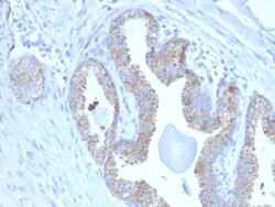
Biotium PMEPA1 / TMEPAI (Tumor Suppressor Oncoprotein)(PMEPA1/2697), CF568 conjugate, 0.1mg/mL
PMEPA1 (prostate transmembrane protein, androgen induced 1 is a 287 amino acid single-pass membrane protein that contains WW-binding motifs and localizes to the cell membrane. Expressed at high levels in prostate, kidney and ovary, PMEPA1 interacts with NEDD4 and may play a role in regulating AR (androgen receptor) levels, specifically in prostate cells. Down regulation of PMEPA1 is observed in prostate tumors, suggesting that PMEPA1 may exhibit activity as a tumor suppressor. Overexpression of this protein may play a role in multiple types of cancer.Primary antibodies are available purified, or with a selection of fluorescent CF Dyes and other labels. CF Dyes offer exceptional brightness and photostability. Note: Conjugates of blue fluorescent dyes like CF405S and CF405M are not recommended for detecting low abundance targets, because blue dyes have lower fluorescence and can give higher non-specific background than other dye colors.