
Mouse Secondary Antibodies


Biotium Rb1 (Tumor Suppressor Protein) (RB1/1754), 0.2mg/mL
This antibody recognizes a 105 kDa phosphoprotein, identified as retinoblastoma (Rb) gene product. It shows no cross reaction with p107 or p130. It reacts with the hyper-phosphorylated as well as the un (under) phosphorylated form of the Rb protein. Retinoblastoma gene product plays a key role in cell cycle control. It has been identified as a tumor suppressor gene whose loss of its function leads to tumor development. It is widely expressed in a variety of human tissues including breast, esophageal, squamous cell and cervical carcinoma.Primary antibodies are available purified, or with a selection of fluorescent CF Dyes and other labels. CF Dyes offer exceptional brightness and photostability. Note: Conjugates of blue fluorescent dyes like CF405S and CF405M are not recommended for detecting low abundance targets, because blue dyes have lower fluorescence and can give higher non-specific background than other dye colors.

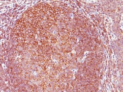
Biotium BCL10 (MALT-Lymphoma Marker)(151), CF568 conjugate, 0.1mg/mL
B-cell lymphoma/leukemia 10 (BCL10) is a signaling protein that promotes apoptosis, pro-caspase-9 maturation, and activation of NF-kappa-B via NIK and IKK. BCL10 contains an N-terminal caspase recruitment domain (CARD), which is found in a number of apoptotic regulatory molecules. BCL10 is involved in the adaptive immune response, and may be an adapter protein between upstream TNFR1-TRADD-RIP complex and the downstream NIK-IKK-IKAP complex. A BCL10 translocation is recurrent in low-grade mucosa-associated lymphoid tissue (MALT lymphoma). Primary antibodies are available purified, or with a selection of fluorescent CF Dyes and other labels. CF Dyes offer exceptional brightness and photostability. Note: Conjugates of blue fluorescent dyes like CF405S and CF405M are not recommended for detecting low abundance targets, because blue dyes have lower fluorescence and can give higher non-specific background than other dye colors.
Biotium Chromogranin A / CHGA (Neuroendocrine Marker) (rCHGA/798), CF488A conjugate, 0.1mg/mL
Chromogranin A is present in neuroendocrine cells throughout the body, including the neuroendocrine cells of the large and small intestine, adrenal medulla and pancreatic islets. It is an excellent marker for carcinoid tumors, pheochromocytomas, paragangliomas, and other neuroendocrine tumors. Co-expression of chromogranin A and neuron specific enolase (NSE) is common in neuroendocrine neoplasms. Reportedly, co-expression of certain keratins and chromogranin indicates neuroendocrine lineage. The presence of strong anti-chromogranin staining and absence of anti-keratin staining should raise the possibility of paraganglioma. The co-expression of chromogranin and NSE is typical of neuroendocrine neoplasms. Most pituitary adenomas and prolactinomas readily express chromogranin.Primary antibodies are available purified, or with a selection of fluorescent CF Dyes and other labels. CF Dyes offer exceptional brightness and photostability. Note: Conjugates of blue fluorescent dyes
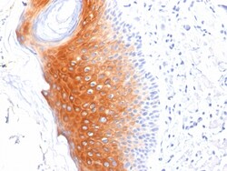
Biotium Cytokeratin 1 (Suprabasal Keratinocyte Marker)(LHK1), 1mg/mL
Keratins are a family of intermediate filament proteins that assemble into filaments through forming heterodimers of one type I keratin (keratins 9 to 23) and one type II keratin (keratins 1 to 8). Keratins demonstrate tissue and differentiation specific expression profiles. Cytokeratin 1 is a hetero-tetramer of two type I as well as two type II keratins, keratin-10 (KRT10). Keratin 1 is differentiation specific keratin that is one of the predominant keratins in suprabasal keratinocytes in stratified epithelia. Mutations in keratin 1 cause epidermolytic hyperkeratosis. Primary antibodies are available purified, or with a selection of fluorescent CF Dyes and other labels. CF Dyes offer exceptional brightness and photostability. Note: Conjugates of blue fluorescent dyes like CF405S and CF405M are not recommended for detecting low abundance targets, because blue dyes have lower fluorescence and can give higher non-specific background than other dye colors.
Biotium Rb1 (Tumor Suppressor Protein) (1F8), CF488A conjugate, 0.1mg/mL
This antibody recognizes a 105 kDa phosphoprotein, identified as retinoblastoma (Rb) gene product. Its epitope is localized between aa 703-772. It shows no cross reaction with p107 or p130. It specifically stains the nuclei of BT-20 cells and primary human foreskin fibroblast (HFF) cells. It does not stain the Rb-negative BT549 cells. It reacts with the hyperphosphorylated as well as the un (under) phosphorylated form of the Rb protein. Retinoblastoma gene product plays a key role in cell cycle control. It has been identified as a tumor suppressor gene whose loss of its function leads to tumor development. It is widely expressed in a variety of human tissues including breast, esophageal, squamous cell and cervical carcinoma.Primary antibodies are available purified, or with a selection of fluorescent CF Dyes and other labels. CF Dyes offer exceptional brightness and photostability. Note: Conjugates of blue fluorescent dyes like CF405S and CF405M are not recommended for d
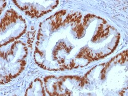
Biotium Estrogen Receptor, alpha (Marker of Estrogen Dependence) (ESR1/1935), Biotin conjugate, 0.1mg/mL
This monoclonal antibody is specific to estrogen receptor alpha (ER alpha) and shows minimal cross-reaction with other members of the family. ER is an important regulator of growth and differentiation in the mammary gland. Presence of ER in breast tumors indicates an increased likelihood of response to anti-estrogen (e.g. tamoxifen) therapy. It strongly stains nuclei of epithelial cells in breast carcinomas.Primary antibodies are available purified, or with a selection of fluorescent CF Dyes and other labels. CF Dyes offer exceptional brightness and photostability. Note: Conjugates of blue fluorescent dyes like CF405S and CF405M are not recommended for detecting low abundance targets, because blue dyes have lower fluorescence and can give higher non-specific background than other dye colors.
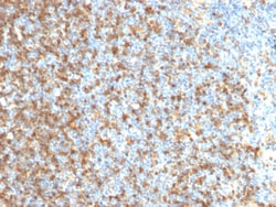
Biotium LMO2 (B-Cell Marker)(LMO2/1971), 0.2mg/mL
The LMO2 protein has a central and crucial role in hematopoietic development and is highly conserved. It has a particular function in normal and lymphatic endothelial cells involving the regulation of angiogenesis and lymph-angiogenesis. Immunohistochemical studies have also demonstrated expression of LMO2 in both normal germinal center B-cells and germinal center-derived B-cell lymphomas, including follicular lymphoma and diffuse large B-cell lymphoma. The use of anti-LMO2 is valuable as a tool in the identification of lymphomas of B-cell origin. LMO2 is useful in differentiating follicular lymphoma (LMO2 ) from nodal marginal zone lymphoma (LM02-). It also is positive in Hodgkin's and Burkitt's lymphomas. Primary antibodies are available purified, or with a selection of fluorescent CF Dyes and other labels. CF Dyes offer exceptional brightness and photostability. Note: Conjugates of blue fluorescent dyes like CF405S and CF405M are not recommended for detecting low abun
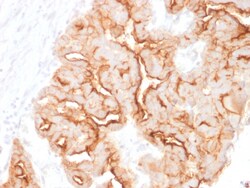
Biotium MUC16 / CA125 (Ovarian Carcinoma Marker)(MUC16/1860), Biotin conjugate, 0.1mg/mL
The mucins are a family of highly glycosylated, secreted proteins with a basic structure consisting of a variable number of tandem repeats (VNTRs). Membrane-associated and secretory Mucins are high molecular weight glycoproteins of the glycocalyx (polysaccharide biofilm) that protects mucosal epithelium from particulate matter and microorganisms. Epithelial Mucins are large, secreted and cell surface glycoproteins crucial for adhesion modulation, signaling and epithelial cell protection. The number of repeats is highly polymorphic and varies among different alleles. The Mucin family consists of Mucins 1-4, Mucin 5 (AC and B), Mucins 6-8, Mucins 11-13 and Mucins 15-17. The Mucin 16 protein (also commonly referred to as CA125), encoded for by the gene MUC16, is a very high molecular weight tumor antigen consisting of three domains: a carboxy terminal domain, an extracellular domain and an amino terminal domain. Mucin 16, an ovarian cancer-associated antigen, is used as a marker
Biotium CDX2 (GI Epithelial Marker) (CDX2/1690), CF488A conjugate, 0.1mg/mL
The intestine-specific transcription factors CDX1 and CDX2 are important for directing intestinal development, differentiation, proliferation and maintenance of the intestinal phenotype. CDX2 protein expression has been seen in GI carcinomas. Anti-CDX2 has been useful to establish GI origin of metastatic adenocarcinomas and carcinoids and is especially useful to distinguish metastatic colorectal adenocarcinoma from lung adenocarcinoma. However, mucinous carcinomas of the ovary also express CDX2 protein. It limits the usefulness of this marker in the distinction of metastatic colorectal adenocarcinoma from mucinous carcinoma of the ovary.Primary antibodies are available purified, or with a selection of fluorescent CF Dyes and other labels. CF Dyes offer exceptional brightness and photostability. Note: Conjugates of blue fluorescent dyes like CF405S and CF405M are not recommended for detecting low abundance targets, because blue dyes have lower fluorescence and can give hi
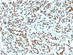
Biotium Histone H1 (Nuclear Marker) (HH1/1784R), CF647 conjugate, 0.1mg/mL
Eukaryotic histones are basic and water-soluble nuclear proteins that form hetero-octameric nucleosome particles by wrapping 146 base pairs of DNA in a left-handed supealpha-helicalturn sequentially to form chromosomal fiber. Two molecules of each of the four core histones (H2A, H2B, H3, and H4) form the octamer; formed of two H2A-H2B dimers and two H3-H4 dimers, forming two nearly symmetrical halves by tertiary structure. Over 80% of nucleosomes contain the linker Histone H1, derived from an intronless gene that interacts with linker DNA between nucleosomes and mediates compaction into higher order chromatin. Histones are subject to posttranslational modification by enzymes primarily on their N-terminal tails, but also in their globular domains. Such modifications include methylation, citrullination, acetylation, phosphorylation, sumoylation, ubiquitination and ADP-ribosylation.Primary antibodies are available purified, or with a selection of fluorescent CF Dyes and other l
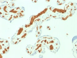
Biotium Spectrin Alpha 1 (Erythrocyte Marker) (SPTA1/1832), CF488A conjugate, 0.1mg/mL
Spectrin is an actin crosslinking and molecular scaffold protein that links the plasma membrane to the actin cytoskeleton, and functions in the determination of cell shape, arrangement of transmembrane proteins, and organization of organelles. Spectrins function as membrane organizers and stabilizers, composed of non-homologous α andβ chains, which aggregate side-to-side in an anti-parallel fashion to form dimers, tetramers and higher polymers. Spectrin α I and spectrinβ I are present in erythrocytes, whereas spectrin α II (also designated fodrin α) and spectrinβ II (also designated fodrinβ) are present in other somatic cells. The spectrin tetramers in erythrocytes act as barriers to lateral diffusion, but spectrin dimers seem to lack this function.Primary antibodies are available purified, or with a selection of fluorescent CF Dyes and other labels. CF Dyes offer exceptional brightness and photostability. Note: Conjugates of blu
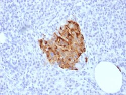
Biotium Chromogranin A / CHGA (Neuroendocrine Marker) (CHGA/1773R), CF740 conjugate, 0.1mg/mL
Chromogranin A is present in neuroendocrine cells throughout the body, including the neuroendocrine cells of the large and small intestine, adrenal medulla and pancreatic islets. It is an excellent marker for carcinoid tumors, pheochromocytomas, paragangliomas, and other neuroendocrine tumors. Co-expression of chromogranin A and neuron specific enolase (NSE) is common in neuroendocrine neoplasms. Reportedly, co-expression of certain keratins and chromogranin indicates neuroendocrine lineage. The presence of strong anti-chromogranin staining and absence of anti-keratin staining should raise the possibility of paraganglioma. The co-expression of chromogranin and NSE is typical of neuroendocrine neoplasms. Most pituitary adenomas and prolactinomas readily express chromogranin.Primary antibodies are available purified, or with a selection of fluorescent CF Dyes and other labels. CF Dyes offer exceptional brightness and photostability. Note: Conjugates of blue fluorescent dyes
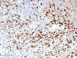
Biotium HSV1 (Herpes Simplex Virus Type I) (HSV1/1934), Biotin conjugate, 0.1mg/mL
The antibody reacts with HSV type 1 specific antigen. It is suitable for detection of HSV in human cellular material obtained from superficial lesions or biopsies and for the early identification of HSV in infected tissue cultures. The herpes simplex virus (HSV) (also known as cold sore, night fever or fever blister) is a virus that causes a contagious disease. There are two main types of Herpes Simplex Virus (HSV), 1 and 2. The HSV-1 strain generally appears in the orafacial organs. HSV2 usually resides in the sacral ganglion at the base of the spine. All herpes viruses are morphologically identical: they have a large double-stranded DNA genome and the virion consists of an icosahedral nucleo-capsid, which is surrounded by a lipid bilayer envelope.Primary antibodies are available purified, or with a selection of fluorescent CF Dyes and other labels. CF Dyes offer exceptional brightness and photostability. Note: Conjugates of blue fluorescent dyes like CF405S and CF405M
Biotium von Willebrand Factor / Factor VIII Related-Ag (Endothelial Marker) (VWF/1465), 0.2mg/mL
von Willebrand Factor (vWF) is a multimeric glycoprotein that is found in endothelial cells, plasma and platelets. It acts as a carrier protein for Factor VIII and promotes platelet adhesion and aggregation. vWF undergoes a variety of posttranslational modifications that influence the affinity and availability for Factor VIII, including cleavage of the propeptide and formation of N-terminal disulfide bonds. This antibody helps to establish the endothelial nature of some lesions of disputed histogenesis, e.g. Kaposi's sarcoma and cardiac myxoma. It is widely used for differentiating vascular lesions from those of other tissue differentiation within a panel of other vascular markers although not all tumors of endothelial differentiation contain this antigen.Primary antibodies are available purified, or with a selection of fluorescent CF Dyes and other labels. CF Dyes offer exceptional brightness and photostability. Note: Conjugates of blue fluorescent dyes like CF405S and C
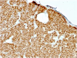
Biotium Parathyroid Hormone (PTH) (N-Terminal)(rPTH/911), CF568 conjugate, 0.1mg/mL
Epitope of this MAb maps in the N-terminus of PTH, a hormone produced by the parathyroid gland that regulates the concentration of calcium and phosphorus in extracellular fluid. This hormone elevates blood Ca2 levels by dissolving the salts in bone and preventing their renal excretion. It is produced in the parathyroid gland as an 84 amino acid single chain polypeptide. It can also be secreted as N-terminal truncated fragments or C-terminal fragments after intracellular degradation, as in case of hypercalcemia. Defects in this gene are a cause of familial isolated hypoparathyroidism (FIH); also called autosomal dominant hypoparathyroidism or autosomal dominant hypocalcemia. FIH is characterized by hypocalcemia and hyperphosphatemia due to inadequate secretion of parathyroid hormone. Symptoms are seizures, tetany and cramps. FIH exist both as autosomal dominant and recessive forms of hypoparathyroidism. Primary antibodies are available purified, or with a selection of fluores