
Mouse Secondary Antibodies

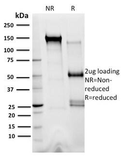
Biotium XRCC3(10F1/6), CF740 conjugate, 0.1mg/mL
The x-ray repair cross-complementing (XRCC) proteins are responsible for efficiently repairing and maintaining genetic stability following DNA base damage. These genes share sequence similarity with the yeast DNA repair protein Rad51. XRCC1 is a protein that facilitates the DNA base excision repair pathway by interacting with DNA ligase III and DNA polymerase to repair DNA single-strand breaks. XRCC2 and XRCC3 are both involved in maintaining chromosome stability during cell division. XRCC2 is required for efficient repair of DNA double-strand breaks by homologous recombination between sister chromatids, and XRCC3 interacts directly with Rad51 to cooperate with Rad51 during recombinational repair. XRCC4 is an accessory factor of DNA Ligase IV that preferentially binds DNA with nicks or broken ends. XRCC4 binds to DNA Ligase IV and enhances its joining activity, and it is also involved in V(D)J recombination. Any defect in one of the known components of the DNA repair/V(D)J re

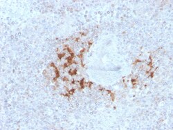
Biotium TRAF1 (TNFR-Associated Factor 1) (Lymphomatoid Papulosis Marker)(TRAF1/3298), CF594 conjugate, 0.1mg/mL
This MAb recognizes a protein of 52 kDa, which is identified as TNFR1 (TNFR-associated factor 1). CD30-positive lymphoproliferations of the skin comprise 30% of all primary cutaneous T-cell lymphomas (CTCLs). Besides borderline cases this group includes lymphomatoid papulosis (LyP) and primary cutaneous anaplastic large T-cell lymphoma (cALCL). Although the two entities overlap clinically, histopathologically, immunopathologically and genetically, they differ considerably in their prognosis. In particular, common feature of both cases is histologically the presence of atypical lymphoid CD30-positive T blasts and genetically a clonal T-cell-receptor rearrangement. However, both cases differ considerably in their clinical course: Lesions of LyP regress spontaneously, whereas those of cALCL persist and may progress and spread. Moreover, LyP patients do not benefit from an aggressive radio- and/or chemotherapeutic approach, in contrast to patients with cALCL. Besides, LyP and cAL

Biotium Ki-67 (Proliferating Cell Marker) (MKI67/2462), CF740 conjugate, 0.1mg/mL
Ki-67 antigen is a nuclear, non-histone protein that is present in all stages of the cell cycle except G0. This characteristic makes Ki-67 an excellent marker for proliferating cells and is commonly used as one of the prognostic factors in cancer studies. A correlation has been demonstrated between Ki-67 index and the histo-pathological grade of neoplasms. Assessment of Ki-67 expression in renal and ureter tumors shows a correlation between tumor proliferation and disease progression, thus making it possible to differentiate high-risk patients. Ki-67 expression may also prove to be important for distinguishing between malignant and benign peripheral nerve sheath tumors. Ki-67 labeling index has been shown to be a prognostic marker in a number of neoplasms including grade II astrocytoma, oligodendroglioma, colon carcinoma, and breast carcinoma. In general, Ki-67 is a good marker of proliferating cell populations.Primary antibodies are available purified, or with a selection of
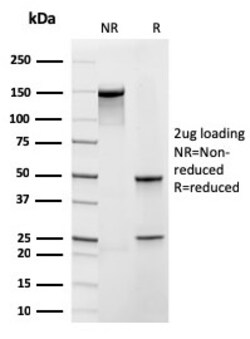
Biotium Frataxin (Mitochondrial Marker)(rFXN/2124), Biotin conjugate, 0.1mg/mL
Frataxin is a nuclear-encoded mitochondrial protein implicated in Friedreich's ataxia (FRDA), a human autosomal recessive neurodegenerative disease. Lack of Frataxin causes iron to accumulate in the mitochondrial matrix suggesting that Frataxin is involved in mitochondrial iron homeostasis and possibly in iron transport. Frataxin has an alpha-beta fold consisting of two helices flanking an antiparallel beta sheet. Primary antibodies are available purified, or with a selection of fluorescent CF Dyes and other labels. CF Dyes offer exceptional brightness and photostability. Note: Conjugates of blue fluorescent dyes like CF405S and CF405M are not recommended for detecting low abundance targets, because blue dyes have lower fluorescence and can give higher non-specific background than other dye colors.
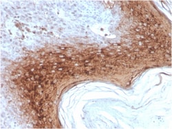
Biotium Involucrin (Squamous Cell Terminal Differentiation Marker)(rIVRN/827), CF488A conjugate, 0.1mg/mL
Recognizes a protein of 66 kDa-170 kDa, identified as involucrin. In Western blotting of cultured human keratinocytes, this MAb reacts with a 120 kDa protein. It stains the involucrin in a variety of sizes: 170 kDa in MCF-7 cells, a doublet of ~115 kDa and 150 kDa in gorilla and owl monkey, 66 kDa in dog, and a doublet of 105 kDa in pig. Its epitope maps between codon 421-568 of human involucrin. Involucrin is expressed in a range of stratified squamous epithelia, including the cornea, which lacks a distinct cornified layer. In normal epidermis, it is first expressed in the upper spinous layers, and in keratinocyte cultures, all cells that have left the basal layer express it. Involucrin expression is altered in pathological conditions: in psoriasis and other benign epidermal hyperplasias, involucrin expression begins closer to the basal layer than normal; expression is abnormal in squamous cell carcinomas and premalignant lesions, and is reduced in severe dysplasias of the l

Biotium Nucleolar / Nucleoli(NM95), CF740 conjugate, 0.1mg/mL
This MAb is an excellent marker for human cells in xenographic model research. It reacts specifically with human cells. This monoclonal antibody is part of a new panel of reagents, which recognizes subcellular organelles or compartments of human cells. These markers may be useful in identification of these organelles in cells, tissues, and biochemical preparations. MAb NM95 recognizes an antigen associated with the nucleoli in human cells. It can be used to stain the nucleoli in cell or tissue preparations and can be used as a marker of the nucleoli in subcellular fractions. It produces a speckled pattern in the nuclei of cells of normal and malignant cells and may be used to stain the nucleoli of cells in fixed or frozen tissue sections. It can be used with paraformaldehyde fixed frozen tissue or cell preparations and formalin fixed, paraffin-embedded tissue sections.Primary antibodies are available purified, or with a selection of fluorescent CF Dyes and other labels. CF

Biotium S100B(4C4.9), CF740 conjugate, 0.1mg/mL
S100 belongs to the family of calcium binding proteins. S100A and S100B proteins are two members of the S100 family. S100A is composed of an alpha and a beta chain whereas S100B is composed of two beta chains. This antibody is specific against an epitope located on the beta-chain (i.e. in S-100A and S-100B) but not on the alpha-chain of S-100 (i.e. in S-100A and S100A0). This antibody can be used to localize S-100A and S-100B in various tissue sections. S-100 protein has been found in normal melanocytes, Langerhans cells, histiocytes, chondrocytes, lipocytes, skeletal and cardiac muscle, Schwann cells, epithelial and myoepithelial cells of the breast, salivary and sweat glands, as well as in glial cells. Neoplasms derived from these cells also express S-100 protein, albeit non-uniformly. A large number of well-differentiated tumors of the salivary gland, adipose and cartilaginous tissue, and Schwann cell-derived tumors express S-100 protein. Almost all malignant melanomas and
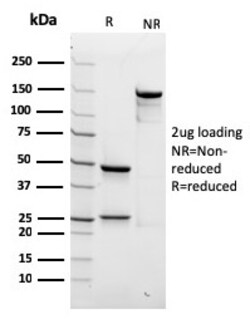
Biotium Spectrin beta III (SPTBN2)(rSPTBN2/1778), Biotin conjugate, 0.1mg/mL
Spectrin is an actin binding protein that is a major component of the plasma membrane skeleton. Spectrins function as membrane organizers and stabilizers by forming dimers, tetramers and higher polymers. Vertebrate spectrins have two alpha-subunits (alpha-I/alpha-II) four beta-subunits (beta-I-beta-IV) and a beta-H subunit creating diversity and specialization of function. Spectrin and spectrin are present in erythrocytes, whereas spectrin II (also designated fodrin ) and spectrin I (also designated fodrin ) are present in other somatic cells. The spectrin tetramers in erythrocytes act as barriers to lateral diffusion, but spectrin dimers seem to lack this function. Spectrin III is highly homologous to both spectrin I and spectrin II. Spectrin III is highly expressed in brain, kidney, pancreas and liver, and at lower levels in lung and placenta. Spectrin beta 3 is primarily expressed in nervous tissues with highest expression levels in the cerebellum, where it is found in Pur
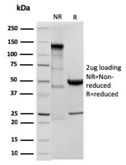
Biotium Ornithine Decarboxylase-1 (ODC-1)(ODC1/3636R), 0.2mg/mL
Recognizes a 53 kDa protein, identified as the Ornithine Decarboxylase (ODC-1). ODC is the initial and rate-limiting enzyme in the biosynthetic pathway of polyamines and is involved in the conversion of ornithine to putrescine. The biological activity of ODC-1 is rapidly induced in response to virtually all agents known to promote cell proliferation including hormones, drugs, growth factors, mitogens, and tumor promoters. Reportedly, ODC mRNA levels are elevated in lung carcinomas as well as in colon adenomas and carcinomas. ODC activity in colorectal carcinomas is greater than those in adenomas and normal mucosa. Primary antibodies are available purified, or with a selection of fluorescent CF Dyes and other labels. CF Dyes offer exceptional brightness and photostability. Note: Conjugates of blue fluorescent dyes like CF405S and CF405M are not recommended for detecting low abundance targets, because blue dyes have lower fluorescence and can give higher non-specific backg
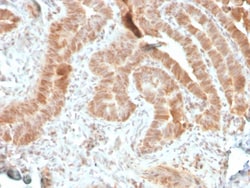
Biotium AKT1 (Prognostic Marker for Neuroendocrine Tumors)(rAKT1/2491), CF488A conjugate, 0.1mg/mL
This antibody recognizes a protein of 62 kDa, which is identified as AKT1. The serine/threonine kinase Akt family contains several members, including Akt1 (also designated PKB or RacPK), Akt2 (also designated PKBbeta or RacPK-beta) and Akt3 (also designated PKB-gamma or thymoma viral proto-oncogene 3), which exhibit sequence homology with the protein kinase A and C families and are encoded by the c-Akt proto-oncogene. All members of the Akt family have a Pleckstrin homology domain. Akt1 and Akt2 are activated by PDGF stimulation. This activation is dependent on PDGFR-beta tyrosine residues 740 and 751, which bind the subunit of the phosphatidylinositol 3-kinase (PI 3-kinase) complex. Activation of Akt1 by insulin or insulin-growth factor-1 (IGF-1) results in phosphorylation of both Thr 308 and Ser 473. Akt proteins become phosphorylated and activated in insulin/IGF-1-stimulated cells by an upstream kinase(s), and the activation of Akt1 and Akt2 is inhibited by the PI kinase i
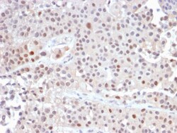
Biotium p21WAF1 (Tumor Suppressor Protein)(CIP1/2489R), CF568 conjugate, 0.1mg/mL
This MAb recognizes a 21 kDa protein, identified as the p21WAF1 tumor suppressor protein. This MAb is highly specific to p21 and shows no cross-reaction with other closely related mitotic inhibitors. p21WAF1 is a specific inhibitor of cdk's and a tumor suppressor involved in the pathogenesis of a variety of malignancies. The expression of this gene acts as an inhibitor of the cell cycle during G1 phase and is tightly controlled by the tumor suppressor protein p53. Its expression is induced by the wild type, but not mutant, p53 suppressor protein. Normal cells generally display a rather intense nuclear p21 expression. Loss of p21 expression has been reported in many carcinomas (gastric carcinoma, non-small cell lung carcinoma, thyroid carcinoma). Primary antibodies are available purified, or with a selection of fluorescent CF Dyes and other labels. CF Dyes offer exceptional brightness and photostability. Note: Conjugates of blue fluorescent dyes like CF405S and CF405M are

Biotium SOX2 (Embryonic Stem Cell Marker)(SOX2/3169R), CF488A conjugate, 0.1mg/mL
SOX2 is required for stem cell maintenance in the central nervous system, and it also regulates gene expression in the stomach. SOX2 is necessary for regulating multiple transcription factors that affect Oct 3/4 expression. An essential function of SOX2 is to stabilize embryonic stem cells in a pluripotent state by maintaining the requisite level of Oct 3/4 expression. Reportedly, SOX2 is associated with aggressive phenotypes of breast, head and neck, gastric, colorectal, bladder, and small cell lung cancers. However, SOX2 is expressed in a high percentage of lung squamous cell carcinomas and has been shown to be an independent favorable prognostic marker. Primary antibodies are available purified, or with a selection of fluorescent CF Dyes and other labels. CF Dyes offer exceptional brightness and photostability. Note: Conjugates of blue fluorescent dyes like CF405S and CF405M are not recommended for detecting low abundance targets, because blue dyes have lower fluoresc

Biotium CD45RB(BRA-11G), CF740 conjugate, 0.1mg/mL
CD45R, also designated CD45 and PTPRC, has been identified as a transmembrane glycoprotein, broadly expressed among hematopoietic cells. Multiple isoforms of CD45R are distributed throughout the immune system according to cell type. These isoforms arise because of alternative splicing of exons 4, 5, and 6. The corresponding protein domains are characterized by the binding of monoclonal antibodies specific for CD45RA (exon 4), CD45RB (exon 5), CD45RC (exon 6) and CD45RO (exons 4 to 6 spliced out). The variation in these isoforms is localized to the extracellular domain of CD45R, while the intracellular domain is conserved. CD45RB is expressed on mature B-lymphocytes and the majority of lymphomas and leukemias of B-cell origin.Primary antibodies are available purified, or with a selection of fluorescent CF Dyes and other labels. CF Dyes offer exceptional brightness and photostability. Note: Conjugates of blue fluorescent dyes like CF405S and CF405M are not recommended for
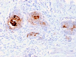
Biotium TAG-72 / CA72.4 (Tumor-Associated Glycoprotein)(rB72.3), CF568 conjugate, 0.1mg/mL
Recognizes an oncofetal antigen of 220 kDa, identified as a tumor-associated glycoprotein (TAG-72) with properties of a mucin. This MAb defines the mucin-carried sialylated-Tn epitope. TAG-72 is usually expressed by adenocarcinomas, but is negative in mesotheliomas. Studies have reported that this antibody has 80% sensitivity and 93% specificity for pulmonary adenocarcinoma. Therefore, TAG-72 is a useful marker to distinguish between mesothelioma and adenocarcinoma. However, false positive reactions can occur so results must be interpreted with the utmost caution. This antibody may be useful in the differentiation of non-small cell carcinomas from small cell carcinomas of the lung. The combined use of anti-TAG-72 and anti-GCDFP-15 is valuable in the diagnosis of apocrine carcinoma. Primary antibodies are available purified, or with a selection of fluorescent CF Dyes and other labels. CF Dyes offer exceptional brightness and photostability. Note: Conjugates of blue fluoresc

Biotium CD6(3F7B5), CF740 conjugate, 0.1mg/mL
CD6 is a type I transmembrane glycoprotein that contains a 24-amino acid signal sequence, three extracellular scavenger receptor cysteine-rich (SRCR) domains, a membrane-spanning domain and a 44-amino acid cytoplasmic domain. The CD6 glycoprotein is tyrosine phosphorylated during TCR-mediated T cell activation. CD6 shows significant homology to CD5. CD6 is present on mature thymocytes, peripheral T cells and a subset of B cells. Antibodies to CD6 are used to deplete T cells from bone marrow transplants to prevent graft versus host disease.Primary antibodies are available purified, or with a selection of fluorescent CF Dyes and other labels. CF Dyes offer exceptional brightness and photostability. Note: Conjugates of blue fluorescent dyes like CF405S and CF405M are not recommended for detecting low abundance targets, because blue dyes have lower fluorescence and can give higher non-specific background than other dye colors.