
Mouse Secondary Antibodies


Biotium CD50 / ICAM3(ICO-60), CF568 conjugate, 0.1mg/mL
Recognizes the N-glycosylated glycoprotein of 120 kDa with intra-chain disulfide bonds, identified as CD50 or ICAM-3. CD50 is the major ligand for LFA-1 (CD11a/CD18) and may have signaling role to increase adhesion. It is expressed on thymocytes and T lymphocytes and is resistant to treatment with phosphatidylinositol (PI) phospholipase C.

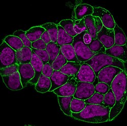
Biotium CD47(IAP/964 + B6H12.2), CF488A conjugate, 0.1mg/mL
This antibody reacts with Ig domain of CD47 protein. CD47, originally named integrin-associated protein (IAP), is a 50 kDa protein containing five membrane-spanning sequences and a short cytoplasmic tail. CD47 plays a role in both cell adhesion by acting as an adhesion receptor for THBS1 on platelets, and in the modulation of integrins. It is important in memory formation and synaptic plasticity in the hippocampus. CD47 may play a role in membrane transport and/or integrin dependent signal transduction.Primary antibodies are available purified, or with a selection of fluorescent CF Dyes and other labels. CF Dyes offer exceptional brightness and photostability. Note: Conjugates of blue fluorescent dyes like CF405S and CF405M are not recommended for detecting low abundance targets, because blue dyes have lower fluorescence and can give higher non-specific background than other dye colors.
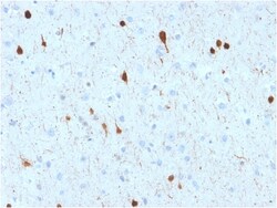
Biotium Calretinin / Calbindin 2 (Mesothelioma Marker)(CALB2/2602), CF488A conjugate, 0.1mg/mL
Calretinin (also known as Calbindin 2) is a member of the troponin C superfamily. Proteins in this family have 6 EF-hand domains which bind to calcium. Calretinin is a vitamin D-dependent calcium-binding protein involved in calcium signaling. It is present in subsets of neurons throughout the brain and spinal chord, including sensory ganglia. Antibodies to calretinin are useful in differentiating mesothelioma from adenocarcinomas of the lung. They can also aid in differentiating adrenal cortical neoplasms from pheochromocytomas Primary antibodies are available purified, or with a selection of fluorescent CF Dyes and other labels. CF Dyes offer exceptional brightness and photostability. Note: Conjugates of blue fluorescent dyes like CF405S and CF405M are not recommended for detecting low abundance targets, because blue dyes have lower fluorescence and can give higher non-specific background than other dye colors.
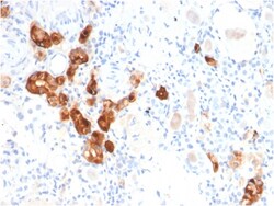
Biotium Calbindin 1 (CALB1)(CALB1/2782), CF568 conjugate, 0.1mg/mL
Calbindin 1, also known as calbindin D28K, is a member of a family of calcium binding proteins. This protein family also includes calmodulin, calbindin 2, S-100, and troponin C. Calbindin 1 is a 261-amino acid protein with 6 EF-hand domains, 4 of which are active calcium-binding domains. Expressed in brain, ovary, uterus, testis, pancreas, liver, kidney and intestine, calbindin 1 acts as a calcium-buffering agent and alters the activity of the plasma membrane ATPase. In neuronal cells, calbindin 1 modulates calcium channel activity, calcium transients and intrinsic neuronal firing activity. Also, calbindin 1 has been implicated to play a role in apoptosis and microtubule function. Primary antibodies are available purified, or with a selection of fluorescent CF Dyes and other labels. CF Dyes offer exceptional brightness and photostability. Note: Conjugates of blue fluorescent dyes like CF405S and CF405M are not recommended for detecting low abundance targets, because blue
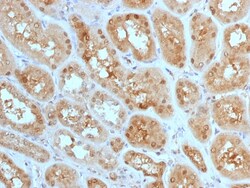
Biotium MTAP (Tumor Suppressor Marker) (MTAP/1813), CF488A conjugate, 0.1mg/mL
This antibody recognizes a protein of 31 kDa, which is identified as MTAP (5'-deoxy-5'-methylthioadenosine phosphorylase). It catalyzes the reversible phosphorolysis of methylthioadenosine, which is important in polyamine metabolism and for the salvage of adenine and methionine. The gene encoding MTAP is linked to the tumor suppressor gene, p16INK4A. Deficient levels of MTAP can occur in cancers primarily through co-deletion of the MTAP gene and the p16INK4A gene. Cells expressing MTAP and possessing adenine salvage pathway activity may be less susceptible to malignancy due to growth-inhibitory actions of agents (e.g. antifolates), whose mechanism of action, in part, involves this de novo purine pathway.Primary antibodies are available purified, or with a selection of fluorescent CF Dyes and other labels. CF Dyes offer exceptional brightness and photostability. Note: Conjugates of blue fluorescent dyes like CF405S and CF405M are not recommended for detecting low abundanc
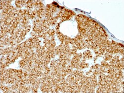
Biotium Parathyroid Hormone (PTH) (N-Terminal)(rPTH/911), CF568 conjugate, 0.1mg/mL
Epitope of this MAb maps in the N-terminus of PTH, a hormone produced by the parathyroid gland that regulates the concentration of calcium and phosphorus in extracellular fluid. This hormone elevates blood Ca2 levels by dissolving the salts in bone and preventing their renal excretion. It is produced in the parathyroid gland as an 84 amino acid single chain polypeptide. It can also be secreted as N-terminal truncated fragments or C-terminal fragments after intracellular degradation, as in case of hypercalcemia. Defects in this gene are a cause of familial isolated hypoparathyroidism (FIH); also called autosomal dominant hypoparathyroidism or autosomal dominant hypocalcemia. FIH is characterized by hypocalcemia and hyperphosphatemia due to inadequate secretion of parathyroid hormone. Symptoms are seizures, tetany and cramps. FIH exist both as autosomal dominant and recessive forms of hypoparathyroidism. Primary antibodies are available purified, or with a selection of fluores
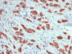
Biotium MUC1 / CA15-3 / EMA / CD227 (Epithelial Marker) (MUC1/1887R), CF647 conjugate, 0.1mg/mL
This MAb reacts with MUC1, a large transmembrane glycoprotein expressed on the ductal surface of normal glandular epithelia. It is used as tracer agent in CA15.3 assays. The extracellular domain of MUC1 largely consists of a highly conserved, O-glycosylated 20 amino acids tandem repeat which can occur 30-100 times per molecule depending on the length of the allele involved. In the vast majority of human carcinomas this protein is up-regulated and poorly glycosylated and appears on the cell surface in a non-polarized fashion. The dominant epitope of this MAb involves both amino acids as well as sugar moieties. Its epitope is destroyed by desialylation i.e. treatment with Neuraminidase.Primary antibodies are available purified, or with a selection of fluorescent CF Dyes and other labels. CF Dyes offer exceptional brightness and photostability. Note: Conjugates of blue fluorescent dyes like CF405S and CF405M are not recommended for detecting low abundance targets, because b
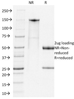
Biotium PD1, Mouse(RMP1-14), Biotin conjugate, 0.1mg/mL
PD-1 is expressed on a subset of CD4-CD8-thymocytes, and on activated T and B cells. PD-1 is thought to be involved in lymphocyte clonal selection and peripheral tolerance. The PD-1 ligands, PD-L1 (also known as B7-H1) and PD-L2 (B7-DC), are members of the B7 immunoglobulin superfamily. PD-1 is transiently expressed on CD4 and CD8 thymocytes as well as activated T and B-lymphocytes and myeloid cells. Additionally, Pdcd1mRNA is expressed in developing B-lymphocytes during the pro-B-cell stage. The RMP1-14 antibody has been reported to block the binding of PD-1 to its ligands (B7-H1 and B7-DC) and to inhibit T cell proliferation and cytokine production co-stimulated by macrophages (but not by dendritic cells and B cells).Primary antibodies are available purified, or with a selection of fluorescent CF Dyes and other labels. CF Dyes offer exceptional brightness and photostability. Note: Conjugates of blue fluorescent dyes like CF405S and CF405M are not recommended for de
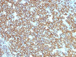
Biotium CD20 / MS4A1 (B-Cell Marker) (rIGEL/773), Biotin conjugate, 0.1mg/mL
This antibody recognizes a protein of 30-33 kDa, which is identified as CD20. It is a non-Ig differentiation antigen of B-cells and its expression is restricted to normal and neoplastic B-cells, being absent from all other leukocytes and tissues. CD20 is expressed by pre B-cells and persists during all stages of B-cell maturation but is lost upon terminal differentiation into plasma cells. This MAb can be used for immunophenotyping of leukemia and malignant cells, B lymphocyte detection in peripheral blood and B cell localization in tissues. It reacts with the majority of B-cells present in peripheral blood and lymphoid tissues and their derived lymphomas. In lymphoid tissue, germinal center blasts and B-immunoblasts are particularly reactive. It is a reliable antibody for ascribing a B-cell phenotype in known lymphoid tissues. Rarely, CD20-positive T-cell lymphomas have been reported. Reactivity has also been noted with Reed-Sternberg cells in cases of Hodgkin's disease, par
Biotium TTF-1 / NKX2.1 (Thyroid & Lung Epithelial Marker) (rNX2.1/690), Biotin conjugate, 0.1mg/mL
This antibody recognizes a protein of 40 kDa, identified as Thyroid transcription factor-1 (TTF-1). TTF-1 is a member of the NKx2 family of homeodomain transcription factors. It is expressed in epithelial cells of the thyroid gland and the lung. Nuclei from liver, stomach, pancreas, small intestine, colon, kidney, breast, skin, testes, pituitary, prostate, and adrenal glands are unreactive. Anti-TTF-1 is useful in differentiating primary adenocarcinoma of the lung from metastatic carcinomas originating in the breast, mediastinal germ cell tumors, and malignant mesothelioma. It can also be used to differentiate small cell lung carcinoma from lymphoid infiltrates. Loss of TTF-1 expression in non-small cell lung carcinoma has been associated with aggressive behavior of such neoplasms. TTF-1 reactivity is also seen in thyroid malignancies.Primary antibodies are available purified, or with a selection of fluorescent CF Dyes and other labels. CF Dyes offer exceptional brightness
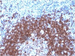
Biotium SOX2 (Transcription Factor) (SOX2/1792), CF488A conjugate, 0.1mg/mL
SOX2 is a member of the SRY-related HMG-box (SOX) family of transcription factors involved in the regulation of embryonic development and in the determination of cell fate. At present, 30 Sox genes have been identified. SOX2 is required for stem cell maintenance in the central nervous system, and it also regulates gene expression in the stomach. SOX2 is necessary for regulating multiple transcription factors that affect Oct 3/4 expression. An essential function of SOX2 is to stabilize embryonic stem cells in a pluripotent state by maintaining the requisite level of Oct 3/4 expression.Primary antibodies are available purified, or with a selection of fluorescent CF Dyes and other labels. CF Dyes offer exceptional brightness and photostability. Note: Conjugates of blue fluorescent dyes like CF405S and CF405M are not recommended for detecting low abundance targets, because blue dyes have lower fluorescence and can give higher non-specific background than other dye colors.
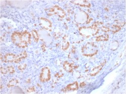
Biotium TTF-1 / NKX2.1 (Thyroid & Lung Epithelial Marker) (rNX2.1/690), 0.2mg/mL
This antibody recognizes a protein of 40 kDa, identified as Thyroid transcription factor-1 (TTF-1). TTF-1 is a member of the NKx2 family of homeodomain transcription factors. It is expressed in epithelial cells of the thyroid gland and the lung. Nuclei from liver, stomach, pancreas, small intestine, colon, kidney, breast, skin, testes, pituitary, prostate, and adrenal glands are unreactive. Anti-TTF-1 is useful in differentiating primary adenocarcinoma of the lung from metastatic carcinomas originating in the breast, mediastinal germ cell tumors, and malignant mesothelioma. It can also be used to differentiate small cell lung carcinoma from lymphoid infiltrates. Loss of TTF-1 expression in non-small cell lung carcinoma has been associated with aggressive behavior of such neoplasms. TTF-1 reactivity is also seen in thyroid malignancies.Primary antibodies are available purified, or with a selection of fluorescent CF Dyes and other labels. CF Dyes offer exceptional brightness
Biotium Cytochrome C (Mitochondrial Marker)(7H8.2C12 + CYCS/1010), 0.2mg/mL
Cytochrome C is a well-characterized mobile electron transport protein that is essential to energy conversion in all aerobic organisms. In mammalian cells, this highly conserved protein is normally localized to the mitochondrial inter-membrane space. More recent studies have identified cytosolic cytochrome c as a factor necessary for activation of apoptosis. During apoptosis, cytochrome c is trans-located from the mitochondrial membrane to the cytosol, where it is required for activation of caspase-3 (CPP32). Overexpression of Bcl-2 has been shown to prevent the translocation of cytochrome c, thereby blocking the apoptotic process. Overexpression of Bax has been shown to induce the release of cytochrome c and to induce cell death. The release of cytochrome c from the mitochondria is thought to trigger an apoptotic cascade, whereby Apaf-1 binds to Apaf-3 (caspase-9) in a cytochrome c-dependent manner, leading to caspase-9 cleavage of caspase-3. This MAb recognizes total cytoch
