
Mouse Secondary Antibodies

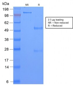
Biotium TAG-72 / CA72.4 (Tumor-Associated Glycoprotein)(CA72/2869R), Biotin conjugate, 0.1mg/mL
Recognizes an oncofetal antigen of 220 kDa, identified as a tumor-associated glycoprotein (TAG-72) with properties of a mucin. This MAb defines the mucin-carried sialylated-Tn epitope. TAG-72 is usually expressed by adenocarcinomas, but is negative in mesotheliomas. Studies have reported that this antibody has 80% sensitivity and 93% specificity for pulmonary adenocarcinoma. Therefore, TAG-72 is a useful marker to distinguish between mesothelioma and adenocarcinoma. However, false positive reactions can occur so results must be interpreted with the utmost caution. This antibody may be useful in the differentiation of non-small cell carcinomas from small cell carcinomas of the lung. The combined use of anti-TAG-72 and anti-GCDFP-15 is valuable in the diagnosis of apocrine carcinoma. Primary antibodies are available purified, or with a selection of fluorescent CF Dyes and other labels. CF Dyes offer exceptional brightness and photostability. Note: Conjugates of blue fluoresc


Biotium Alpha-1-Antichymotrypsin (SERPINA3) (Histiocytoma Marker) (AACT/1451), CF568 conjugate, 0.1mg/mL
Alpha-1 Antichymotrypsin (AACT) is a plasma protease inhibitor synthesized in the liver as a single glycopeptide chain. In human, the normal serum level of AACT is about one-tenth that of alpha-1-antitrypsin (AAT), with which it shares nucleic acid and protein sequence homology. Both are major acute phase reactants; their concentrations in plasma increase in response to trauma, surgery and infection. Elevated levels of AACT are widely, but not universally, reported in the cerebrospinal fluid and plasma of AD patients. Prostate-specific antigen (PSA) and its SDS-stable complex with AACT are in widespread use as markers for the diagnosis of prostate cancer. AACT deficiency may also be a possible cause of chronic liver disease. AACT antibody reacts with histiocytes and histiocytic neoplasms. It is widely used to identify histiocytes and tumors derived from them. Acinar tumors of the pancreas and salivary gland may also exhibit AACT positivity. Primary antibodies are available
Biotium MAML3 (Mastermind Like Transcriptional Coactivator 3) (MAML3/1303), CF647 conjugate, 0.1mg/mL
MAML3 (mastermind-like protein 3) is a nuclear speckle protein that acts as a transcriptional coactivator for Notch receptors. The Notch signaling pathway influences cell fate by regulating the ability of precursor cells to properly respond to developmental signals. MAML3 is a member of the mastermind-like family of proteins that are human homologs of the Drosophila melanogaster mastermind protein. Through its N-terminal region, MAML3 interacts with the ankyrin repeats of the Notch proteins Notch 1, Notch 2, Notch 3 and Notch 4.This interaction leads to formation of a DNA-binding complex with the Notch proteins and RBP-J, a complex that can then induce HES1 gene expression. While the N-terminal domain of MAML3 is essential for proper Notch binding, the C-terminal domain of MAML3 is essential for transcriptional activation. Due to its involvement in cell signaling and transcriptional activation, upregulation of MAML3 is thought to be involved in oncogenesis.Primary antibodies
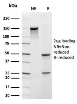
Biotium Frataxin (Mitochondrial Marker)(rFXN/2124), CF488A conjugate, 0.1mg/mL
Frataxin is a nuclear-encoded mitochondrial protein implicated in Friedreich's ataxia (FRDA), a human autosomal recessive neurodegenerative disease. Lack of Frataxin causes iron to accumulate in the mitochondrial matrix suggesting that Frataxin is involved in mitochondrial iron homeostasis and possibly in iron transport. Frataxin has an alpha-beta fold consisting of two helices flanking an antiparallel beta sheet. Primary antibodies are available purified, or with a selection of fluorescent CF Dyes and other labels. CF Dyes offer exceptional brightness and photostability. Note: Conjugates of blue fluorescent dyes like CF405S and CF405M are not recommended for detecting low abundance targets, because blue dyes have lower fluorescence and can give higher non-specific background than other dye colors.
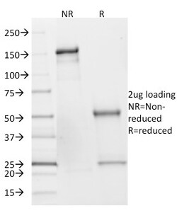
Biotium SDHB (Succinate Dehydrogenase B) (Pheochromocytoma Marker)(SDHB/2382), CF568 conjugate, 0.1mg/mL
Succinate dehydrogenase (SDH) is Complex II in the mitochondria, vital for mitochondrial electron transport, as well as Krebs cycle function. Four subunits comprise the SDH protein complex: a flavochrome subunit (SDHA), an iron-sulfur protein (SDHB) and two membrane-bound subunits (SDHC and SDHD) anchored to the inner mitochondrial membrane. The SDH complex functions as a tumor suppressor. Loss of any subunit proteins lead to destabilization of the complex and tumor formation. Antibody to SDHB is helpful in the identification of phaeochromocytomas, paragangliomas and GIST. Primary antibodies are available purified, or with a selection of fluorescent CF Dyes and other labels. CF Dyes offer exceptional brightness and photostability. Note: Conjugates of blue fluorescent dyes like CF405S and CF405M are not recommended for detecting low abundance targets, because blue dyes have lower fluorescence and can give higher non-specific background than other dye colors.
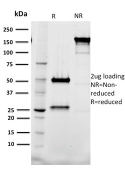
Biotium Tal1(BTL73), CF740 conjugate, 0.1mg/mL
Activation of TAL1 characterizes up to 60% of cases of human T cell acute lymphoblastic leukemia, making it the most frequent gain-of-function mutation observed in this disorder. TAL1 (also designated SCL) is a serine phosphoprotein and basic helix-loop-helix transcription factor known to regulate embryonic hematopoiesis. This transcription factor binds as a heterodimer with E2A and HEB/HTF4 to a nucleotide sequence motif termed the E-box. In addition, leukemogenesis is accelerated dramatically by transgenic co-expression of TAL1 and the catalytic subunit of casein kinase IIHLH transcription factors. Primary antibodies are available purified, or with a selection of fluorescent CF Dyes and other labels. CF Dyes offer exceptional brightness and photostability. Note: Conjugates of blue fluorescent dyes like CF405S and CF405M are not recommended for detecting low abundance targets, because blue dyes have lower fluorescence and can give higher non-specific background than oth
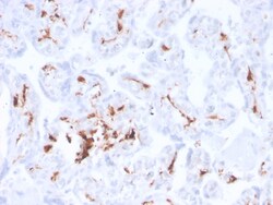
Biotium Factor XIIIa (Coagulation Factor XIIIA Chain)(F13A1/3772R), CF488A conjugate, 0.1mg/mL
Recognizes a protein of 83 kDa, which is identified as Factor XIIIa. It has been identified in platelets, megakaryocytes, and fibroblast-like mesenchymal or histiocytic cells in the placenta, uterus, and prostate, monocytes and macrophages and dermal dendritic cells. Anti-factor XIIIa has been found to be useful in differentiating between dermatofibroma (almost all cases are positive), dermatofibrosarcoma protuberans (-/ ) and desmoplastic malignant melanoma (-). Anti-factor XIIIa positivity is also seen in capillary hemagioblastoma, hemangioendothelioma, hemangiopericytoma, xanthogranuloma, xanthoma, hepatocellular carcinoma, glomus tumor, and meningioma. Primary antibodies are available purified, or with a selection of fluorescent CF Dyes and other labels. CF Dyes offer exceptional brightness and photostability. Note: Conjugates of blue fluorescent dyes like CF405S and CF405M are not recommended for detecting low abundance targets, because blue dyes have lower fluoresc
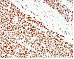
Biotium MSH6 (DNA Mismatch Repair Protein)(MSH6/3086), CF488A conjugate, 0.1mg/mL
The finding that mutations in DNA mismatch repair genes are associated with hereditary nonpolyposis colorectal cancer (HNPCC) has resulted in considerable interest in the understanding of the mechanism of DNA mismatch repair. Initially, inherited mutations in the MSH2 and MLH1 homologs of the bacterial DNA mismatch repair genes mutS and mutL were demonstrated at high frequency in HNPCC and were shown to be associated with microsatellite instability. A member of the mismatch repair family, GTBP (also designated MSH6), is an MSH2-related protein that binds to DNA containing G/T mismatches. Findings suggest that the mismatch-binding factor in human cells is composed of a heterodimer of GTBP and MSH2. Primary antibodies are available purified, or with a selection of fluorescent CF Dyes and other labels. CF Dyes offer exceptional brightness and photostability. Note: Conjugates of blue fluorescent dyes like CF405S and CF405M are not recommended for detecting low abundance targ
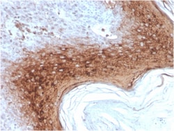
Biotium Involucrin (Squamous Cell Terminal Differentiation Marker)(rIVRN/827), CF488A conjugate, 0.1mg/mL
Recognizes a protein of 66 kDa-170 kDa, identified as involucrin. In Western blotting of cultured human keratinocytes, this MAb reacts with a 120 kDa protein. It stains the involucrin in a variety of sizes: 170 kDa in MCF-7 cells, a doublet of ~115 kDa and 150 kDa in gorilla and owl monkey, 66 kDa in dog, and a doublet of 105 kDa in pig. Its epitope maps between codon 421-568 of human involucrin. Involucrin is expressed in a range of stratified squamous epithelia, including the cornea, which lacks a distinct cornified layer. In normal epidermis, it is first expressed in the upper spinous layers, and in keratinocyte cultures, all cells that have left the basal layer express it. Involucrin expression is altered in pathological conditions: in psoriasis and other benign epidermal hyperplasias, involucrin expression begins closer to the basal layer than normal; expression is abnormal in squamous cell carcinomas and premalignant lesions, and is reduced in severe dysplasias of the l
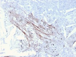
Biotium Tenascin C (Stromal Marker for Epithelial Malignancy)(TNC/3635), CF568 conjugate, 0.1mg/mL
In Western blotting, this antibody reacts with two bands of ~MW of 210 kDa and 300 kDa, identified as two isoforms of Tenascin C. Specificity of this MAb is validated by sequential immunoprecipitation with a PAb against Tenascin C. Tenascin C is a multifunctional, disulfide-linkedhexameric extracellular matrix glycoprotein expressed in association with mesenchymal epithelial interactions during development and in the neo-vasculature and stroma of undifferentiated tumors. In adults, it is restricted to certain epithelial-stromal interfaces and increases markedly in hyper-proliferative diseases and in stroma of many neoplasms, including gliomas, breast, squamous and lung carcinomas. Primary antibodies are available purified, or with a selection of fluorescent CF Dyes and other labels. CF Dyes offer exceptional brightness and photostability. Note: Conjugates of blue fluorescent dyes like CF405S and CF405M are not recommended for detecting low abundance targets, because blue
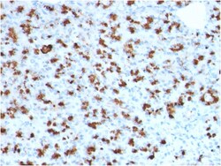
Biotium BARX1 (Prognostic Biomarker in Hepatocellular Carcinoma)(BARX1/2760), 0.2mg/mL
BarX1 is a member of the Bar subclass of homeobox transcription factors. Studies of the Mouse and chick homolog suggest the encoded protein may play a role in developing teeth and craniofacial mesenchyme of neural crest origin. The protein may also be associated with differentiation of stomach epithelia. Down-regulation of Barx1 promotes hepatocellular carcinoma cell (HCC) migration, invasion and metastasis, whereas up-regulation of Barx1 inhibits HCC migration, invasion and metastasis. The loss of Barx1 expression represents a prognostic biomarker in human HCC. Primary antibodies are available purified, or with a selection of fluorescent CF Dyes and other labels. CF Dyes offer exceptional brightness and photostability. Note: Conjugates of blue fluorescent dyes like CF405S and CF405M are not recommended for detecting low abundance targets, because blue dyes have lower fluorescence and can give higher non-specific background than other dye colors.
Biotium STAT5B(STAT5B/2611), CF568 conjugate, 0.1mg/mL
Signal transducer and activator of transcription 5A (Stat5a) and Stat5b, which share 96% homology, undergo receptor tyrosine kinase or G protein-coupled receptor-dependent phosphorylation in response to cytokines or growth factors, and then form homo- or heterodimers that translocate to the nucleus, where they initiate transcription. Activation of Stat5a via IL-2, IL-3, IL-7/ GM-CSF, erythropoietin, thrombopoietin and growth hormones influences proliferation, differentiation and apoptosis in lymphohematopoietic cells. Phosphorylation of Stat5a at Ser127/Ser128 and Ser779 are contigent on ErbB-4-mediated activation of Stat5a. Activation of Stat5b via IL-2, IL-4, CSF-1 and growth hormones influences TCR signaling, apoptosis, adult mammary gland development and sexual dimorphism of liver gene expression. Stat5b is the major liver-expressed Stat5 form that has been shown to fuse with the retinoic acid receptor a gene in acute promyelocytic leukemias (APLL). Stat5a/b null mice hav
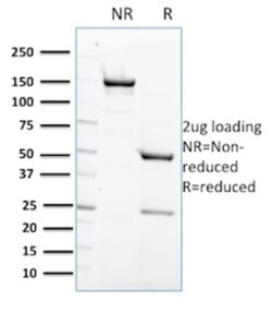
Biotium Protein Tyrosine Phosphatase, non-receptor type 6(CPTC-PTPN6-2), CF740 conjugate, 0.1mg/mL
PTPN6, which is also designated as SHP-1, is expressed primarily in hematopoietic cells and characterized by the presence of two SH2 domains N-terminal to the PTP domain. It maps to 12p13, a region commonly involved in leukemia-associated chromosomal abnormalities. SHP-1 plays a role in modulating intracellular signaling for various molecules. In T-cell lymphomas and other malignancies, SHP-1 phosphatase expression is lost, by a thus far undetermined mechanism. Preliminary studies suggest that it may be helpful in differentiating mantle/marginal zone lymphomas (SHP-1 ) from follicular lymphomas (SHP-1 negative). Primary antibodies are available purified, or with a selection of fluorescent CF Dyes and other labels. CF Dyes offer exceptional brightness and photostability. Note: Conjugates of blue fluorescent dyes like CF405S and CF405M are not recommended for detecting low abundance targets, because blue dyes have lower fluorescence and can give higher non-specific background t
Biotium EGFR (Epidermal Growth Factor Receptor) (GFR/1708), CF568 conjugate, 0.1mg/mL
This MAb recognizes a protein of 170 kDa, identified as EGFR. EGFR is type I receptor tyrosine kinase with sequence homology to erbB-1, -2, -3 -4 or HER-1, -2, -3 -4. It binds to Epidermal Growth Factor (EGF), Transforming Growth Factor-a (TGF-a), Heparin-binding EGF (HB-EGF), amphiregulin, betacellulin and epiregulin. EGFR is overexpressed in tumors of breast, brain, bladder, lung, gastric, head & neck, esophagus, cervix, vulva, ovary, and endometrium. It is predominantly present in squamous cell carcinomas.Primary antibodies are available purified, or with a selection of fluorescent CF Dyes and other labels. CF Dyes offer exceptional brightness and photostability. Note: Conjugates of blue fluorescent dyes like CF405S and CF405M are not recommended for detecting low abundance targets, because blue dyes have lower fluorescence and can give higher non-specific background than other dye colors.
Biotium Ki-67 (Proliferating Cell Marker) (MKI67/2462), CF568 conjugate, 0.1mg/mL
Ki-67 antigen is a nuclear, non-histone protein that is present in all stages of the cell cycle except G0. This characteristic makes Ki-67 an excellent marker for proliferating cells and is commonly used as one of the prognostic factors in cancer studies. A correlation has been demonstrated between Ki-67 index and the histo-pathological grade of neoplasms. Assessment of Ki-67 expression in renal and ureter tumors shows a correlation between tumor proliferation and disease progression, thus making it possible to differentiate high-risk patients. Ki-67 expression may also prove to be important for distinguishing between malignant and benign peripheral nerve sheath tumors. Ki-67 labeling index has been shown to be a prognostic marker in a number of neoplasms including grade II astrocytoma, oligodendroglioma, colon carcinoma, and breast carcinoma. In general, Ki-67 is a good marker of proliferating cell populations.Primary antibodies are available purified, or with a selection of