
Mouse Secondary Antibodies

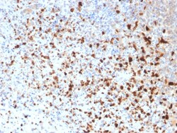
Biotium HSV1 (Herpes Simplex Virus Type I)(10A3), 0.2mg/mL
The antibody reacts with HSV type 1 specific antigen. It is suitable for detection of HSV in human cellular material obtained from superficial lesions or biopsies and for the early identification of HSV in infected tissue cultures. The herpes simplex virus (HSV) (also known as cold sore, night fever or fever blister) is a virus that causes a contagious disease. There are two main types of Herpes Simplex Virus (HSV), 1 and 2. The HSV-1 strain generally appears in the orafacial organs. HSV2 usually resides in the sacral ganglion at the base of the spine. All herpes viruses are morphologically identical: they have a large double-stranded DNA genome and the virion consists of an icosahedral nucleo-capsid, which is surrounded by a lipid bilayer envelope. Primary antibodies are available purified, or with a selection of fluorescent CF Dyes and other labels. CF Dyes offer exceptional brightness and photostability. Note: Conjugates of blue fluorescent dyes like CF405S and CF405M

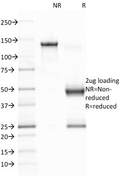
Biotium Epstein-Barr Virus (LMP-1)(CS1), CF740 conjugate, 0.1mg/mL
This antibody is a mixture of four different monoclonal antibodies. This antibody is specific to 60 kDa latent membrane protein (LMP-1) encoded by the BNLF1 gene of the EBV. Each clone reacts with different epitopes on the hydrophilic C-terminus of the cytoplasmic domain of LMP-1. This antibody stains strongly with EBV-positive lymphoblastoid cell lines and EBV infected B cell immunoblasts in infectious mononucleosis. EBV, also designated human herpesvirus 4 (HHV-4), is a member of the herpesvirus family and is one of the most common human viruses. EBV infects B cells and, though often asymptomatic, it can cause infectious mononucleosis, a disease characterized by fatigue, fever, sore throat and muscle soreness. Primary antibodies are available purified, or with a selection of fluorescent CF Dyes and other labels. CF Dyes offer exceptional brightness and photostability. Note: Conjugates of blue fluorescent dyes like CF405S and CF405M are not recommended for detecting low
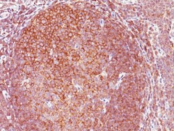
Biotium BCL10 (MALT-Lymphoma Marker)(151), CF568 conjugate, 0.1mg/mL
B-cell lymphoma/leukemia 10 (BCL10) is a signaling protein that promotes apoptosis, pro-caspase-9 maturation, and activation of NF-kappa-B via NIK and IKK. BCL10 contains an N-terminal caspase recruitment domain (CARD), which is found in a number of apoptotic regulatory molecules. BCL10 is involved in the adaptive immune response, and may be an adapter protein between upstream TNFR1-TRADD-RIP complex and the downstream NIK-IKK-IKAP complex. A BCL10 translocation is recurrent in low-grade mucosa-associated lymphoid tissue (MALT lymphoma). Primary antibodies are available purified, or with a selection of fluorescent CF Dyes and other labels. CF Dyes offer exceptional brightness and photostability. Note: Conjugates of blue fluorescent dyes like CF405S and CF405M are not recommended for detecting low abundance targets, because blue dyes have lower fluorescence and can give higher non-specific background than other dye colors.
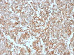
Biotium DOG-1 / TMEM16A (Gastrointestinal Stromal Tumor Marker)(rDG1/447), CF740 conjugate, 0.1mg/mL
Expression of DOG-1 protein is elevated in the gastrointestinal stromal tumors (GIST's), c-kit signaling-driven mesenchymal tumors of the GI tract. DOG-1 is rarely expressed in other soft tissue tumors, which, due to appearance, may be difficult to diagnose. Immunoreactivity for DOG-1 has been reported in 97. 8 percent of scorable GIST's, including all c-kit negative GIST's. Overexpression of DOG-1 has been suggested to aid in the identification of GISTs, including Platelet-Derived Growth Factor Receptor Alpha mutants that fail to express c-kit antigen. The overall sensitivity of DOG1 and c-kit in GIST's is nearly identical: 94. 4% vs. 94. 7%. Primary antibodies are available purified, or with a selection of fluorescent CF Dyes and other labels. CF Dyes offer exceptional brightness and photostability. Note: Conjugates of blue fluorescent dyes like CF405S and CF405M are not recommended for detecting low abundance targets, because blue dyes have lower fluorescence and can

Biotium Prohibitin (Mitochondrial Marker)(PHB/1881), CF740 conjugate, 0.1mg/mL
Recognizes a protein of 30 kDa which is identified as Prohibitin, an evolutionarily conserved protein with homologues found in yeast to man. It is located in the inner membrane of mitochondria. Although prohibitin mRNA and protein expression occurs throughout the cell cycle, maximum levels are detected during the G1/S phase transition and minimum levels are seen in S phase and the G2/mitosis boundary. Prohibitin is located exclusively in the mitochondria with the highest concentration on the inner membrane. Prohibitin is an ideal mitochondrial marker. It shows antiproliferative activity and has been proposed to play a role in normal cell cycle regulation, replicative senescence, cellular immortalization, and tumor suppression. Primary antibodies are available purified, or with a selection of fluorescent CF Dyes and other labels. CF Dyes offer exceptional brightness and photostability. Note: Conjugates of blue fluorescent dyes like CF405S and CF405M are not recommended fo
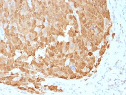
Biotium S100A1 (Melanoma Marker)(S100A1/1942), CF488A conjugate, 0.1mg/mL
The specificity of this monoclonal antibody to its intended target was validated by HuProt™ Array, containing more than 19, 000, full-length human proteins. S100 belongs to the family of calcium binding proteins. S100A and S100B proteins are two members of the S100 family. S100A is composed of an alpha and a beta chain whereas S100B is composed of two beta chains. This antibody is specific against an epitope located on the alpha-chain (i. e. in S-100A and S-100B) but not on the beta-chain of S-100 (i. e. in S-100B). This antibody can be used to localize S-100A in various tissue sections. S-100 protein has been found in normal melanocytes, Langerhans cells, histiocytes, chondrocytes, lipocytes, skeletal and cardiac muscle, epithelial and myoepithelial cells of the breast, salivary and sweat glands. Neoplasms derived from these cells also express S-100 protein. Almost all malignant melanomas and cases of histiocytosis X are positive for S-100 protein. Primary antibodies
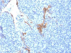
Biotium Cytokeratin 16 (KRT16) (Suprabasal Keratinocyte Marker)(LL025), 0.2mg/mL
Cytokeratins are a family of intermediate filament proteins that assemble into filaments through forming heterodimers of one type I Cytokeratins (Cytokeratins 9 to 23) and one type II Cytokeratins (keratins 1 to 8). The cytokeratin proteins play a critical role in differentiation, as well as tissue specialization and function, to maintain the overall structural integrity of epithelial cells. Cytokeratins are also useful markers in identifying the origin of metastatic tumors. Cytokeratin 16 is expressed in benign stratified squamous epithelium and squamous cell carcinoma of the head and neck, as well as luminal cells of mammary gland and sweat ducts. It is absent in non-invasive breast carcinomas and normal breast tissue. Primary antibodies are available purified, or with a selection of fluorescent CF Dyes and other labels. CF Dyes offer exceptional brightness and photostability. Note: Conjugates of blue fluorescent dyes like CF405S and CF405M are not recommended for dete
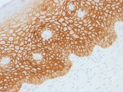
Biotium CD44v4 (Marker of Tumor Metastasis), 0.2mg/mL
This antibody recognizes an epitope encoded by exon v4 on the variant portion of human CD44. The CD44 molecule belongs to a family of cellular adhesion molecules found on a wide range of normal and malignant cells in epithelial, mesothelial and hemopoiesis tissues. CD44 is a single gene with 20 exons, of which 10 are normally expressed to encode the basic CD44 (H-CAM) molecule. The additional 10 exons (v1 to v10) are only expressed by alternative splicing of the nuclear RNA. The expression of specific cell adhesion molecule CD44 splice variants has been reported to be associated with metastasis in certain human malignancies. Primary antibodies are available purified, or with a selection of fluorescent CF Dyes and other labels. CF Dyes offer exceptional brightness and photostability. Note: Conjugates of blue fluorescent dyes like CF405S and CF405M are not recommended for detecting low abundance targets, because blue dyes have lower fluorescence and can give higher non-spe
Biotium Cytokeratin 10 (KRT10) (Suprabasal Epithelial Marker) (KRT10/1990R), CF740 conjugate, 0.1mg/mL
This MAb recognizes a protein of 56.5 kDa, identified as cytokeratin 10 (CK10). CK10 is expressed in all suprabasal layers of the epidermis. In the epidermis, expression of CK10 strictly parallels the extent of differentiation; it is absent in the basal layer, appears in the first suprabasal layers and increases in concentration towards the granular layer. However, CK10 is rarely detected in early stages of vulvar squamous carcinomas (tumors less than 2 cm, clinical stage I) regardless of the tumor grade. In larger and more advanced tumors (greater than 2 cm, clinical stages II and III), CK10 is detected very frequently. Expression of CK10 is related to maturation of malignant keratinocytes, being preferentially detected in more-differentiated parts.Primary antibodies are available purified, or with a selection of fluorescent CF Dyes and other labels. CF Dyes offer exceptional brightness and photostability. Note: Conjugates of blue fluorescent dyes like CF405S and CF405M
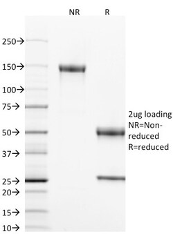
Biotium CD40 / TNFRSF5 / CD40L-Receptor(T8P2G4*A6), CF405S conjugate, 0.1mg/mL
This antibody is specific for the soluble form of CD40. It is a receptor on antigen-presenting cells of the immune system and is essential for mediating a broad variety of immune and inflammatory responses including T cell-dependent immunoglobulin class switching, memory B cell development, and germinal center formation. AT-hook transcription factor AKNA is reported to coordinately regulate the expression of this receptor and its ligand, which may be important for homotypic cell interactions. Adaptor protein TNFR2 interacts with this receptor and serves as a mediator of the signal transduction. The interaction of this receptor and its ligand is found to be necessary for amyloid-beta-induced microglial activation, and thus is thought to be an early event in Alzheimer disease pathogenesis. CD40 is expressed on B-lymphocytes, follicular dendritic cells, bone marrow-derived dendritic cells, thymic epithelium, and interdigitating cells in the T-cell zones of secondary lymphoid org
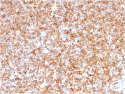
Biotium HLA-DP/-DQ/-DR (MHC II)(CR3/43), 1mg/mL
Reacts with a common epitope of human major histocompatibility (MHC) class II antigens, HLA-DP, -DQ and -DR. Human MHC class II antigens are transmembrane glycoproteins composed of an chain (36 kDa) and a chain (27 kDa). They are expressed primarily on antigen presenting cells such as B lymphocytes, monocytes, macrophages, and thymic epithelial cells and are also present on activated T lymphocytes. Human MHC class II genes are located in the HLA-D region that encodes at least six and ten chain genes. Three loci, DR, DQ and DP, encode the major expressed products of the human class II region. The human MHC class II molecules bind intracellularly processed peptides and present them to T-helper cells. They, therefore, have a critical role in the initiation of the immune response. It has been shown that some autoimmune diseases are associated with certain class II alleles.Primary antibodies are available purified, or with a selection of fluorescent CF Dyes and other labels.
Biotium Parathyroid Hormone (PTH) (N-Terminal) (PTH/1717R), 1mg/mL
Epitope of this MAb maps in the N-terminus of PTH, a hormone produced by the parathyroid gland that regulates the concentration of calcium and phosphorus in extracellular fluid. This hormone elevates blood calcium levels by dissolving the salts in bone and preventing their renal excretion. It is produced in the parathyroid gland as an 84 amino acid single chain polypeptide. It can also be secreted as N-terminal truncated fragments or C-terminal fragments after intracellular degradation, as in case of hypercalcemia. Defects in this gene are a cause of familial isolated hypoparathyroidism (FIH); also called autosomal dominant hypoparathyroidism or autosomal dominant hypocalcemia. FIH is characterized by hypocalcemia and hyperphosphatemia due to inadequate secretion of parathyroid hormone. Symptoms are seizures, tetany and cramps. FIH exist both as autosomal dominant and recessive forms of hypoparathyroidism.Primary antibodies are available purified, or with a selection of fluo

Biotium Desmin (Muscle Cell Marker)(D33), 1mg/mL
Cytoskeletal intermediate filaments (IFs) constitute a diverse group of proteins that are expressed in a highly tissue-specific manner. IFs are constructed from two-chain -helical coiled-coil molecules arranged on an imperfect helical lattice, and have been widely used as markers for distinguishing individual cell types within a tissue and identifying the origins of metastatic tumors. Vimentin is an IF general marker of cells originating in the mesenchyme. Vimentin and Desmin, a related class III IF, are both expressed during skeletal muscle development. Desmin, a 469 amino acid protein found near the Z line in sarcomeres, is expressed more frequently in adult differentiated state tissues. Anti-desmin detects cells of normal smooth, skeletal, and cardiac muscles. Antibody reacts with leiomyomas, leiomyosarcoma, rhabdomyomas, rhabdomyosarcoma, and perivascular cells of glomus tumors of the skin. Primary antibodies are available purified, or with a selection of fluorescent CF
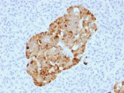
Biotium Chromogranin A / CHGA (Neuroendocrine Marker) (CHGA/1731R), 1mg/mL
Chromogranin A is present in neuroendocrine cells throughout the body, including the neuroendocrine cells of the large and small intestine, adrenal medulla and pancreatic islets. It is an excellent marker for carcinoid tumors, pheochromocytomas, paragangliomas, and other neuroendocrine tumors. Co-expression of chromogranin A and neuron specific enolase (NSE) is common in neuroendocrine neoplasms. Reportedly, co-expression of certain keratins and chromogranin indicates neuroendocrine lineage. The presence of strong anti-chromogranin staining and absence of anti-keratin staining should raise the possibility of paraganglioma. The co-expression of chromogranin and NSE is typical of neuroendocrine neoplasms. Most pituitary adenomas and prolactinomas readily express chromogranin.Primary antibodies are available purified, or with a selection of fluorescent CF Dyes and other labels. CF Dyes offer exceptional brightness and photostability. Note: Conjugates of blue fluorescent dyes
Biotium GFAP (Astrocyte & Neural Stem Cell Marker) (ASTRO/1974R), 0.2mg/mL
This MAb recognizes a protein of ~50 kDa which is identified as Glial Fibrillary Acidic Protein (GFAP). It shows no cross-reaction with other intermediate filament proteins. GFAP is specifically found in astroglia. GFAP is a very popular marker for localizing benign astrocyte and neoplastic cells of glial origin in the central nervous system. Antibody to GFAP is useful in differentiating primary gliomas from metastatic lesions in the brain and for documenting astrocytic differentiation in tumors outside the CNS.Primary antibodies are available purified, or with a selection of fluorescent CF Dyes and other labels. CF Dyes offer exceptional brightness and photostability. Note: Conjugates of blue fluorescent dyes like CF405S and CF405M are not recommended for detecting low abundance targets, because blue dyes have lower fluorescence and can give higher non-specific background than other dye colors.