
Mouse Secondary Antibodies

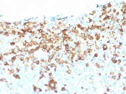
Biotium NGF-Receptor (p75) / CD271 (Soft Tissue Tumor Marker) (NGFR/1964), 0.2mg/mL
This antibody recognizes a glycoprotein of 75 kDa, identified as low affinity Nerve Growth Factor (NGF) Receptor (p75NGFR) or Neurotrophin Receptor (p75NTR). NGFR is expressed in various neural crest cells and their tumors such as melanocytes, melanomas, neuroblastomas, pheochromocytomas and neurofibromas. Reportedly, anti-NGFR is a reliable marker for desmoplastic and neurotropic melanomas. NGFR is expressed in mature non-neural cells such as perivascular cells, dental pulp cells, lymphoidal follicular dendritic cells, basal epithelium of oral mucosa and hair follicles, prostate basal cells, and myoepithelial cells. Anti-NGFR stains the myoepithelial cells of breast ducts and intra-lobular fibroblasts of breast ducts.Primary antibodies are available purified, or with a selection of fluorescent CF Dyes and other labels. CF Dyes offer exceptional brightness and photostability. Note: Conjugates of blue fluorescent dyes like CF405S and CF405M are not recommended for det

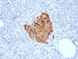
Biotium Chromogranin A / CHGA (Neuroendocrine Marker) (CHGA/1773R), CF740 conjugate, 0.1mg/mL
Chromogranin A is present in neuroendocrine cells throughout the body, including the neuroendocrine cells of the large and small intestine, adrenal medulla and pancreatic islets. It is an excellent marker for carcinoid tumors, pheochromocytomas, paragangliomas, and other neuroendocrine tumors. Co-expression of chromogranin A and neuron specific enolase (NSE) is common in neuroendocrine neoplasms. Reportedly, co-expression of certain keratins and chromogranin indicates neuroendocrine lineage. The presence of strong anti-chromogranin staining and absence of anti-keratin staining should raise the possibility of paraganglioma. The co-expression of chromogranin and NSE is typical of neuroendocrine neoplasms. Most pituitary adenomas and prolactinomas readily express chromogranin.Primary antibodies are available purified, or with a selection of fluorescent CF Dyes and other labels. CF Dyes offer exceptional brightness and photostability. Note: Conjugates of blue fluorescent dyes
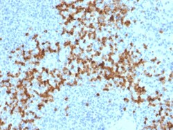
Biotium MMP9 (Matrix Metalloproteinase 9)(2C3), CF647 conjugate, 0.1mg/mL
The matrix metalloproteinases (MMP) are a family of peptidase enzymes responsible for the degradation of extracellular matrix components, including collagen, gelatin, fibronectin, laminin and proteoglycan. Transcription of MMP genes is differentially activated by phorbol ester, lipopolysaccharide (LPS) or staphylococcal enterotoxin B (SEB). MMP catalysis requires both calcium and zinc. MMP-9 (also designated 92 kDa type IV collagenase or gelatinase B) has been shown to degrade bone collagens in concert with MMP-1 (also designated interstitial collagenase, fibroblast collagenase or collagenase-1), and cysteine proteases and may play a role in bone osteoclastic resorption. MMP-1 is down-regulated by p53, and abnormality of p53 expression may contribute to joint degradation in rheumatoid arthritis by regulating MMP-1 expression. Primary antibodies are available purified, or with a selection of fluorescent CF Dyes and other labels. CF Dyes offer exceptional brightness and photo
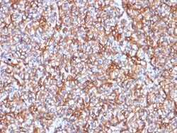
Biotium Spectrin beta III (SPTBN2) (SPTBN2/1778), CF740 conjugate, 0.1mg/mL
Spectrin is an actin binding protein that is a major component of the plasma membrane skeleton. Spectrins function as membrane organizers and stabilizers by forming dimers, tetramers and higher polymers. Vertebrate spectrins have two alpha-subunits (alpha-I/alpha-II), four beta-subunits (beta-I-beta-IV) and a beta-H subunit creating diversity and specialization of function. Spectrin α and spectrinβ are present in erythrocytes, whereas spectrin α II (also designated fodrin α) and spectrinβ I (also designated fodrinβ) are present in other somatic cells. The spectrin tetramers in erythrocytes act as barriers to lateral diffusion, but spectrin dimers seem to lack this function. Spectrinβ III is highly homologous to both spectrinβ I and spectrinβ II. Spectrinβ III is highly expressed in brain, kidney, pancreas and liver, and at lower levels in lung and placenta. Spectrin beta 3 is primarily expressed in nervous tissues with high
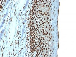
Biotium Histone H1 (Nuclear Marker) (r1415-1), 0.2mg/mL
Eukaryotic histones are basic and water-soluble nuclear proteins that form hetero-octameric nucleosome particles by wrapping 146 base pairs of DNA in a left-handed supealpha-helicalturn sequentially to form chromosomal fiber. Two molecules of each of the four core histones (H2A, H2B, H3, and H4) form the octamer; formed of two H2A-H2B dimers and two H3-H4 dimers, forming two nearly symmetrical halves by tertiary structure. Over 80% of nucleosomes contain the linker Histone H1, derived from an intronless gene that interacts with linker DNA between nucleosomes and mediates compaction into higher order chromatin. Histones are subject to posttranslational modification by enzymes primarily on their N-terminal tails, but also in their globular domains. Such modifications include methylation, citrullination, acetylation, phosphorylation, sumoylation, ubiquitination and ADP-ribosylation.Primary antibodies are available purified, or with a selection of fluorescent CF Dyes and other l
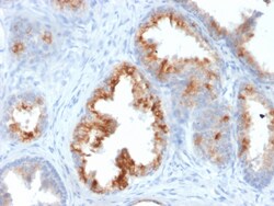
Biotium AMACR / p504S (Prostate Cancer Marker)(AMACR/1864), 0.2mg/mL
This antibody recognizes a protein of 42 kDa, which is identified as Alpha-methylacyl-CoA Racemase (AMACR), also known as p504S. It is an enzyme that is involved in bile acid biosynthesis and -oxidation of branched-chain fatty acids. AMACR is essential in lipid metabolism. It is expressed in cells of premalignant high-grade prostatic intraepithelial neoplasia (HGPIN) and prostate adenocarcinoma. The majority of the carcinoma cells show a distinct granular cytoplasmic staining reaction. AMACR is present at low or undetectable levels in glandular epithelial cells of normal prostate and benign prostatic hyperplasia. A spotty granular cytoplasmic staining is seen in a few cells of the benign glands. AMACR is expressed in normal liver (hepatocytes), kidney (tubular epithelial cells) and gall bladder (epithelial cells). Expression has also been found in lung (bronchial epithelial cells) and colon (colonic surface epithelium). AMACR expression can also be found in hepatocellular car
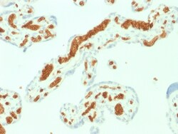
Biotium Spectrin Alpha 1 (Erythrocyte Marker) (SPTA1/1832), CF568 conjugate, 0.1mg/mL
Spectrin is an actin crosslinking and molecular scaffold protein that links the plasma membrane to the actin cytoskeleton, and functions in the determination of cell shape, arrangement of transmembrane proteins, and organization of organelles. Spectrins function as membrane organizers and stabilizers, composed of non-homologous α andβ chains, which aggregate side-to-side in an anti-parallel fashion to form dimers, tetramers and higher polymers. Spectrin α I and spectrinβ I are present in erythrocytes, whereas spectrin α II (also designated fodrin α) and spectrinβ II (also designated fodrinβ) are present in other somatic cells. The spectrin tetramers in erythrocytes act as barriers to lateral diffusion, but spectrin dimers seem to lack this function.Primary antibodies are available purified, or with a selection of fluorescent CF Dyes and other labels. CF Dyes offer exceptional brightness and photostability. Note: Conjugates of blu

Biotium CELA3B / ELA3B (Pancreatic Function Marker) (CELA3B/1757), CF740 conjugate, 0.1mg/mL
This MAb recognizes a protein of ~30 kDa, identified as CELA3B (Chymotrypsin like elastase family member 3B). Elastases form a subfamily of serine proteases that hydrolyze many proteins in addition to elastin. Humans have six elastase genes which encode the structurally similar proteins elastase 1, 2, 2A, 2B, 3A, and 3B. Unlike other elastases, elastase 3B has little elastolytic activity. Like most of the human elastases, elastase 3B is secreted from the pancreas as a zymogen and, like other serine proteases such as trypsin, chymotrypsin and kallikrein; it has a digestive function in the intestine. Elastase 3B preferentially cleaves proteins after alanine residues. Elastase 3B may also function in the intestinal transport and metabolism of cholesterol. Both elastase 3A and elastase 3B have been referred to as protease E and as elastase 1, and excretion of this protein in fecal material is frequently used as a measure of pancreatic function.Primary antibodies are available purified
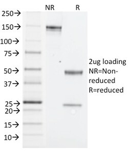
Biotium NK1.1 / CD161c / Klrb1c, Mouse(PK136), Biotin conjugate, 0.1mg/mL
NK1. 1 is a type II integral membrane glycoprotein with a C-type lectin domain and is encoded by theKlrb1c/NKR-P1Cgene. It is predominantly expressed as a disulfide-linked homodimer on NK cells however; it is also expressed on NK-T cells, a rare population of T lymphocytes. NK1. 1 is expressed in some mouse strains, including C57BL/6, FVB/N, and NZB, but not AKR, BALB/c, CBA/J, C3H, DBA/1, DBA/2, NOD, SJL, and 129. Expression of NKR-P1C antigen has been correlated with lysis of tumor cells in vitro and rejection of bone marrow allograftsin vivo. NK-1. 1 has also been shown to play a role in NK cell activation, IFN- production, and cytotoxic granule release. NK-1. 1 is commonly used as a mouse NK cell marker. Primary antibodies are available purified, or with a selection of fluorescent CF Dyes and other labels. CF Dyes offer exceptional brightness and photostability. Note: Conjugates of blue fluorescent dyes like CF405S and CF405M are not recommended for detecting low abu
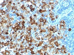
Biotium gp100 / Melanosome / PMEL17 / SILV (Melanoma Marker) (PMEL/1825R), CF568 conjugate, 0.1mg/mL
Cytotoxic T lymphocytes (CTLs) recognize melanoma-associated antigens, which belong to three main groups. These groups include tumor-associated testis-specific antigens, melanocyte differentiation antigens and mutated or aberrantly expressed antigens, which are routinely used as markers to identify melanomas based on their binding to specific monoclonal antibodies. gp100, also designated ME20-M, ME20-S and PMEL 17, is classified as a melanocyte differentiation antigen and is expressed at low levels in normal cell lines and tissues, but is upregulated in melanocytes. gp100 is a highly glycosylated protein. It is also the product of proteolytic cleavage, which results in a secreted protein.Primary antibodies are available purified, or with a selection of fluorescent CF Dyes and other labels. CF Dyes offer exceptional brightness and photostability. Note: Conjugates of blue fluorescent dyes like CF405S and CF405M are not recommended for detecting low abundance targets, becau
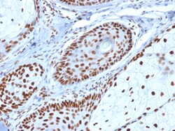
Biotium Nucleophosmin (Acute Myeloid Leukemia Marker)(NPM1/1902), 0.2mg/mL
Recognizes a 33 kDa glycoprotein, identified as Nucleophosmin (NPM). It is predominantly localized in the nucleus of cells in most tissues. NPM is involved in ribosomal assembly and rRNA transport. It is an abundant protein that is highly phosphorylated by Cdc2 kinase during mitosis. This phosphoprotein moves between the nucleus and the cytoplasm. It is thought to be involved in several processes including regulation of the ARF/p53 pathway. A number of genes are fusion partners, in particular the anaplastic lymphoma kinase gene on chromosome 2. Mutations in exon 12 affecting the C-terminus of the protein are associated with an aberrant cytoplasmic location. Mutations in this gene are associated with acute myeloid leukemia. The antibody may be a useful aid for classification of acute myeloid leukemia. Primary antibodies are available purified, or with a selection of fluorescent CF Dyes and other labels. CF Dyes offer exceptional brightness and photostability. Note: Conjugat
Biotium Rb1 (Tumor Suppressor Protein) (RB1/1754), CF740 conjugate, 0.1mg/mL
This antibody recognizes a 105 kDa phosphoprotein, identified as retinoblastoma (Rb) gene product. It shows no cross reaction with p107 or p130. It reacts with the hyper-phosphorylated as well as the un (under) phosphorylated form of the Rb protein. Retinoblastoma gene product plays a key role in cell cycle control. It has been identified as a tumor suppressor gene whose loss of its function leads to tumor development. It is widely expressed in a variety of human tissues including breast, esophageal, squamous cell and cervical carcinoma.Primary antibodies are available purified, or with a selection of fluorescent CF Dyes and other labels. CF Dyes offer exceptional brightness and photostability. Note: Conjugates of blue fluorescent dyes like CF405S and CF405M are not recommended for detecting low abundance targets, because blue dyes have lower fluorescence and can give higher non-specific background than other dye colors.
Biotium Rb1 (Tumor Suppressor Protein) (RB1/1754), CF740 conjugate, 0.1mg/mL
This antibody recognizes a 105 kDa phosphoprotein, identified as retinoblastoma (Rb) gene product. It shows no cross reaction with p107 or p130. It reacts with the hyper-phosphorylated as well as the un (under) phosphorylated form of the Rb protein. Retinoblastoma gene product plays a key role in cell cycle control. It has been identified as a tumor suppressor gene whose loss of its function leads to tumor development. It is widely expressed in a variety of human tissues including breast, esophageal, squamous cell and cervical carcinoma.Primary antibodies are available purified, or with a selection of fluorescent CF Dyes and other labels. CF Dyes offer exceptional brightness and photostability. Note: Conjugates of blue fluorescent dyes like CF405S and CF405M are not recommended for detecting low abundance targets, because blue dyes have lower fluorescence and can give higher non-specific background than other dye colors.
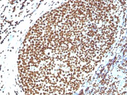
Biotium Histone H1 (Pan Nuclear Marker)(N/A), CF568 conjugate, 0.1mg/mL
Eukaryotic histones are basic and water-soluble nuclear proteins that form hetero-octameric nucleosome particles by wrapping 146 base pairs of DNA in a left-handed super-helical turn sequentially to form chromosomal fiber. Two molecules of each of the four core histones (H2A, H2B, H3, and H4) form the octamer; formed of two H2A-H2B dimers and two H3-H4 dimers, forming two nearly symmetrical halves by tertiary structure. Over 80% of nucleosomes contain the linker Histone H1, derived from an intronless gene that interacts with linker DNA between nucleosomes and mediates compaction into higher order chromatin. Histones are subject to posttranslational modification by enzymes primarily on their N-terminal tails, but also in their globular domains. Such modifications include methylation, citrullination, acetylation, phosphorylation, sumoylation, ubiquitination and ADP-ribosylation. Primary antibodies are available purified, or with a selection of fluorescent CF Dyes and other lab
Biotium Cytokeratin, pan (Epithelial Marker), 0.2mg/mL
Twenty human keratins are resolved with two-dimensional gel electrophoresis into acidic (pI 6.0) subfamilies. This antibody cocktail recognizes acidic (Type I or LMW) and basic (Type II or HMW) cytokeratins, which 67 kDa (CK1); 64 kDa (CK3); 59 kDa (CK4); 58 kDa (CK5); 56 kDa (CK6); 52 kDa (CK8); 56.5 kDa (CK10); 50 kDa (CK14); 50 kDa (CK15); 48 kDa (CK16); 40 kDa (CK19). Many studies have shown the usefulness of keratins as markers in cancer research and tumor diagnosis. It is a broad spectrum anti pan-cytokeratin antibody, which differentiates epithelial tumors from non-epithelial tumors e.g. squamous vs. adenocarcinoma of the lung, liver carcinoma, breast cancer, and esophageal cancer. It may be useful to characterize the source of various neoplasms and to study the distribution of cytokeratin containing cells in epithelia during normal development and during the development of epithelial neoplasms. This antibody stains cytokeratins present in normal and abnormal human tis