
Antibody and Dye Based Assays

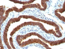
Biotium Cytokeratin 19(Ks19.1), CF488A conjugate, 0.1mg/mL
This MAb reacts with the rod domain of human cytokeratin-19 (CK19), a polypeptide of 40 kDa. Its epitope maps between amino acid 312-335. CK19 is expressed in sweat gland, mammary gland ductal and secretory cells, bile ducts, gastrointestinal tract, bladder urothelium, oral epithelia, esophagus, and ectocervical epithelium. Anti-CK19 reacts with a wide variety of epithelial malignancies including adenocarcinomas of the colon, stomach, pancreas, biliary tract, liver, and breast. Perhaps the most useful application is the identification of thyroid carcinoma of the papillary type, although 50%-60% of follicular carcinomas are also labeled. Anti-CK19 is a useful marker for detection of tumor cells in lymph nodes, peripheral blood, bone marrow and breast cancer.Primary antibodies are available purified, or with a selection of fluorescent CF Dyes and other labels. CF Dyes offer exceptional brightness and photostability. Note: Conjugates of blue fluorescent dyes like CF405S and

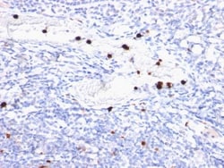
Biotium Granulocyte Marker(BM-2), CF488A conjugate, 0.1mg/mL
Recognizes 183 kDa protein with DNA-binding characteristics, which is identified as a myeloid specific antigen. It reacts with myeloid precursor cells and granulocytes in bone marrow. Its antigen appears to be restricted to M2 and M3 acute myelogenous leukemia (AML) subtypes. Markers of myeloid cells are useful in the identification of different levels of cellular differentiation. This MAb reacts with early precursor and mature forms of human myeloid cells. It is useful in the identification of myelogenous leukemias, distinguishing granulocytic sarcomas from lymphoid malignancies and also in the study of differentiation and transformation of human myeloid cells. The biological function of this antigen is not clear, although it has been proposed that it may play a role in the differentiation of myeloid cells.Primary antibodies are available purified, or with a selection of fluorescent CF Dyes and other labels. CF Dyes offer exceptional brightness and photostability. Note: C

Biotium MART-1 / Melan-A(M2-7C10), CF488A conjugate, 0.1mg/mL
This antibody recognizes a protein doublet of 20-22 kDa, identified as MART-1 (Melanoma Antigen Recognized by T cells 1) or Melan-A. MART-1 is a newly identified melanocyte differentiation antigen recognized by autologous cytotoxic T lymphocytes. Seven other melanoma associated antigens recognized by autologous cytotoxic T cells include MAGE-1, MAGE-3, tyrosinase, gp100, gp75, BAGE-1, and GAGE-1. Subcellular fractionation shows that MART-1 is present in melanosomes and endoplasmic reticulum. This MAb labels melanomas and other tumors showing melanocytic differentiation. It is also a useful positive-marker for angiomyolipomas. It does not stain tumor cells of epithelial, lymphoid, glial, or mesenchymal origin.Primary antibodies are available purified, or with a selection of fluorescent CF Dyes and other labels. CF Dyes offer exceptional brightness and photostability. Note: Conjugates of blue fluorescent dyes like CF405S and CF405M are not recommended for detecting low abu
Biotium CD19(CD19/366), CF555 conjugate, 0.1mg/mL
CD19 is a transmembrane glycoprotein that contains two extracellular immunoglobulin-like domains. CD19 is present in both benign and malignant B-cells and is considered to be the most reliable surface marker of this lineage over a wide range of maturational stages. In normal lymphoid tissue, CD19 is observed in germinal centers, in mantle zone cells, and in scattered cells of the inter-follicular areas. Anti-CD19 exhibits an overall immunoreactivity pattern similar to those of the antibodies against CD20 and CD22. However, in contrast to CD20, expression of CD19 is continuous throughout B-cell development and through terminal differentiation of B-cells into plasma cells. Anti-CD19 positivity is seen in the vast majority of B-cell neoplasms commonly at a lower intensity than normal B-cell counterparts. Plasma cell neoplasms are nearly always negative, as are T-cell neoplasms.Primary antibodies are available purified, or with a selection of fluorescent CF Dyes and other labels
Biotium CD8a(Rabbit PAb), CF488A conjugate, 0.1mg/mL
CD8 is a cell surface receptor expressed either as a heterodimer with the CD8 beta chain (CD8 alpha/beta) or as a homodimer (CD8 alpha/alpha). A majority of thymocytes and a subpopulation of mature T cells and NK cell express CD8a. CD8 binds to MHC class 1 and through its association with protein tyrosine kinase p56lck plays a role in T cell development and activation of mature T cells. For mature T-cells, CD4 and CD8 are mutually exclusive, so anti-CD8, generally used in conjunction with anti-CD4. It is a useful marker for distinguishing helper/inducer T-lymphocytes, and most peripheral T-cell lymphomas are CD4 /CD8-. Anaplastic large cell lymphoma is usually CD4 and CD8-, and in T-lymphoblastic lymphoma/leukemia, CD4 and CD8 are often co-expressed. CD8 is also found in littoral cell angioma of the spleen.Primary antibodies are available purified, or with a selection of fluorescent CF Dyes and other labels. CF Dyes offer exceptional brightness and photostability. Note:
Biotium CD3e (T-Cell Marker)(OKT3), CF488A conjugate, 0.1mg/mL
Recognizes the epsilon-chain of CD3, which consists of five different polypeptide chains (designated as gamma, delta, epsilon, zeta, and eta) with MW ranging from 16-28 kDa. CD3, also known as T3, is a member of the immunoglobulin superfamily that plays a role in antigen recognition, signal transduction, and T cell activation. It is found on all mature T-lymphocytes, NK, T-cells, and some thymocytes, and is expressed, typically at high levels, on peripheral T cells and majority of T cell neoplasms. Thymocytes express CD3 at different level on the cell surface in the course of differentiation and CD3 is predominantly intracytoplasmic in cortical thymus.Anti-CD3 clone OKT3 has been reported to have potent immunosuppressive properties in vivo and has been proven effective in the treatment of renal, heart and liver allograft rejection. In the crystal structure of the antibody-receptor complex, clone OKT3 was found to bind a conformational epitope on CD3e dominated by a linear
Biotium Calnexin (Endoplasmic Reticulum Marker) (CANX/1541), Biotin conjugate, 0.1mg/mL
Calnexin is a type-I integral membrane protein of the endoplasmic reticulum (ER). It is a calcium-binding chaperone protein that interacts transiently with newly synthesized N-linked glycoproteins, facilitating protein folding and assembly. It may also play a central role in the quality control of protein folding by retaining incorrectly folded protein subunits within the ER for degradation.Primary antibodies are available purified, or with a selection of fluorescent CF Dyes and other labels. CF Dyes offer exceptional brightness and photostability. Note: Conjugates of blue fluorescent dyes like CF405S and CF405M are not recommended for detecting low abundance targets, because blue dyes have lower fluorescence and can give higher non-specific background than other dye colors.
Biotium Ep-CAM / CD326(VU-1D9), CF488A conjugate, 0.1mg/mL
This antibody reacts with the first EGF repeat in the extracellular domain of Ep-CAM. It is a 40-43 kDa transmembrane epithelial glycoprotein, also identified as epithelial specific antigen (ESA), or epithelial cellular adhesion molecule (Ep-CAM). It is expressed on baso-lateral cell surface in most simple epithelia and a vast majority of carcinomas with the exception of adult squamous epithelium, hepatocytes and gastric epithelial cells. This antibody has been used to distinguish adenocarcinoma from pleural mesothelioma and hepatocellular carcinoma. This antibody is also useful in distinguishing serous carcinomas of the ovary from mesothelioma.Primary antibodies are available purified, or with a selection of fluorescent CF Dyes and other labels. CF Dyes offer exceptional brightness and photostability. Note: Conjugates of blue fluorescent dyes like CF405S and CF405M are not recommended for detecting low abundance targets, because blue dyes have lower fluorescence and
Biotium Estrogen Receptor(AER314), CF488A conjugate, 0.1mg/mL
This MAb is specific to ER alpha and shows minimal cross-reaction with other members of the family. Epitope of this MAb is mapped between aa120-170. ER is an important regulator of growth and differentiation in the mammary gland. Presence of ER in breast tumors indicates an increased likelihood of response to anti-estrogen (e.g. tamoxifen) therapy.Primary antibodies are available purified, or with a selection of fluorescent CF Dyes and other labels. CF Dyes offer exceptional brightness and photostability. Note: Conjugates of blue fluorescent dyes like CF405S and CF405M are not recommended for detecting low abundance targets, because blue dyes have lower fluorescence and can give higher non-specific background than other dye colors.
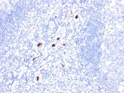
Biotium Granulocyte Marker(BM-1), CF488A conjugate, 0.1mg/mL
Recognizes 183 kDa protein with DNA-binding characteristics, which is identified as a myeloid specific antigen. It reacts with myeloid precursor cells and granulocytes in bone marrow. Its antigen appears to be restricted to M2 and M3 acute myelogenous leukemia (AML) subtypes. Markers of myeloid cells are useful in the identification of different levels of cellular differentiation. This MAb reacts with early precursor and mature forms of human myeloid cells. It is useful in the identification of myelogenous leukemias, distinguishing granulocytic sarcomas from lymphoid malignancies and also in the study of differentiation and transformation of human myeloid cells. The biological function of this antigen is not clear, although it has been proposed that it may play a role in the differentiation of myeloid cells.Primary antibodies are available purified, or with a selection of fluorescent CF Dyes and other labels. CF Dyes offer exceptional brightness and photostability. Note: C
Biotium CD31 / PECAM-1(C31.7), CF488A conjugate, 0.1mg/mL
CD31 (PECAM-1) is a transmembrane glycoprotein member of the immunoglobulin supergene family of adhesion molecules. CD31 is expressed by stem cells of the hematopoietic system and is primarily used to identify and concentrate these cells for experimental studies as well as for bone marrow transplantation. Anti-CD31 has shown to be highly specific and sensitive for vascular endothelial cells. Staining of nonvascular tumors (excluding hematopoietic neoplasms) is rare. CD31 MAb reacts with normal, benign, and malignant endothelial cells which make up blood vessel lining. The level of CD31 expression can help to determine the degree of tumor angiogenesis, and a high level of CD31 expression may imply a rapidly growing tumor and potentially a predictor of tumor recurrence.Primary antibodies are available purified, or with a selection of fluorescent CF Dyes and other labels. CF Dyes offer exceptional brightness and photostability. Note: Conjugates of blue fluorescent dyes li
Biotium Calnexin (Endoplasmic Reticulum Marker) (CANX/1541), CF568 conjugate, 0.1mg/mL
Calnexin is a type-I integral membrane protein of the endoplasmic reticulum (ER). It is a calcium-binding chaperone protein that interacts transiently with newly synthesized N-linked glycoproteins, facilitating protein folding and assembly. It may also play a central role in the quality control of protein folding by retaining incorrectly folded protein subunits within the ER for degradation.Primary antibodies are available purified, or with a selection of fluorescent CF Dyes and other labels. CF Dyes offer exceptional brightness and photostability. Note: Conjugates of blue fluorescent dyes like CF405S and CF405M are not recommended for detecting low abundance targets, because blue dyes have lower fluorescence and can give higher non-specific background than other dye colors.
Biotium Desmocollin-2/3 (7G6), CF488A conjugate, 0.1mg/mL
Desmosomes are intercellular adhering junctions that represent cell surface attachment sites for intermediate filament. The desmosome is subdivided into two regions. The plaque region lies adjacent to the plasma, and is believed to contain molecules that attach the intermediate filament cytoskeleton to the desmosome. The core region is composed of transmembrane glycoproteins that are thought to mediate cell-cell adhesion. Desmogleins and desmocollins are the main desmosomal transmembrane proteins. These desmosomal glycoproteins belong to the members of the cadherin family of adhesion molecules. Three different isoforms of both desmogleins and desmocollins have been identified, named as desmoglein 1-3 and desmocollins. Desmosomal cadherins showed differentiation-specific expression in the human epidermis, although the functional significance of this differential expression is not fully understood. Desmocollin-1 can be found in the upper layers. The expression of desmocollin-2
Biotium CD18(68-5A5), CF488A conjugate, 0.1mg/mL
This antibody recognizes a transmembrane glycoprotein of 95 kDa, identified as CD18 or integrin -2 (Workshop III). It complexes non-covalently with either L, M, or X integrin (CD11a, b, or c) to form the heterodimers, LFA-1, Mac-1, and p150,95, respectively. LFA-1 is the receptor for three members of the Ig supergene family of proteins, ICAM-1 (CD54), ICAM-2 (CD102), and Mac-1 and p150,95 bind to ICAM-1, fibrinogen, and iC3b. ICAM-3 (CD50). CD18/CD11 heterodimeric molecules are involved with cell/cell and cell/extracellular adhesion in immune and inflammatory responses. This MAb blocks these cellular interactions.Primary antibodies are available purified, or with a selection of fluorescent CF Dyes and other labels. CF Dyes offer exceptional brightness and photostability. Note: Conjugates of blue fluorescent dyes like CF405S and CF405M are not recommended for detecting low abundance targets, because blue dyes have lower fluorescence and can give higher non-specific b
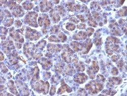
Biotium Golgi Complex(371-4), CF488A conjugate, 0.1mg/mL
This MAb recognizes an antigen associated with the Golgi complex in human cells only. It can be used to stain the Golgi complex in cell or tissue preparations and can be used as a Golgi marker in subcellular fractions. It produces a diffuse staining pattern of the Golgi zone in normal and malignant cells. This MAb is an excellent marker for human cells in xenographic model research. It reacts specifically with human cells. The Golgi apparatus is an organelle present in all eukaryotic cells that forms a part of the endomembrane system. The primary function of the Golgi apparatus is to process and package macromolecules synthesized by the cell for exocytosis or use within the cell. The Golgi is made up of a stack of flattened, membrane-bound sacs known as cisternae, with three functional regions: the cis face, medial region and trans face. Each region consists of various enzymes that selectively modify the macromolecules passing though them, depending on where they are destined