
Antibody and Dye Based Assays

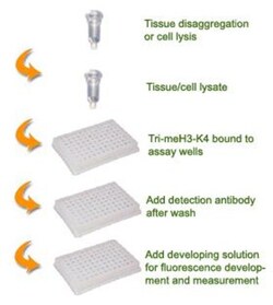
Epigentek Inc EpiQuik Global Tri-Methyl Histone H3K4 Quantification Kit (Fluorometric) (96 assays)
For fluorescence-based measurement of total H3K4me3 amounts

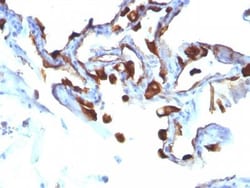
Biotium Cytokeratin 8/18(C-51), CF488A conjugate, 0.1mg/mL
Cytokeratin 8 (CK8) belongs to the type II (or B or basic) subfamily of high molecular weight cytokeratins and exists in combination with cytokeratin 18 (CK18). This MAb recognizes all simple epithelia including glandular epithelium, for example thyroid, female breast, gastrointestinal tract, respiratory tract, and urogenital tract including transitional epithelium. All adenocarcinomas and most squamous carcinomas are positive but keratinizing squamous carcinomas are usually negative. This antibody is useful in demonstrating the presence of Paget cells; there is very little keratin 18 in the normal epidermis so only Paget cells are stained.Primary antibodies are available purified, or with a selection of fluorescent CF Dyes and other labels. CF Dyes offer exceptional brightness and photostability. Note: Conjugates of blue fluorescent dyes like CF405S and CF405M are not recommended for detecting low abundance targets, because blue dyes have lower fluorescence and can give
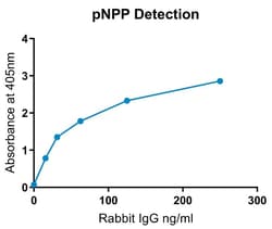
Abcam Alkaline Phosphatase Conjugation Kit (Animal Free) - Lightning-Link, 100UG
Alkaline Phosphatase conjugation / labeling in < 4 hrs with 30 secs hands-on time using Alkaline Phosphatase Conjugation Kit (Animal Free) - Lightning-Link ab269901. 100% antibody recovery.
The product is subject to the following: Abcam Restricted Use Statement


Medchemexpress LLC (E)-9-amino-10-methyl-2-(naphthalen-1-ylmethylene)-1-oxo-1,2,3,4-tetrahydroacridin-10-ium iodide | 2252162-81-3 | 98.1% | C25H21IN2O | 10MG

Aβ/tau aggregation-IN-1 is a small-molecule research inhibitor that reduces aggregation of amyloid-β (Aβ1-42) and tau by preventing β-sheet formation and disrupting aggregate interactions. It demonstrates activity in cell-based tau aggregation assays and has documented physicochemical and storage properties for preclinical use.
- Inhibits aggregation of Aβ1-42 and tau, reducing β-sheet formation.
- Reported KD values ≈160 μM for Aβ1-42 and ≈337 μM for tau.
- Reduces tau aggregation foci at 1 μM in HEK-293T cellular assays.
- High purity (98.1%) suitable for research applications.
- Soluble in DMSO (~33.3 mg/mL); in vivo formulation examples provided.
- Solid storage at 4°C; in solvent store at -80°C for long-term stability.
- For research use only; not for human use.
Revvity Health Sciences Inc HTRF TDP-43 Aggregation Kit, 100,000 Assay Points
HTRF TDP-43 Aggregation Kit, 100,000 Assay Points

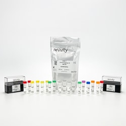
Revvity Health Sciences Inc HTRF TDP-43 Aggregation Kit, 10,000 Assay Points
HTRF TDP-43 Aggregation Kit, 10,000 Assay Points
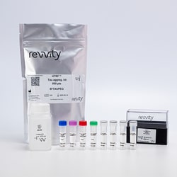
Revvity Health Sciences Inc HTRF Tau Aggregation Kit, 100,000 Assay Points
HTRF Tau Aggregation Kit, 100,000 Assay Points
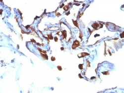
Biotium Cytokeratin 8/18(C-51), CF488A conjugate, 0.1mg/mL
Cytokeratin 8 (CK8) belongs to the type II (or B or basic) subfamily of high molecular weight cytokeratins and exists in combination with cytokeratin 18 (CK18). This MAb recognizes all simple epithelia including glandular epithelium, for example thyroid, female breast, gastrointestinal tract, respiratory tract, and urogenital tract including transitional epithelium. All adenocarcinomas and most squamous carcinomas are positive but keratinizing squamous carcinomas are usually negative. This antibody is useful in demonstrating the presence of Paget cells; there is very little keratin 18 in the normal epidermis so only Paget cells are stained.Primary antibodies are available purified, or with a selection of fluorescent CF Dyes and other labels. CF Dyes offer exceptional brightness and photostability. Note: Conjugates of blue fluorescent dyes like CF405S and CF405M are not recommended for detecting low abundance targets, because blue dyes have lower fluorescence and can give

Biotium CD3e(Rabbit PAb), CF488A conjugate, 0.1mg/mL
The immunogen for this Mab is derived from the cytoplasmic domain of CD3 epsilon chain. CD3 consists of five different polypeptide chains (designated as gamma, delta, epsilon, zeta, and eta) with MW ranging from 16-28 kDa. The CD3 complex is closely associated at the lymphocyte cell surface with the T cell antigen receptor (TCR). Reportedly, CD3 complex is involved in signal transduction to the T cell interior following antigen recognition. The CD3 antigen is first detectable in early thymocytes and probably represents one of the earliest signs of commitment to the T cell lineage. In cortical thymocytes, CD3 is predominantly intra-cytoplasmic. However, in medullary thymocytes, it appears on the T cell surface. CD3 antigen is a highly specific marker for T cells, and is present in majority of T cell neoplasms.Primary antibodies are available purified, or with a selection of fluorescent CF Dyes and other labels. CF Dyes offer exceptional brightness and photostability. No
Biotium MART-1 / Melan-A(M2-7C10), CF488A conjugate, 0.1mg/mL
This antibody recognizes a protein doublet of 20-22 kDa, identified as MART-1 (Melanoma Antigen Recognized by T cells 1) or Melan-A. MART-1 is a newly identified melanocyte differentiation antigen recognized by autologous cytotoxic T lymphocytes. Seven other melanoma associated antigens recognized by autologous cytotoxic T cells include MAGE-1, MAGE-3, tyrosinase, gp100, gp75, BAGE-1, and GAGE-1. Subcellular fractionation shows that MART-1 is present in melanosomes and endoplasmic reticulum. This MAb labels melanomas and other tumors showing melanocytic differentiation. It is also a useful positive-marker for angiomyolipomas. It does not stain tumor cells of epithelial, lymphoid, glial, or mesenchymal origin.Primary antibodies are available purified, or with a selection of fluorescent CF Dyes and other labels. CF Dyes offer exceptional brightness and photostability. Note: Conjugates of blue fluorescent dyes like CF405S and CF405M are not recommended for detecting low abu
Biotium CD19(CD19/366), CF555 conjugate, 0.1mg/mL
CD19 is a transmembrane glycoprotein that contains two extracellular immunoglobulin-like domains. CD19 is present in both benign and malignant B-cells and is considered to be the most reliable surface marker of this lineage over a wide range of maturational stages. In normal lymphoid tissue, CD19 is observed in germinal centers, in mantle zone cells, and in scattered cells of the inter-follicular areas. Anti-CD19 exhibits an overall immunoreactivity pattern similar to those of the antibodies against CD20 and CD22. However, in contrast to CD20, expression of CD19 is continuous throughout B-cell development and through terminal differentiation of B-cells into plasma cells. Anti-CD19 positivity is seen in the vast majority of B-cell neoplasms commonly at a lower intensity than normal B-cell counterparts. Plasma cell neoplasms are nearly always negative, as are T-cell neoplasms.Primary antibodies are available purified, or with a selection of fluorescent CF Dyes and other labels
Biotium Cytokeratin 8(K8/383), CF488A conjugate, 0.1mg/mL
Cytokeratin 8 (CK8) belongs to the type II (or B or basic) subfamily of high molecular weight cytokeratins and exists in combination with cytokeratin 18 (CK18). CK8 is primarily found in the non-squamous epithelia and is present in majority of adenocarcinomas and ductal carcinomas. It is absent in squamous cell carcinomas. Hepatocellular carcinomas are defined by the use of antibodies that recognize only cytokeratin 8 and 18. CK8 exists on several types of normal and neoplastic epithelia, including many ductal and glandular epithelia such as colon, stomach, small intestine, trachea, and esophagus as well as in transitional epithelium. Anti-CK8 does not react with skeletal muscle or nerve cells. Epithelioid sarcoma, chordoma, and adamantinoma show strong positivity corresponding to that of simple epithelia (with antibodies against CK8, CK18 and CK19). Reportedly, anti-CK8 is useful for the differentiation of lobular (ring-like, perinuclear) from ductal (peripheral-predominant)
Miltenyi Biotec, Inc. Labeling Check Reagent-PE, AC146, 100 t
Staining of cells separated by MACS Technology
Biotium CD43(84-3C1), CF488A conjugate, 0.1mg/mL
This antibody recognizes a cell surface glycoprotein of 95/115/135 kDa (depending upon the extent of glycosylation), identified as CD43 (Workshop III). 70-90% of T-cell lymphomas and from 22-37% of B-cell lymphomas express CD43. No reactivity has been observed with reactive B-cells. So a B-lineage population that co-expresses CD43 is highly likely to be a malignant lymphoma, especially a low-grade lymphoma, rather than a reactive B-cell population. When CD43 antibody is used in combination with anti-CD20, effective immunophenotyping of the lymphomas in formalin-fixed tissues can be obtained. Co-staining of a lymphoid infiltrate with anti-CD20 and anti-CD43 argues against a reactive process and favors a diagnosis of lymphoma.Primary antibodies are available purified, or with a selection of fluorescent CF Dyes and other labels. CF Dyes offer exceptional brightness and photostability. Note: Conjugates of blue fluorescent dyes like CF405S and CF405M are not recommended f