
Antibody and Dye Based Assays

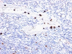
Biotium Granulocyte Marker(BM-2), CF488A conjugate, 0.1mg/mL
Recognizes 183 kDa protein with DNA-binding characteristics, which is identified as a myeloid specific antigen. It reacts with myeloid precursor cells and granulocytes in bone marrow. Its antigen appears to be restricted to M2 and M3 acute myelogenous leukemia (AML) subtypes. Markers of myeloid cells are useful in the identification of different levels of cellular differentiation. This MAb reacts with early precursor and mature forms of human myeloid cells. It is useful in the identification of myelogenous leukemias, distinguishing granulocytic sarcomas from lymphoid malignancies and also in the study of differentiation and transformation of human myeloid cells. The biological function of this antigen is not clear, although it has been proposed that it may play a role in the differentiation of myeloid cells.Primary antibodies are available purified, or with a selection of fluorescent CF Dyes and other labels. CF Dyes offer exceptional brightness and photostability. Note: C

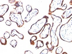
Biotium HCG-beta(HCGb/54), CF488A conjugate, 0.1mg/mL
This MAb reacts with a protein of 22 kDa, identified as β sub-unit of HCG. It does not cross react with the α sub-unit. HCG is a glycoprotein, which is secreted in large quantities by normal trophoblasts. It is present only in trace amounts in non-pregnant urine and sera but rises sharply during pregnancy. HCG is composed of two non-identical, non-covalently linked polypeptide chains designated as the alpha and beta subunits. The alpha subunit is identical to that of thyroid stimulating hormone (TSH), follicle stimulating hormone (FSH), and luteinizing hormone (LH). hCG MAb detects cells and tumors of trophoblastic origin such as choriocarcinoma. Large cell carcinoma and adenocarcinoma of the lung demonstrate anti-hCG positivity in 90% and 60% of cases respectively. 20% of lung squamous cell carcinomas are positive. hCG expression by non-trophoblastic tumors may indicate aggressive behavior.Primary antibodies are available purified, or with a selection of fluoresce

Biotium CD43(DF-T1), CF488A conjugate, 0.1mg/mL
This antibody recognizes a cell surface glycoprotein of 95/115/135 kDa (depending upon the extent of glycosylation), identified as CD43 . Epitope of MAb Bra7G is clearly different from that of MAb DF-T1, called b as opposed to a for DF-T1. 70-90% of T-cell lymphomas and from 22-37% of B-cell lymphomas express CD43. No reactivity has been observed with reactive B-cells. So a B-lineage population that co-expresses CD43 is highly likely to be a malignant lymphoma, especially a low-grade lymphoma, rather than a reactive B-cell population. When CD43 antibody is used in combination with anti-CD20, effective immunophenotyping of the lymphomas in formalin-fixed tissues can be obtained. Co-staining of a lymphoid infiltrate with anti-CD20 and anti-CD43 argues against a reactive process and favors a diagnosis of lymphoma.Primary antibodies are available purified, or with a selection of fluorescent CF Dyes and other labels. CF Dyes offer exceptional brightness and photostability. Note
Biotium Cytokeratin 18(DC10), CF488A conjugate, 0.1mg/mL
This MAb reacts with a wide variety of simple epithelia. It does not react with stratified squamous epithelia. It reacts with epithelial tumors of the gastrointestinal tract, lung, breast, pancreas, ovary, and thyroid. Cytokeratin 18, which belongs to the type A (acidic) subfamily of low molecular weight keratins, exists in combination with cytokeratin 8. It is reported that tissues from gastrointestinal tract are positive for both cytokeratin 8 and 18 but do not contain cytokeratin 14. Tissues from gastrointestinal tract, respiratory tract and urogenital tract, as well as endocrine and exocrine tissues and mesothelial cells are positive for cytokeratin 18.Primary antibodies are available purified, or with a selection of fluorescent CF Dyes and other labels. CF Dyes offer exceptional brightness and photostability. Note: Conjugates of blue fluorescent dyes like CF405S and CF405M are not recommended for detecting low abundance targets, because blue dyes have lower fluoresc
Biotium Desmocollin-2/3 (7G6), CF488A conjugate, 0.1mg/mL
Desmosomes are intercellular adhering junctions that represent cell surface attachment sites for intermediate filament. The desmosome is subdivided into two regions. The plaque region lies adjacent to the plasma, and is believed to contain molecules that attach the intermediate filament cytoskeleton to the desmosome. The core region is composed of transmembrane glycoproteins that are thought to mediate cell-cell adhesion. Desmogleins and desmocollins are the main desmosomal transmembrane proteins. These desmosomal glycoproteins belong to the members of the cadherin family of adhesion molecules. Three different isoforms of both desmogleins and desmocollins have been identified, named as desmoglein 1-3 and desmocollins. Desmosomal cadherins showed differentiation-specific expression in the human epidermis, although the functional significance of this differential expression is not fully understood. Desmocollin-1 can be found in the upper layers. The expression of desmocollin-2
Biotium Estrogen Receptor(AER314), CF488A conjugate, 0.1mg/mL
This MAb is specific to ER alpha and shows minimal cross-reaction with other members of the family. Epitope of this MAb is mapped between aa120-170. ER is an important regulator of growth and differentiation in the mammary gland. Presence of ER in breast tumors indicates an increased likelihood of response to anti-estrogen (e.g. tamoxifen) therapy.Primary antibodies are available purified, or with a selection of fluorescent CF Dyes and other labels. CF Dyes offer exceptional brightness and photostability. Note: Conjugates of blue fluorescent dyes like CF405S and CF405M are not recommended for detecting low abundance targets, because blue dyes have lower fluorescence and can give higher non-specific background than other dye colors.
Biotium CD18(68-5A5), CF488A conjugate, 0.1mg/mL
This antibody recognizes a transmembrane glycoprotein of 95 kDa, identified as CD18 or integrin -2 (Workshop III). It complexes non-covalently with either L, M, or X integrin (CD11a, b, or c) to form the heterodimers, LFA-1, Mac-1, and p150,95, respectively. LFA-1 is the receptor for three members of the Ig supergene family of proteins, ICAM-1 (CD54), ICAM-2 (CD102), and Mac-1 and p150,95 bind to ICAM-1, fibrinogen, and iC3b. ICAM-3 (CD50). CD18/CD11 heterodimeric molecules are involved with cell/cell and cell/extracellular adhesion in immune and inflammatory responses. This MAb blocks these cellular interactions.Primary antibodies are available purified, or with a selection of fluorescent CF Dyes and other labels. CF Dyes offer exceptional brightness and photostability. Note: Conjugates of blue fluorescent dyes like CF405S and CF405M are not recommended for detecting low abundance targets, because blue dyes have lower fluorescence and can give higher non-specific b
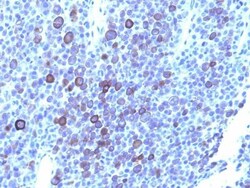
Biotium Glypican-3(GPC3/863 + 1G12), CF488A conjugate, 0.1mg/mL
Glypican-3 (GPC3) is an integral membrane protein that is mutated in the Simpson-Golabi-Behmel syndrome (SGBS). SGBS is characterized by pre- and post-natal overgrowth and is a recessive X-linked condition. GPC3 may also be found in a secreted form. Anti-GPC3 has been identified as a useful tumor marker for the diagnosis of hepatocellular carcinoma (HCC), hepatoblastoma, melanoma, testicular germ cell tumors, and Wilm's tumor. In patients with HCC, GPC3 is overexpressed in neoplastic liver tissue and elevated in serum, but is undetectable in normal liver, benign liver, and the serum of healthy donors. GPC3 expression is also found to be higher in HCC liver tissue than in cirrhotic liver or liver with focal lesions such as dysplastic nodules and areas of hepatic adenoma (HA) with malignant transformation. In the context of testicular germ cell tumors, GPC3 expression is up regulated in certain histologic subtypes, specifically yolk sac tumors and choriocarcinoma. A high level
Biotium Chromogranin A(CGA/414), CF488A conjugate, 0.1mg/mL
Chromogranin A is present in neuroendocrine cells throughout the body, including the neuroendocrine cells of the large and small intestine, adrenal medulla and pancreatic islets. It is an excellent marker for carcinoid tumors, pheochromocytomas, paragangliomas, and other neuroendocrine tumors. Co-expression of chromogranin A and neuron specific enolase (NSE) is common in neuroendocrine neoplasms. Reportedly, co-expression of certain keratins and chromogranin indicates neuroendocrine lineage. The presence of strong anti-chromogranin staining and absence of anti-keratin staining should raise the possibility of paraganglioma. The co-expression of chromogranin and NSE is typical of neuroendocrine neoplasms. Most pituitary adenomas and prolactinomas readily express chromogranin.Primary antibodies are available purified, or with a selection of fluorescent CF Dyes and other labels. CF Dyes offer exceptional brightness and photostability. Note: Conjugates of blue fluorescent dyes
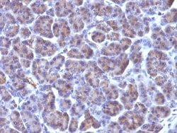
Biotium Golgi Complex(371-4), CF488A conjugate, 0.1mg/mL
This MAb recognizes an antigen associated with the Golgi complex in human cells only. It can be used to stain the Golgi complex in cell or tissue preparations and can be used as a Golgi marker in subcellular fractions. It produces a diffuse staining pattern of the Golgi zone in normal and malignant cells. This MAb is an excellent marker for human cells in xenographic model research. It reacts specifically with human cells. The Golgi apparatus is an organelle present in all eukaryotic cells that forms a part of the endomembrane system. The primary function of the Golgi apparatus is to process and package macromolecules synthesized by the cell for exocytosis or use within the cell. The Golgi is made up of a stack of flattened, membrane-bound sacs known as cisternae, with three functional regions: the cis face, medial region and trans face. Each region consists of various enzymes that selectively modify the macromolecules passing though them, depending on where they are destined
Biotium HER-2 / CD340(HRB2/282), CF488A conjugate, 0.1mg/mL
Recognizes a protein of 185 kDa, which is identified as c-erbB-2/HER-2/neu. Its epitope is localized in the extracellular domain. C-erbB-2/HER-2 is a member of the EGFR family. This MAb is specific and shows minimal cross-reaction with other members of the EGFR-family. Receptors of this family are located on the plasma membrane and consist of an extracellular ligand-binding domain that is connected to a large intracellular domain by a single transmembrane sequence. c-erbB-2/HER-2 protein is over-expressed in a variety of carcinomas especially those of breast and ovary.Primary antibodies are available purified, or with a selection of fluorescent CF Dyes and other labels. CF Dyes offer exceptional brightness and photostability. Note: Conjugates of blue fluorescent dyes like CF405S and CF405M are not recommended for detecting low abundance targets, because blue dyes have lower fluorescence and can give higher non-specific background than other dye colors.
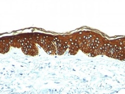
Biotium Cytokeratin, HMW(AE-3), CF488A conjugate, 0.1mg/mL
This MAb recognizes basic (Type II or HMW) cytokeratins, which include 67 kDa (CK1); 64 kDa (CK3); 59 kDa (CK4); 58 kDa (CK5); 56 kDa (CK6); 52 kDa (CK8). Twenty human keratins are resolved with two-dimensional gel electrophoresis into acidic (pI 6.0) subfamilies. The acidic keratins have molecular weights (MW) of 56.5, 55, 51, 50, 50 , 48, 46, 45, and 40 kDa. MAb AE3 recognizes the 65-67, 64, 59, 58, 56, and 52 kDa keratins of basic subfamily. Many studies have shown the usefulness of keratins as markers in cancer research and tumor diagnosis. AE1/AE3 is a broad spectrum anti pan-keratin antibody cocktail, which differentiates epithelial tumors from non-epithelial tumors e.g. squamous vs. adenocarcinoma of the lung, liver carcinoma, breast cancer, and esophageal cancer.Primary antibodies are available purified, or with a selection of fluorescent CF Dyes and other labels. CF Dyes offer exceptional brightness and photostability. Note: Conjugates of blue fluorescent dyes lik
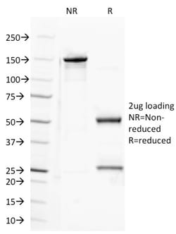
Biotium CD100 (Semaphorin-4D)(A8), CF488A conjugate, 0.1mg/mL
CD100 (also known as Semaphorin 4D), is a homodimeric protein comprised of 50 kDa subunits. It is expressed on resting T cells and weakly on other lymphocytes, and its expression is increased on B and T cells after stimulation. CD100 is a cell surface receptor that binds to CD72, Plexin B1 and Plexin B2. It promotes B cell aggregation, cell survival, and cell proliferation.Primary antibodies are available purified, or with a selection of fluorescent CF Dyes and other labels. CF Dyes offer exceptional brightness and photostability. Note: Conjugates of blue fluorescent dyes like CF405S and CF405M are not recommended for detecting low abundance targets, because blue dyes have lower fluorescence and can give higher non-specific background than other dye colors.
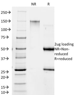
Biotium Giardia lamblia(BB1.1E5), CF488A conjugate, 0.1mg/mL
Giardiasisis a diarrheal illness caused by a single celled microscopic protozoan parasite, Giardia lamblia, also known as Giardia intestinalis. Giardia lamblia exists in two forms, an active form called a trophozoite, and an inactive form called a cyst. The active trophozoite attaches to the lining of the small intestine and is responsible for causing the signs and symptoms of giardiasis. The trophozoite cannot live long outside of the body and spread of infection is via the cyst, which is excreted in the host's feces. When it is ingested, stomach acid activates the cyst, and the cyst develops into the disease causing trophozoite in the new host. Giardiasis is diagnosed by finding cysts or trophozoites in the feces. Primary antibodies are available purified, or with a selection of fluorescent CF Dyes and other labels. CF Dyes offer exceptional brightness and photostability. Note: Conjugates of blue fluorescent dyes like CF405S and CF405M are not recommended for detecting
GENOVIS INC FabRICATOR MagIC | 2mL
FabRICATOR MagIC employs magnetic beads for fast automated processing of up to 96 antibodies in parallel. The popular cysteine protease FabRICATOR (IdeS) immobilized on magnetic agarose beads delivers rapid and consistent subunit generation for multiple antibodies in parallel, perfect for fast automated analysis of CQAs in a middle-level LC/MS workflow.
