
Primary Antibody Matched Pairs


Biotium CD209 / DC-SIGN (Pathogen Receptor on Dendritic Cells) (C209/1781), CF640R conjugate, 0.1mg/mL
DC-SIGN is a transmembrane receptor that is expressed on the surface of dendritic cells and macrophages. It is involved in the innate immune system and recognizes numerous evolutionarily divergent pathogens ranging from parasites to viruses. The protein is organized into three distinct domains: an N-terminal transmembrane domain, a tandem-repeat neck domain and C-type lectin carbohydrate recognition domain. The extracellular region consisting of the C-type lectin and neck domains has a dual function as a pathogen recognition receptor and a cell adhesion receptor by binding carbohydrate ligands on the surface of microbes and endogenous cells. The neck region is important for homo-oligomerization, which allows the receptor to bind multivalent ligands with high avidity.Primary antibodies are available purified, or with a selection of fluorescent CF Dyes and other labels. CF Dyes offer exceptional brightness and photostability. Note: Conjugates of blue fluorescent dyes like CF


Biotium CD56 / NCAM1 / NKH1 (Neuronal Cell Marker) (NCAM1/1496), CF488A conjugate, 0.1mg/mL
This MAb reacts with an extracellular domain (close to transmembrane) of CD56/NCAM. Three isoforms of neural cell adhesion molecule (NCAM) are produced by differential splicing of the RNA transcript from a single gene. The 135 kDa isoform is the basic molecule, which is glycosylated or sialylated to produce the mature species. Anti-CD56 recognizes two proteins of the neural cell adhesion molecule, the basic molecule expressed on most neuroectodermally derived tissues and neoplasms (e.g. retinoblastoma, medulloblastomas, astrocytomas, neuroblastomas, and small cell carcinomas). It is also expressed on some mesodermally derived tumors (rhabdomyosarcoma). Anti-CD56 plays an important role in the diagnosis of nodal and nasal NK/T-cell lymphomas.Primary antibodies are available purified, or with a selection of fluorescent CF Dyes and other labels. CF Dyes offer exceptional brightness and photostability. Note: Conjugates of blue fluorescent dyes like CF405S and CF405M are not
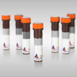
Miltenyi Biotec, Inc. MACSpep HiAg CKS2 (KYFDEHYEY) HLA-C*07
MACSpep single peptides developed for loading onto MHC multimers using the MHC MACSimer FleX Kits.
This product is subject to the following: Miltenyi Biotec, Inc. Terms and Conditions of Sale For Products and Services
Genscript Corporation CD55 His Cynomolgus
Decay Accelerating Factor (or CD55) is a major regulator of the alternative and classical pathways of complement activation and is expressed on all serum-exposed cells It is commonly hijacked by invading pathogens including many enteroviruses and uropathogenic Escherichia coli to promote cellular attachment prior to infection

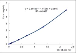
AdipoGen Erythroferrone (human) Matched Pair Detection Set (1 Set)
Matched Pair Detection Set. Detects human Erythroferrone [Myonectin; CTRP15] in serum, plasma and cell culture supernatant. Colorimetric sandwich assay. Sample types: Cell Culture Supernatant, Plasma, Serum. Range: 0.156 to 10ng/ml. Erythroferrone plays two different regulatory role.s: i) As a myokine, Erythroferrone/Myonectin acts as an endurance exercise-driven myokine which protects the heart from ischemic injury by reducing cardiomyocyte apoptosis and macrophage inflammatory response, and ii) Erythroferrone is produced by erythroblasts after bleeding or EPO treatment and acts on hepatocytes to suppress hepcidin expression, resulting in increased release of iron from cellular iron stores. Erythroferrone acts on hepcidin by inhibiting members of the BMP-5, BMP-6 and BMP-7 subgroup of BMPs. Erythroferrone functions as erythroid modulator of iron metabolism and hemoglobin synthesis.
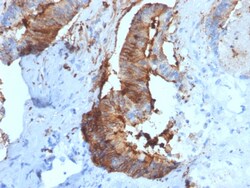
Biotium Cathepsin D (Tumor Marker)(CTSD/2781), CF568 conjugate, 0.1mg/mL
Cathepsin D is a ubiquitously expressed lysosomal aspartyl protease involved in the normal degradation of proteins. It is synthesized as an inactive 43 kDa preprocathepsin D that is cleaved and glycosylated to form a 46 kDa procathepsin D and then further cleaved to produce 28 kDa and 15 kDa subunits (heavy and light chains, respectively). Cathepsin D exhibits pepsin-like activity and plays a role in protein turnover and in the proteolytic activation of hormones and growth factors. Mutations in this gene play a causal role in neuronal ceroid lipofuscinosis-10 and may be involved in the pathogenesis of several other diseases, including breast cancer and possibly Alzheimer's disease. Primary antibodies are available purified, or with a selection of fluorescent CF Dyes and other labels. CF Dyes offer exceptional brightness and photostability. Note: Conjugates of blue fluorescent dyes like CF405S and CF405M are not recommended for detecting low abundance targets, because blu
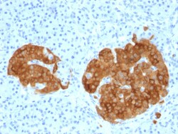
Biotium GAD2 / GAD65 (GABAergic Neuronal Marker) (GAD2/1960), CF568 conjugate, 0.1mg/mL
This MAb recognizes a protein of 65 kDa, which is identified as glutamic acid decarboxylase 2 (GDA2). It is responsible for catalyzing the production of gamma-aminobutyric acid from L-glutamic acid. There are two forms of glutamic acid decarboxylases (GAD s) that are found in the brain: GAD2 (also known as GAD65) and GAD1 (also known as GAD67). GAD1 and GAD2 are members of the group II decarboxylase family of proteins and are responsible for catalyzing the rate-limiting step in the production of GABA (ÃŽ³-aminobutyric acid) from L-glutamic acid. Although both GAD s are found in the brain, GAD2 localizes to synaptic vesicle membranes in nerve terminals, while GAD1 is distributed throughout the cell. A pathogenic role for GAD2 is identified in the human pancreas since it has been identified as an autoantibody and an auto-reactive T cell target in insulin-dependent diabetes.Primary antibodies are available purified, or with a selection of fluorescent CF Dyes and other label
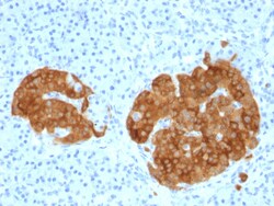
Biotium GAD2 / GAD65 (GABAergic Neuronal Marker)(GAD2/1960), 1mg/mL
This MAb recognizes a protein of 65 kDa, which is identified as glutamic acid decarboxylase 2 (GDA2). It is responsible for catalyzing the production of gamma-aminobutyric acid from L-glutamic acid. There are two forms of glutamic acid decarboxylases (GAD s) that are found in the brain: GAD2 (also known as GAD65) and GAD1 (also known as GAD67). GAD1 and GAD2 are members of the group II decarboxylase family of proteins and are responsible for catalyzing the rate-limiting step in the production of GABA (ÃŽ³-aminobutyric acid) from L-glutamic acid. Although both GAD s are found in the brain, GAD2 localizes to synaptic vesicle membranes in nerve terminals, while GAD1 is distributed throughout the cell. A pathogenic role for GAD2 is identified in the human pancreas since it has been identified as an autoantibody and an auto-reactive T cell target in insulin-dependent diabetes.Primary antibodies are available purified, or with a selection of fluorescent CF Dyes and other label

Ferret TNF ELISA Pair Set 15PP (Sino Biological)
Each vial contains 200 ng of recombinant Ferret TNF-alpha / TNFA ELISA Pair Set. Reconstitute with 1 mL Dilution Buffer. After reconstitution, store at -20℃ to -80℃ in a manual defrost freezer. A seven-point standard curve using 2-fold serial dilutions in Dilution Buffer, and a high standard of 5000 pg/mL is recommended.
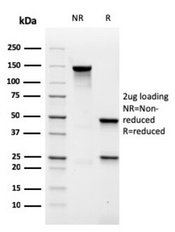
Biotium MALT1 (MALT-Lymphoma Marker)(rMT1/410), CF488A conjugate, 0.1mg/mL
Mucosa associated lymphoid tissue lymphoma translocation gene 1 (MALT1) is found in extranodal low-grade B cell lymphomas. MALT1 encodes two Ig-like C2-type domains and fuses with an API2 gene, which is highly expressed in adult lymphoid tissue. The translocation of this MALT1 gene, which maps to human chromosome 18q21, and the apoptosis-inhibiting API2 gene results in an increased development of MALT lymphomas and apoptosis inhibition. Sites at which this API2-MALT1 (11;18)(q21;q21) translocation commonly occurs are within human lung and kidney tissue. MALT lymphoma expresses nuclear Bcl10, which mediates the oligomerization and activation of a MALT1 caspase-like domain. MALT1 mRNA is found in pre-B cells, mature B cells and plasma cells. Primary antibodies are available purified, or with a selection of fluorescent CF Dyes and other labels. CF Dyes offer exceptional brightness and photostability. Note: Conjugates of blue fluorescent dyes like CF405S and CF405M are not r
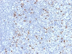
Biotium Perforin-1 (Pore Forming Protein) (Apoptosis Marker)(PRF1/2470), CF568 conjugate, 0.1mg/mL
Perforin is a pore-forming protein that leads to osmotic lysis of the target cells and subsequently enables granzymes to enter the target cells and activate apoptosis. Perforin has structural and functional similarities to complement component 9 (C9). Like C9, this protein creates transmembrane tubules and is capable of lysing non-specifically a variety of target cells. It is one of the main cytolytic proteins of cytolytic granules, and is known to be a key effector molecule for T-cell- and natural killer-cell-mediated cytolysis. Defects in this gene cause familial hemophagocytic lymphohistiocytosis type 2 (HPLH2), a rare and lethal autosomal recessive disorder of early childhood. The expression of perforin is reportedly upregulated in activated CD8 T-cells, natural killer cells and some CD4 T-cells. Primary antibodies are available purified, or with a selection of fluorescent CF Dyes and other labels. CF Dyes offer exceptional brightness and photostability. Note: Conjug
Miltenyi Biotec, Inc. CD69-PerCP-Vio700, mouse, H1.2F3, 100 t
Identification and enumeration of CD69+ cells by flow cytometry.
This product is subject to the following: Miltenyi Biotec, Inc. Terms and Conditions of Sale For Products and Services
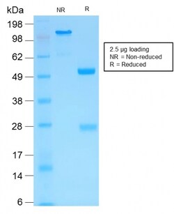
Biotium MMP3 (Marker of Metastasis and Rheumatoid Arthritis)(MMP3/1994R), CF740 conjugate, 0.1mg/mL
The matrix metalloproteinases (MMP) are a family of peptidase enzymes responsible for the degradation of extracellular matrix components, including collagen, gelatin, fibronectin, laminin and proteoglycan. Transcription of MMP genes is differentially activated by phorbol ester, lipopolysaccharide (LPS) or staphylococcal enterotoxin B (SEB). MMP catalysis requires both calcium and zinc. MMP-3, MMP-10 and MMP-11 (also designated stromelysin-1, 2 and 3, respectively) activate procollagenase. MMP-3 activation of procollagenase can occur via two pathways. Direct activation by MMP-3 is slow and activation by MMP-3 in conjunction with tissue or plasma proteinases is rapid. MMP-10 is expressed in small intestine, and at lower levels in lung and heart. MMP-11 is specifically expressed in stromal cells of breast carcinomas and contributes to epithelial cell malignancies. Primary antibodies are available purified, or with a selection of fluorescent CF Dyes and other labels. CF Dyes of
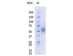
Sino Biological Recombinant Mouse uPAR/PLAUR Protein (His Tag), HPLC-verified, Endotoxin-Free
A DNA sequence encoding the extracellular domain of mouse PLAUR (NP_035243.1) precursor (Met 1-Thr 297) was expressed, fused with a polyhistidine tag at the C-terminus.
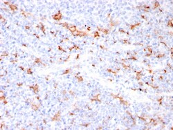
Biotium S100A9 + Calprotectin (S100A8/A9 Complex)(MAC3157R), 0.2mg/mL
Recognizes the L1 or Calprotectin molecule, an intra-cytoplasmic antigen comprising of a 12 kDa alpha chain and a 14 kDa beta chain expressed by granulocytes, monocytes and by tissue macrophages. Macrophages usually arise from hematopoietic stem cells in the bone marrow. Under migration into tissues, the monocytes undergo further differentiation to become multifunctional tissue macrophages. They are classified into normal and inflammatory macrophages. Normal macrophages include macrophages in connective tissue (histiocytes), liver (Kupffer's cells), lung (alveolar macrophages), lymph nodes (free and fixed macrophages), spleen (free and fixed macrophages), bone marrow (fixed macrophages), serous fluids (pleural and peritoneal macrophages), skin (histiocytes, Langerhans's cell) and in other tissues. Inflammatory macrophages are present in various exudates. Macrophages are part of the innate immune system, recognizing, engulfing and destroying many potential pathogens including