
Donkey Secondary Antibodies

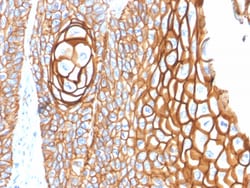
Biotium CD9 (TSPAN29) (Motility-Related Protein-1)(P1/33/2), CF647 conjugate, 0.1mg/mL
CD9 is a type IV transmembrane glycoprotein with four transmembrane domains. CD9 on pre-B cells may play a role in cell-cell adhesion. In addition, CD9 may play a role in signal transduction mediated by interaction with low molecular weight GTP binding proteins. CD9 is expressed on early B cells, eosinophils, basophils and activated T cells and is a major component of the platelet cell surface. It is also expressed on most non-T acute lymphoblastic leukemia cells and on some acute myeloid and chronic lymphoid leukemias.Primary antibodies are available purified, or with a selection of fluorescent CF Dyes and other labels. CF Dyes offer exceptional brightness and photostability. Note: Conjugates of blue fluorescent dyes like CF405S and CF405M are not recommended for detecting low abundance targets, because blue dyes have lower fluorescence and can give higher non-specific background than other dye colors.


Biotium IDH1(IDH1/1152), CF647 conjugate, 0.1mg/mL
This antibody recognizes a 45 kDa protein, which is identified as isocitrate dehydrogenase (IDH1). It belongs to the isocitrate and isopropylmalate dehydrogenases family. IDH1 catalyzes the third step of the citric acid cycle, which involves the oxidative decarboxylation of isocitrate, forming alpha-ketoglutarate and CO2 in a two-step reaction. The first step involves the oxidation of isocitrate to the intermediate oxalosuccinate, while the second step involves the production of alpha-ketoglutarate. During this process, either NADH or NADPH is produced along with CO2. Recently, an inactivating mutation of IDH1 has been implicated in glioblastoma. IDH1 appears to function as a tumor suppressor that, when mutationally inactivated, contributes to tumorigenesis in part through induction of the HIF-1 pathway.Primary antibodies are available purified, or with a selection of fluorescent CF Dyes and other labels. CF Dyes offer exceptional brightness and photostability. Note: Conjugat
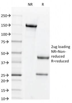
Biotium CD44v6 (Marker of Tumor Metastasis)(2F10), CF647 conjugate, 0.1mg/mL
This antibody recognizes an epitope encoded by exon v6 on the variant portion of human CD44. The CD44 molecule belongs to a family of cellular adhesion molecules found on a wide range of normal and malignant cells in epithelial, mesothelial and hemopoiesis tissues. CD44 is a single gene with 20 exons, of which 10 are normally expressed to encode the basic CD44 (H-CAM) molecule. The additional 10 exons (v1 to v10) are only expressed by alternative splicing of the nuclear RNA. The expression of specific cell adhesion molecule CD44 splice variants has been reported to be associated with metastasis in certain human malignancies.Primary antibodies are available purified, or with a selection of fluorescent CF Dyes and other labels. CF Dyes offer exceptional brightness and photostability. Note: Conjugates of blue fluorescent dyes like CF405S and CF405M are not recommended for detecting low abundance targets, because blue dyes have lower fluorescence and can give higher non-
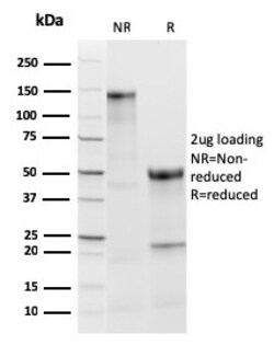
Biotium TPO (Thyroid Peroxidase) (Thyroid Marker)(TPO/3813R), CF647 conjugate, 0.1mg/mL
Thyroperoxidase (TPO) is a 933-amino acid, type I transmembrane glycoprotein that plays a key role in thyroid gland function and autoimmunity. It is present as a dimer on the apical surface of thyroid follicular cells. TPO functions in the iodination of tyrosine residues in thyroglobulin and phenoxy-ester formation between pairs of iodinated tyrosines to generate the thyroid hormones, thyroxine and triiodothyronine. Mutations in this gene are associated with several disorders of thyroid hormonogenesis, including congenital hypothyroidism, congenital goiter, and thyroid hormone organification defect IIA. Malignant thyroid tumors exhibit an anomaly in TPO resulting in lower affinity for anti-TPO. This antibody may aid in the differentiation between benign and malignant thyroid tumors. Primary antibodies are available purified, or with a selection of fluorescent CF Dyes and other labels. CF Dyes offer exceptional brightness and photostability. Note: Conjugates of blue fluores

Jackson Immuno Research Labs Peroxidase-AffiniPure Donkey Anti-Mouse IgM µ Chain Specific
Peroxidase-AffiniPure Donkey Anti-Mouse IgM µ Chain Specific

Jackson Immuno Research Labs Alkaline Phosphatase-AffiniPure F(ab')2 Fragment Donkey Anti-Mouse IgG (H+L) (min X Bov Ck Gt GP Sy Hms Hrs Hu Rb Rat Shp Sr Prot)
Alkaline Phosphatase-AffiniPure F(ab')2 Fragment Donkey Anti-Mouse IgG (H+L) (min X Bov Ck Gt GP Sy Hms Hrs Hu Rb Rat Shp Sr Prot)
Biotium CD11c (Dendritic Cell Marker) (ITGAX/2507), CF647 conjugate, 0.1mg/mL
This antibody recognizes a protein of 145 kDa, identified as CD11c. CD11c (ITGAX), a member of the leukointegrin family, shares the same beta subunit with other members of the leukocyte adhesion molecule family, which includes CD11a (LFA-1), CD11b (MAC-1) and CD11d (ITGAD), but has a unique alpha chain. CD11c has been shown to play a role in phagocytosis, cell migration, and cytokine production by monocytes/macrophages as well as induction of T-cell proliferation by Langerhans cells. CD11c is expressed prominently on the plasma membranes of monocytes, tissue macrophages, NK cells, and most dendritic cells (DCs). A lower level of expression is also observed on neutrophils as a result of its high level of expression on most DCs. An antibody to CD11c may aid in identification of lesions with histiocytic origin. It may also been used as a marker for hairy cell leukemia in paraffin-embedded tissues.Primary antibodies are available purified, or with a selection of fluorescent CF Dyes a
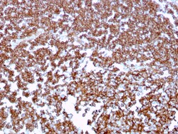
Biotium CD20 / MS4A1 (B-Cell Marker)(MS4A1/3409), CF647 conjugate, 0.1mg/mL
Recognizes a protein of 30-33 kDa, which is identified as CD20. It is a non-Ig differentiation antigen of B-cells and its expression is restricted to normal and neoplastic B-cells, being absent from all other leukocytes and tissues. CD20 is expressed by pre-B-cells and persists during all stages of B-cell maturation but is lost upon terminal differentiation into plasma cells. This MAb can be used for immunophenotyping of leukemia and malignant cells, B lymphocyte detection in peripheral blood and B cell localization in tissues. It reacts with the majority of B-cells present in peripheral blood and lymphoid tissues and their derived lymphomas. In lymphoid tissue, germinal center blasts and B-immunoblasts are particularly reactive. It is a reliable antibody for ascribing a B-cell phenotype in known lymphoid tissues. Rarely, CD20-positive T-cell lymphomas have been reported. Reactivity has also been noted with Reed-Sternberg cells in cases of Hodgkin's disease, particularly of l
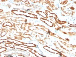
Biotium Erythropoietin (EPO) (Marker of Placentation Disorders)(EPO/3793R), CF647 conjugate, 0.1mg/mL
Recognizes a protein of about 37 kDa, which is identified as Erythropoietin (EPO). Erythropoietin is a secreted, glycosylated cytokine hormone composed of four alpha helical bundles. It is the primary factor responsible for regulating erythropoiesis during steady-state conditions and in response to blood loss and hemorrhage in the adult organism. Erythropoietin is synthesized by the kidney and stimulates the proliferation and maturation of bone marrow erythroid precursor cells. The protein is found in the plasma and regulates red cell production by promoting erythroid differentiation and initiating hemoglobin synthesis. Primary antibodies are available purified, or with a selection of fluorescent CF Dyes and other labels. CF Dyes offer exceptional brightness and photostability. Note: Conjugates of blue fluorescent dyes like CF405S and CF405M are not recommended for detecting low abundance targets, because blue dyes have lower fluorescence and can give higher non-specific
Biotium Cytokeratin 6 (KRT6) (Hyperproliferation-Related Keratin) (KRT6/1702), CF647 conjugate, 0.1mg/mL
This MAb recognizes a protein of 56 kDa, identified as cytokeratin 6 (CK6). In humans, multiple isoforms of Cytokeratin 6 (6A-6F), encoded by several highly homologous genes, have distinct tissue expression patterns, and Cytokeratin 6A is the dominant form in epithelial tissue. The gene encoding human Cytokeratin 6A maps to chromosome 12q13, and mutations in this gene are linked to several inheritable hair and skin pathologies. Keratins 6 and 16 are expressed in keratinocytes, which are undergoing rapid turnover in the suprabasal region (also known as hyper-proliferation-related keratins). Keratin 6 is found in hair follicles, suprabasal cells of a variety of internal stratified epithelia, in epidermis, in both normal and hyper-proliferative situations. Epidermal injury results in activation of keratinocytes, which express CK6 and CK16. CK6 is strongly expressed in about 75% of head and neck squamous cell carcinomas. Expression of CK6 is particularly associated with different
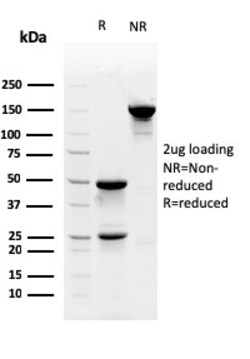
Biotium Myofibroblast Marker(PR 2D3), CF647 conjugate, 0.1mg/mL
Myofibroblasts are a unique group of smooth muscle-like fibroblasts that play an important role in oncogenesis, inflammation, repair, wound contraction and fibrosis. Like smooth muscle (SM) cells, myofibroblasts contain microfilament bundles and express -SM Actin, the Actin isoform that is present in myoepithelial cells and SM cells and especially abundant in vascular SM cells. Myofibroblasts secrete inflammatory and anti-inflammatory cytokines, chemokines, growth factors and lipid and gaseous inflammatory mediators, as well as extracellular matrix proteins and proteases in most organs and tissues. Besides being temporarily present following tissue injuries and fibrocontractive diseases, myofibroblasts are also present under normal conditions in regions such as the skin, pulmonary septa and periodontal ligaments. Stem cell factor and platelet-derived growth factor (PDGF) are two secreted proteins responsible for differentiating myofibroblasts from embryological stem cells. PR
Immunoreagents Inc DONKEY A-MOUSE IGG AP
Secondary Antibody; Donkey anti-mouse IgG (H&L) - F(ab)'2 fragment, min x w/Bo, Ck, Eq, Gt, Gp, Ha, Hu, Rb, Rt or Sh IgG, Host: Donkey, Format: Liq, Label: None, Applications: WB, ELISA, IHC/ICC, IF, IP, FC
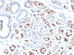
Biotium GATA-3 (Breast and Urothelial Marker)(GATA3/2444), CF647 conjugate, 0.1mg/mL
GATA-3 is a zinc finger transcription factor and plays an important role in promoting and directing cell proliferation, development, and differentiation in many tissues and cell types. GATA-3 expression is primarily seen in breast and urothelial carcinomas. 'Therefore, GATA3 antibody can be useful in the identification of unknown primary carcinoma when carcinomas of the breast or bladder are a possibility. Primary antibodies are available purified, or with a selection of fluorescent CF Dyes and other labels. CF Dyes offer exceptional brightness and photostability. Note: Conjugates of blue fluorescent dyes like CF405S and CF405M are not recommended for detecting low abundance targets, because blue dyes have lower fluorescence and can give higher non-specific background than other dye colors.
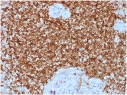
Biotium Bcl-2 (Apoptosis & Follicular Lymphoma Marker) (BCL2/1878R), CF647 conjugate, 0.1mg/mL
This antibody recognizes a protein of 25-26 kDa, identified as Bcl-2. This antibody shows no cross-reaction with Bcl-x or Bax proteins. Bcl-2 suppresses apoptosis in a variety of cell systems including factor-dependent lymphohematopoietic and neural cells. It regulates cell death by controlling the mitochondrial membrane permeability. Bcl-2 appears to function in a feedback loop system with caspases. It inhibits caspase activity either by preventing the release of cytochrome c from the mitochondria and/or by binding to the apoptosis-activating factor (APAF-1). Expression of Bcl-2 inhibits programmed cell death (apoptosis). In most follicular lymphomas, neoplastic germinal centers express high levels of Bcl-2 protein, whereas the normal or hyperplastic germinal centers are negative. Consequently, this antibody is valuable when distinguishing between reactive and neoplastic follicular proliferation in lymph node biopsies. It may also be used in distinguishing between those foll