
Mouse Secondary Antibodies


Biotium Human IgM (Immunoglobulin Mu Heavy Chain) (B-Cell Marker)(IGHM/2557R), CF740 conjugate, 0.1mg/mL
Recognizes a protein of 75 kDa, identified as mu heavy chain of human immunoglobulins. It does not cross-react with alpha (IgA), gamma (IgG), epsilon (IgE), or delta (IgD), heavy chains, T-cells, monocytes, granulocytes, or erythrocytes. Monomeric IgM is expressed as a membrane bound antibody on the surface of B cells and as a pentamer when secreted by plasma cells. IgM antibody is prominent in early immune responses to most antigens. Aberrant levels are associated with immune deficiency states, hereditary deficiencies, myeloma, Waldenstrom's macroglobulinemia, chronic infection and hepatocellular disease. This MAb is useful in the identification of leukemias, plasmacytomas, and certain non-Hodgkin's lymphomas. The most common feature of these malignancies is the restricted expression of a single heavy chain class. Demonstration of clonality in lymphoid infiltrates indicates that the infiltrate is clonal and therefore malignant. Primary antibodies are available purified, or w

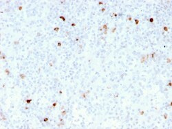
Biotium IgM (Immunoglobulin Mu Heavy Chain) (B-Cell Marker)(rIM260), CF405S conjugate, 0.1mg/mL
Recognizes a protein of 75 kDa, identified as mu heavy chain of human immunoglobulins. It does not cross-react with alpha (IgA), gamma (IgG), epsilon (IgE), or delta (IgD), heavy chains, T-cells, monocytes, granulocytes, or erythrocytes. This MAb is useful in the identification of leukemias, plasmacytomas, and certain non-Hodgkin's lymphomas. The most common feature of these malignancies is the restricted expression of a single heavy chain class. Demonstration of clonality in lymphoid infiltrates indicates that the infiltrate is clonal and therefore malignant. Primary antibodies are available purified, or with a selection of fluorescent CF Dyes and other labels. CF Dyes offer exceptional brightness and photostability. Note: Conjugates of blue fluorescent dyes like CF405S and CF405M are not recommended for detecting low abundance targets, because blue dyes have lower fluorescence and can give higher non-specific background than other dye colors.
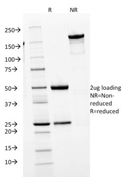
Biotium Human IgM (Immunoglobulin Mu Heavy Chain) (B-Cell Marker)(IGHM/1623), 1mg/mL
Recognizes a protein of 75 kDa, identified as mu heavy chain of human immunoglobulins. It does not cross-react with alpha (IgA), gamma (IgG), epsilon (IgE), or delta (IgD), heavy chains, T-cells, monocytes, granulocytes, or erythrocytes. Monomeric IgM is expressed as a membrane bound antibody on the surface of B cells and as a pentamer when secreted by plasma cells. IgM antibody is prominent in early immune responses to most antigens. Aberrant levels are associated with immune deficiency states, hereditary deficiencies, myeloma, Waldenstrom's macroglobulinemia, chronic infection and hepatocellular disease. This MAb is useful in the identification of leukemias, plasmacytomas, and certain non-Hodgkin's lymphomas. The most common feature of these malignancies is the restricted expression of a single heavy chain class. Demonstration of clonality in lymphoid infiltrates indicates that the infiltrate is clonal and therefore malignant. Primary antibodies are available purified, or w

Miltenyi Biotec, Inc. CD352 (NTB-A)-APC, mouse, REA1097, 100 t
Identification and enumeration of CD352 (NTB-A)+ cells by flow cytometry.
This product is subject to the following: Miltenyi Biotec, Inc. Terms and Conditions of Sale For Products and Services
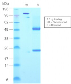
Biotium Human IgM (Immunoglobulin Mu Heavy Chain) (B-Cell Marker)(rIGHM/2558), CF594 conjugate, 0.1mg/mL
Recognizes a protein of 75 kDa, identified as mu heavy chain of human immunoglobulins. It does not cross-react with alpha (IgA), gamma (IgG), epsilon (IgE), or delta (IgD), heavy chains, T-cells, monocytes, granulocytes, or erythrocytes. Monomeric IgM is expressed as a membrane bound antibody on the surface of B cells and as a pentamer when secreted by plasma cells. IgM antibody is prominent in early immune responses to most antigens. Aberrant levels are associated with immune deficiency states, hereditary deficiencies, myeloma, Waldenstrom's macroglobulinemia, chronic infection and hepatocellular disease. This MAb is useful in the identification of leukemias, plasmacytomas, and certain non-Hodgkin's lymphomas. The most common feature of these malignancies is the restricted expression of a single heavy chain class. Demonstration of clonality in lymphoid infiltrates indicates that the infiltrate is clonal and therefore malignant. Primary antibodies are available purified, or w
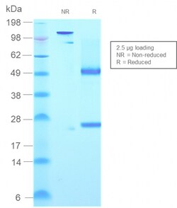
Biotium Human IgM (Immunoglobulin Mu Heavy Chain) (B-Cell Marker)(rIGHM/2558), CF647 conjugate, 0.1mg/mL
Recognizes a protein of 75 kDa, identified as mu heavy chain of human immunoglobulins. It does not cross-react with alpha (IgA), gamma (IgG), epsilon (IgE), or delta (IgD), heavy chains, T-cells, monocytes, granulocytes, or erythrocytes. Monomeric IgM is expressed as a membrane bound antibody on the surface of B cells and as a pentamer when secreted by plasma cells. IgM antibody is prominent in early immune responses to most antigens. Aberrant levels are associated with immune deficiency states, hereditary deficiencies, myeloma, Waldenstrom's macroglobulinemia, chronic infection and hepatocellular disease. This MAb is useful in the identification of leukemias, plasmacytomas, and certain non-Hodgkin's lymphomas. The most common feature of these malignancies is the restricted expression of a single heavy chain class. Demonstration of clonality in lymphoid infiltrates indicates that the infiltrate is clonal and therefore malignant. Primary antibodies are available purified, or w
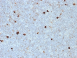
Biotium Human IgM (Immunoglobulin Mu Heavy Chain) (B-Cell Marker)(R1/69), 1mg/mL
Recognizes a protein of 75 kDa, identified as mu heavy chain of human immunoglobulins. It does not cross-react with alpha (IgA), gamma (IgG), epsilon (IgE), or delta (IgD), heavy chains, T-cells, monocytes, granulocytes, or erythrocytes. Monomeric IgM is expressed as a membrane bound antibody on the surface of B cells and as a pentamer when secreted by plasma cells. IgM antibody is prominent in early immune responses to most antigens. Aberrant levels are associated with immune deficiency states, hereditary deficiencies, myeloma, Waldenstrom's macroglobulinemia, chronic infection and hepatocellular disease. This MAb is useful in the identification of leukemias, plasmacytomas, and certain non-Hodgkin's lymphomas. The most common feature of these malignancies is the restricted expression of a single heavy chain class. Demonstration of clonality in lymphoid infiltrates indicates that the infiltrate is clonal and therefore malignant. Primary antibodies are available purified, or with a
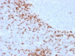
Biotium Human IgD (Immunoglobulin Delta Heavy Chain) (B-Cell Marker)(IgD26), CF488A conjugate, 0.1mg/mL
Immunoglobulins are four-chain, Y-shaped, monomeric structures comprised of two identical heavy chains and two identical light chains held together through interchain disulfide bonds. The chains form two domains, the Fab (antigen binding) fragment and the Fc (constant) fragment. Immunoglobulin D (IgD) exists as a monomer with light chains. It plays a biological role as a transmembrane receptor molecule, co-expressed with IgM on the surface of mature/naive B cells. In particular, it is found on spleen B cell surfaces. Compared to IgM, IgD exists in much lower numbers and is not expressed on immature B cells. IgD surface expression on B cells is regulated in part by IL-27. In mice, the inhibition of this immunoglobulin isotype does not cause a significant change to the immune system. Primary antibodies are available purified, or with a selection of fluorescent CF Dyes and other labels. CF Dyes offer exceptional brightness and photostability. Note: Conjugates of blue fluorescent d
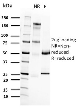
Biotium RET Proto-oncogene(RET/2662), CF488A conjugate, 0.1mg/mL
The Ret proto-oncogene is structurally related to the growing family of tyrosine kinase transmembrane receptors and is involved in GDNF signaling. RET expression is reported in several regions of the central nervous system; in the developing cranial nerve ganglia and a subset of cells within dorsal root ganglia, in motor neurons in the spinal cord and hindbrain, in neuro-retina and the growing tips of the renal collecting ducts in developing kidney. Alterations in RETgene are associated with diseases including papillary thyroid carcinoma, multiple endocrine neoplasia (type 2A and 2B), familial medullary thyroid carcinoma, and a congenital developmental disorder known as Hirschsprung's disease. Primary antibodies are available purified, or with a selection of fluorescent CF Dyes and other labels. CF Dyes offer exceptional brightness and photostability. Note: Conjugates of blue fluorescent dyes like CF405S and CF405M are not recommended for detecting low abundance targets,
Biotium Isotype Control, Mouse IgG1 Kappa (IGG1/1331), CF660R Conjugate, 0.1 mg/mL
This is a non-reactive mouse monoclonal IgG1 kappa antibody labeled with one of our bright and photostable CF Dyes and other labels. This antibody may serve as a control for a mouse monoclonal IgG1 antibody under identical experimental conditions. Biotium also carries mouse monoclonal isotype control antibodies for IgG2a Kappa (MG2a-53) and IgG2b Kappa (IGG1/1333). Isotype control for antibody-based experiments Available with 9 bright and photostable CF Dyes APC, R-PE, RPE-Astral„¢616, APC-Astral„¢813, and biotin conjugates also available Suitable for western, flow cytometry, ELISA, immunofluorescence, and immunohistologyCF Dyes offer exceptional brightness and photostability; learn more about CF Dyes.

Biotium Human IgM (Immunoglobulin Mu Heavy Chain) (B-Cell Marker)(IGHM/3803R), CF740 conjugate, 0.1mg/mL
Recognizes a protein of 75 kDa, identified as mu heavy chain of human immunoglobulins. It does not cross-react with alpha (IgA), gamma (IgG), epsilon (IgE), or delta (IgD), heavy chains, T-cells, monocytes, granulocytes, or erythrocytes. Monomeric IgM is expressed as a membrane bound antibody on the surface of B cells and as a pentamer when secreted by plasma cells. IgM antibody is prominent in early immune responses to most antigens. Aberrant levels are associated with immune deficiency states, hereditary deficiencies, myeloma, Waldenstrom's macroglobulinemia, chronic infection and hepatocellular disease. This MAb is useful in the identification of leukemias, plasmacytomas, and certain non-Hodgkin's lymphomas. The most common feature of these malignancies is the restricted expression of a single heavy chain class. Demonstration of clonality in lymphoid infiltrates indicates that the infiltrate is clonal and therefore malignant. Primary antibodies are available purified, or w

Miltenyi Biotec, Inc. CD9-APC, mouse, REAL501, 100 tests
Identification and enumeration of CD9+ cells by flow cytometry.
This product is subject to the following: Miltenyi Biotec, Inc. Terms and Conditions of Sale For Products and Services

Miltenyi Biotec, Inc. CD64-APC, mouse, REA286, 100 t
Identification and enumeration of CD64+ cells by flow cytometry.
This product is subject to the following: Miltenyi Biotec, Inc. Terms and Conditions of Sale For Products and Services

Cayman Chemical JWH 250 N- 4-hydroxypentyl m
Expected to be a cytochrome P450 phase I metabolite of JWH 250, detectable both in serum and urine; intended for research and forensic applications
