
Protein Labeling Reagents


BIOTIUM INC MALT-1(MT1/410), CF568 conjugate, 0.1mg/mL
Mucosa associated lymphoid tissue lymphoma translocation gene 1 (MALT1) is found in extranodal low-grade B cell lymphomas. MALT1 encodes two Ig-like C2-type domains and fuses with an API2 gene, which is highly expressed in adult lymphoid tissue. The translocation of this MALT1 gene, which maps to human chromosome 18q21, and the apoptosis-inhibiting API2 gene results in an increased development of MALT lymphomas and apoptosis inhibition. Sites at which this API2-MALT1 (11;18)(q21;q21) translocation commonly occurs are within human lung and kidney tissue. MALT lymphoma expresses nuclear Bcl10, which mediates the oligomerization and activation of a MALT1 caspase-like domain. MALT1 mRNA is found in pre-B cells, mature B cells and plasma cells.Primary antibodies are available purified, or with a selection of fluorescent CF Dyes and other labels. CF Dyes offer exceptional brightness and photostability. Note: Conjugates of blue fluorescent dyes like CF405S and CF405M are not re

BIOTIUM INC CD4(C4/206), CF488A conjugate, 0.1mg/mL
Recognizes a protein of 55 kDa, identified as CD4. CD4 is a membrane glycoprotein of T lymphocytes that interacts with major histocompatibility complex class II antigens and is also a receptor for the human immunodeficiency virus. This protein is expressed not only in T lymphocytes, but also in B cells, macrophages, and granulocytes. It is also expressed in specific regions of the brain. The protein functions to initiate or augment the early phase of T-cell activation, and may function as an important mediator of indirect neuronal damage in infectious and immune-mediated diseases of the central nervous system. Multiple alternatively spliced transcript variants encoding different isoforms have been identified.Primary antibodies are available purified, or with a selection of fluorescent CF Dyes and other labels. CF Dyes offer exceptional brightness and photostability. Note: Conjugates of blue fluorescent dyes like CF405S and CF405M are not recommended for detecting low
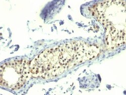
BIOTIUM INC Complement C4d(C4D205), CF488A conjugate, 0.1mg/mL
This MAb is specific to Complement 4d (C4d) and it reacts with the secreted as well as cell-bound C4d.C4d is a degradation product of the activated complement factor C4b. Complement 4b is typically activated by binding of Abs to specific target molecules. Following activation and degradation of the C4 molecule, thio-ester groups are exposed, which allow transient, covalent binding of the degradation product Complement 4d to endothelial cell surfaces and extracellular matrix components of vascular basement membranes near the sites of C4 activation. The presence of C4d in peritubular capillaries is a key indicator for acute humoral (i.e. antibody-mediated) rejection of kidney, heart, pancreas and lung allografts. As an established marker of antibody-mediated acute renal allograft rejection and its proclivity for endothelium, this component can be detected in peritubular capillaries in chronic renal allograft rejection as well as hyperacute rejection, acute vascular rejection, a

CAYMAN CHEMICAL PhotoclIck MyrIstIc AcId 500ug
A derivative of myristic acid; contains a terminal clickable alkyne and a photoactivatable diazirine at carbon 10
BIOTIUM INC CD37(IPO-24), CF488A conjugate, 0.1mg/mL
Recognizes a protein of 33-55 kDa, identified as CD37 (Workshop V; Code CD37.7). CD37 is strongly expressed on normal and neoplastic mature (sIg ) B-lymphocytes. In B-cell ontogeny, CD37 appears after the pre-B-cell stage, is maintained during peripheral B-cell development and is lost upon terminal differentiation into plasma cells.1 CD37 is also present, at low densities, on resting and activated T cells, neutrophils, monocytes, and some myelomonocytic leukemia cells. It is absent from platelets, erythrocytes. CD37 is a member of a family of tetraspan transmembrane proteins, including CD9, CD53, CD63, CD81, and CD82. It associates other tetraspan transmembrane proteins and MHC class II molecules to form a large complex at the surface of B cells and play a role in signal transduction. CD37 is a valuable and stable marker for peripheral mature B-cells and corresponding malignancies like B-cell chronic lymphocytic leukemia (B-CLL), hairy cell leukemia (HCL), and all types of B-
BIOTIUM INC CD33(WM53), CF488A conjugate, 0.1mg/mL
Recognizes a 67 kDa glycoprotein, which is identified as CD33 (HLDA IV; WS Code M-505). CD33 is a transmembrane protein of the sialic acid-binding immunoglobulin-like lectin (Siglec) family. It belongs to the immuno-receptor tyrosine-based inhibitory motif (ITIM)-containing molecules able of recruiting protein tyrosine phosphatases SHP-1 and SHP-2 to signal assemblies; these ITIMs are also used for ubiquitin-mediated removal of the receptor from the cell surface. CD33 is expressed on cells of myelomonocytic lineage, binds sialic acid residues in N- and O-glycans on cell surfaces, and is a therapeutic target for acute myeloid leukemia. CD33 is expressed on myeloid progenitors, monocytes, granulocytes, dendritic cells and mast cells. It is absent on platelets, lymphocytes, erythrocytes and hematopoietic stem cells.Primary antibodies are available purified, or with a selection of fluorescent CF Dyes and other labels. CF Dyes offer exceptional brightness and photostability. No
BIOTIUM INC CD27(203.6), CF488A conjugate, 0.1mg/mL
Recognizes a protein of a disulfide-linked 120 kDa dimer, identified as CD27 (Workshop VI; Code 6T-028). CD27 is expressed on the majority of peripheral T cells, medullary thymocytes, memory-type B cells, and natural killer cells. It is a transmembrane phosphoglycoprotein that belongs to the tumor necrosis factor receptor (TNFR) superfamily. CD27 binds to its ligand CD70, a member of the TNF family, and induces T-cell co-stimulation and B-cell activation. It also interacts with TRAFs and is involved in activation of NFB and SAPK/JNK and induces apoptosis.Primary antibodies are available purified, or with a selection of fluorescent CF Dyes and other labels. CF Dyes offer exceptional brightness and photostability. Note: Conjugates of blue fluorescent dyes like CF405S and CF405M are not recommended for detecting low abundance targets, because blue dyes have lower fluorescence and can give higher non-specific background than other dye colors.
BIOTIUM INC CD19(CVID3/155), CF488A conjugate, 0.1mg/mL
Recognizes a protein of 95 kDa, identified as CD19. CD19 is a transmembrane glycoprotein that contains two extracellular immunoglobulin-like domains. CD19 is present in both benign and malignant B-cells and is considered to be the most reliable surface marker of this lineage over a wide range of maturational stages. In normal lymphoid tissue, CD19 is observed in germinal centers, in mantle zone cells, and in scattered cells of the inter-follicular areas. Anti-CD19 exhibits an overall immunoreactivity pattern similar to those of the antibodies against CD20 and CD22. However, in contrast to CD20, expression of CD19 is continuous throughout B-cell development and through terminal differentiation of B-cells into plasma cells. Anti-CD19 positivity is seen in the vast majority of B-cell neoplasms commonly at a lower intensity than normal B-cell counterparts. Plasma cell neoplasms are nearly always negative, as are T-cell neoplasms.Primary antibodies are available purified, or w
BIOTIUM INC CD13(WM15), CF488A conjugate, 0.1mg/mL
Recognizes an integral membrane glycoprotein of 150 kDa, identified as CD13 (also known as aminopeptidase-N). The antibody recognizes an extracellular epitope. The CD13 antigen is present on most cells of myeloid origin including granulocytes, monocytes, mast cells, and GM-progenitor cells. It is also expressed by the majority of AML, CML in myeloid blast crisis, and in a smaller fraction of lymphoid leukemias. CD13 is absent from normal lymphocytes, platelets and erythrocytes. CD13 is also present on fibroblasts; endothelial cells, epithelial cells from renal proximal tubules and intestinal brush border, bone marrow stromal cells, osteoclasts, and cells lining bile duct canaliculi. CD13 is identical to aminopeptidase N (APN), a prominent membrane-bound metalloprotease present on the surface of intestinal brush border and renal tubules. CD13 plays a role in metabolism of biologically active peptides, in phagocytosis, and in bactericidal/tumoricidal activities. It also serves
BIOTIUM INC CD13(WM15), CF488A conjugate, 0.1mg/mL
Recognizes an integral membrane glycoprotein of 150 kDa, identified as CD13 (also known as aminopeptidase-N). The antibody recognizes an extracellular epitope. The CD13 antigen is present on most cells of myeloid origin including granulocytes, monocytes, mast cells, and GM-progenitor cells. It is also expressed by the majority of AML, CML in myeloid blast crisis, and in a smaller fraction of lymphoid leukemias. CD13 is absent from normal lymphocytes, platelets and erythrocytes. CD13 is also present on fibroblasts; endothelial cells, epithelial cells from renal proximal tubules and intestinal brush border, bone marrow stromal cells, osteoclasts, and cells lining bile duct canaliculi. CD13 is identical to aminopeptidase N (APN), a prominent membrane-bound metalloprotease present on the surface of intestinal brush border and renal tubules. CD13 plays a role in metabolism of biologically active peptides, in phagocytosis, and in bactericidal/tumoricidal activities. It also serves
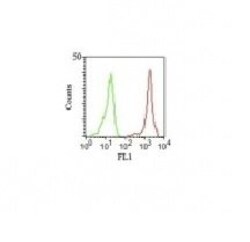
BIOTIUM INC CD11a(Cris-3), CF488A conjugate, 0.1mg/mL
Recognizes a protein of 180 kDa, identified as CD11a (Leucocyte Workshop II; Code N202). CD11a complex with the 2 subunit of the integrin family, CD18, to form the cell surface heterodimer, LFA-1 or CD11a /C18 (aLbL). LFA-1 is expressed on all leukocytes including lymphocytes, monocytes, and granulocytes. It is involved in leukocyte adhesion to its ligands including intercellular adhesion molecule-1 (ICAM-1 or CD54), ICAM-2 (CD102), ICAM-3 (CD50) and Telencephalin (TLN) and play a role in most immune/inflammatory responses.Primary antibodies are available purified, or with a selection of fluorescent CF Dyes and other labels. CF Dyes offer exceptional brightness and photostability. Note: Conjugates of blue fluorescent dyes like CF405S and CF405M are not recommended for detecting low abundance targets, because blue dyes have lower fluorescence and can give higher non-specific background than other dye colors.
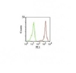
BIOTIUM INC CD11a(Cris-3), CF488A conjugate, 0.1mg/mL
Recognizes a protein of 180 kDa, identified as CD11a (Leucocyte Workshop II; Code N202). CD11a complex with the 2 subunit of the integrin family, CD18, to form the cell surface heterodimer, LFA-1 or CD11a /C18 (aLbL). LFA-1 is expressed on all leukocytes including lymphocytes, monocytes, and granulocytes. It is involved in leukocyte adhesion to its ligands including intercellular adhesion molecule-1 (ICAM-1 or CD54), ICAM-2 (CD102), ICAM-3 (CD50) and Telencephalin (TLN) and play a role in most immune/inflammatory responses.Primary antibodies are available purified, or with a selection of fluorescent CF Dyes and other labels. CF Dyes offer exceptional brightness and photostability. Note: Conjugates of blue fluorescent dyes like CF405S and CF405M are not recommended for detecting low abundance targets, because blue dyes have lower fluorescence and can give higher non-specific background than other dye colors.
BIOTIUM INC S100B(4C4.9), CF488A conjugate, 0.1mg/mL
S100 belongs to the family of calcium binding proteins. S100A and S100B proteins are two members of the S100 family. S100A is composed of an alpha and a beta chain whereas S100B is composed of two beta chains. This antibody is specific against an epitope located on the beta-chain (i.e. in S-100A and S-100B) but not on the alpha-chain of S-100 (i.e. in S-100A and S100A0). This antibody can be used to localize S-100A and S-100B in various tissue sections. S-100 protein has been found in normal melanocytes, Langerhans cells, histiocytes, chondrocytes, lipocytes, skeletal and cardiac muscle, Schwann cells, epithelial and myoepithelial cells of the breast, salivary and sweat glands, as well as in glial cells. Neoplasms derived from these cells also express S-100 protein, albeit non-uniformly. A large number of well-differentiated tumors of the salivary gland, adipose and cartilaginous tissue, and Schwann cell-derived tumors express S-100 protein. Almost all malignant melanomas and
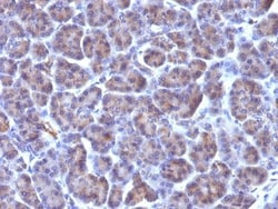
BIOTIUM INC CMV-p65(CMV100), CF488A conjugate, 0.1mg/mL
Ten to forty percent of the patients with acquired immunodeficiency syndrome (AIDS) develop cytomegalovirus (CMV) infections. In some patients with AIDS, CMV is detected in the bronchoalveolar lavage fluid (BALF), urine, and other specimens, even when there are no symptoms of CMV disease. An indicator of active CMV infection is needed to facilitate the diagnosis of CMV disease in patients with AIDS or HIV infection. CMV p65 antigen was detected in the leukocytes of both the peripheral blood and BALF during the early phase of CMV disease.Primary antibodies are available purified, or with a selection of fluorescent CF Dyes and other labels. CF Dyes offer exceptional brightness and photostability. Note: Conjugates of blue fluorescent dyes like CF405S and CF405M are not recommended for detecting low abundance targets, because blue dyes have lower fluorescence and can give higher non-specific background than other dye colors.
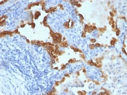
BIOTIUM INC Nuclear Membrane(NM97), CF488A conjugate, 0.1mg/mL
This monoclonal antibody is part of a new panel of reagents, which recognizes subcellular organelles or compartments of human cells. These markers may be useful in identification of these organelles in cells, tissues, and biochemical preparations. It recognizes an antigen associated with the nuclear membrane expressed in human cells. It can be used to stain the nuclear membrane in cell or tissue preparations and can be used as a marker of the nuclear membrane in subcellular fractions. It produces a ring pattern around the nucleus of cells of normal and malignant cells and may be used to stain the nuclear membrane of cells in fixed or frozen tissue sections. ,The nuclear envelope (also known as the perinuclear envelope, nuclear membrane, nucleolemma or karyotheca) is the double membrane of the nucleus that encloses genetic material in eukaryotic cells. It separates the contents of the nucleus (DNA in particular) from the cytosol (cytoplasm). Numerous nuclear pores are present