
Antibody and Dye Based Assays


Biotium CD20(109-3C2), CF488A conjugate, 0.1mg/mL
Recognizes a protein of 30-33 kDa, which is identified as CD20 (Workshop V; Code CD20.12. Workshop IV; Code B17). It recognizes an extracellular domain of CD20. It is a non-Ig differentiation antigen of B-cells and its expression is restricted to normal and neoplastic B-cells, being absent from all other leukocytes and tissues. CD20 is expressed by pre B-cells and persists during all stages of B-cell maturation but is lost upon terminal differentiation into plasma cells. The protein passes through the membrane 4 times with both ends in cytoplasm and exposes one short and one longer loop to the external environment. CD20 is not glycosylated in resting B-cells and its cytoplasmic domains are differentially phosphorylated upon activation. It acts as calcium channel involved in B cell activation and cell cycle progression.Primary antibodies are available purified, or with a selection of fluorescent CF Dyes and other labels. CF Dyes offer exceptional brightness and photostabilit

Biotium Histiocytoma Marker(D11), CF488A conjugate, 0.1mg/mL
In Western blotting, this antibody detects an antigen of 125 kDa in human liver and 135 kDa in tumors of histiocytic origin. Comparative study of this MAb and a standard CD68 MAb showed that their antigens are different. Its antigen in all macrophage types studied is located on the plasma membrane and within cytoplasmic structures including lysosomes. This MAb shows a restricted reactivity to cells of the monocyte/macrophage system. It specifically reacts with blood monocytes and stains resident macrophages in a wide variety of human tissues. This MAb does not stain antigen-presenting cells, e.g., Langerhans cells. Reportedly, its reactivity is restricted to histiocytes and macrophages.Primary antibodies are available purified, or with a selection of fluorescent CF Dyes and other labels. CF Dyes offer exceptional brightness and photostability. Note: Conjugates of blue fluorescent dyes like CF405S and CF405M are not recommended for detecting low abundance targets, because
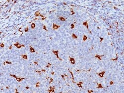
Biotium CD68(KP1), CF555 conjugate, 0.1mg/mL
This antibody recognizes a glycoprotein of 110 kDa, which is identified as CD68. It is important for identifying macrophages in tissue sections. It stains macrophages in a wide variety of human tissues, including Kupffer cells and macrophages in the red pulp of the spleen, in lamina propria of the gut, in lung alveoli, and in bone marrow. It reacts with myeloid precursors and peripheral blood granulocytes. It also reacts with plasmacytoid T cells, which are supposed to be of monocyte/macrophage origin. It shows strong granular cytoplasmic staining of chronic and acute myeloid leukemia and also reacts with rare cases of true histiocytic neoplasia. Lymphomas are negative or show few granules.Primary antibodies are available purified, or with a selection of fluorescent CF Dyes and other labels. CF Dyes offer exceptional brightness and photostability. Note: Conjugates of blue fluorescent dyes like CF405S and CF405M are not recommended for detecting low abundance targets,
Medchemexpress LLC Aβ/tau aggregation-IN-1 | 2252162-81-3 | 98.1% | C25H21IN2O | 50 MG

Aβ/tau aggregation-IN-1 is a potent inhibitor of Aβ1-42 β-sheets formation and tau aggregation, with KD values of 160 μM for Aβ1-42 and 337 μM for tau. This compound can permeate the blood-brain barrier. In vitro studies show it significantly reduces tau aggregation foci and inhibits Aβ1-42 aggregation by 84.7%-99.5%, and tau aggregation by 71.2%-101.8%.
- Potent inhibitor of Aβ1-42 β-sheets formation and tau aggregation
- Permeates the blood-brain barrier
- Significantly reduces tau aggregation foci in HEK-293 T cells
- Inhibits Aβ1-42 aggregation by 84.7%-99.5%
- Inhibits tau aggregation by 71.2%-101.8% through N-methylation of the quinolone ring
- Interacts with Aβ1-42 and tau via noncovalent interactions
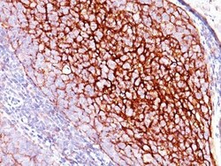
Biotium CD35 / CR1(E11), CF488A conjugate, 0.1mg/mL
Recognizes a protein of 210-220 kDa, which is identified as the complement receptor 1 (CR1)/CD35. This MAb is specific for a site in CR1 that is distal from the C3b-binding site, so that it is unable to block CR1 activity. This MAb is highly specific to CR1 and shows no cross-reaction with CR2. The primary function of CR1 is to serve as the cellular receptor for C3b and C4b, the most important components of the complement system leading to clearance of foreign macromolecules. The Knops blood group system is a system of antigens located on this protein. Follicular dendritic cells (FDC) are restricted to the B-cell regions of secondary lymphoid follicles. They are CD21 /CD35 /CD1a-. This MAb labels follicular dendritic cells and follicular dendritic cell sarcoma.Primary antibodies are available purified, or with a selection of fluorescent CF Dyes and other labels. CF Dyes offer exceptional brightness and photostability. Note: Conjugates of blue fluorescent dyes like CF405S
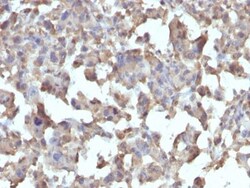
Biotium CD284 / TLR4(TLR4/230), CF488A conjugate, 0.1mg/mL
This MAb reacts with human Toll-like receptor 2 (TLR4). It is a member of the Toll-like receptor (TLR) family, which plays a fundamental role in pathogen recognition and activation of innate immunity. TLRs are highly conserved from Drosophila to humans and share structural and functional similarities. They recognize pathogen-associated molecular patterns that are expressed on infectious agents, and mediate the production of cytokines necessary for the development of effective immunity. The various TLRs exhibit different patterns of expression. This receptor has been implicated in signal transduction events induced by lipopolysaccharide (LPS) found in most gram-negative bacteria. Mutations in this gene have been associated with differences in LPS responsiveness. Multiple transcript variants encoding different isoforms have been found for this gene.Primary antibodies are available purified, or with a selection of fluorescent CF Dyes and other labels. CF Dyes offer exceptional
Biotium CD7(B-F12), CF488A conjugate, 0.1mg/mL
Recognizes a protein of 40 kDa, identified as CD7 (also known as gp40, Leu9). CD7 is a member of the immunoglobulin gene superfamily. Its N-terminal amino acids 1-107 are highly homologous to Ig kappa-L chains whereas the carboxyl-terminal region of the extracellular domain is proline-rich and has been postulated to form a stalk from which the Ig domain projects. CD7 is expressed on the majority of immature and mature T-lymphocytes, and T cell leukemia. It is also found on natural killer cells, a small subpopulation of normal B cells and on malignant B cells. Cross-linking surface CD7 positively modulates T cell and NK cell activity as measured by calcium fluxes, expression of adhesion molecules, cytokine secretion and proliferation. CD7 associates directly with phosphoinositol 3'-kinase. CD7 ligation induces production of D-3 phosphoinositides and tyrosine phosphorylation.Primary antibodies are available purified, or with a selection of fluorescent CF Dyes and other labels.
Biotium Cytokeratin 7(KRT7/1198), CF488A conjugate, 0.1mg/mL
This antibody recognizes an intermediate filament protein (IFP) of 55 kDa, which is identified as cytokeratin 7. This MAb is highly specific to cytokeratin 7 and shows no cross-reaction with other IFPs. Cytokeratin 7 is a basic cytokeratin, which is found in most glandular and transitional epithelia but not in the stratified squamous epithelia. Keratin 7 is expressed in the epithelial cells of ovary, lung, and breast but not of colon, prostate, or gastrointestinal tract. This MAb is highly useful in distinguishing ovarian carcinomas (keratin 7 ) from colon carcinomas (keratin 7-).Primary antibodies are available purified, or with a selection of fluorescent CF Dyes and other labels. CF Dyes offer exceptional brightness and photostability. Note: Conjugates of blue fluorescent dyes like CF405S and CF405M are not recommended for detecting low abundance targets, because blue dyes have lower fluorescence and can give higher non-specific background than other dye colors.
Biotium MART-1 / Melan-A(M2-9E3), CF488A conjugate, 0.1mg/mL
This antibody recognizes a protein doublet of 20-22 kDa, identified as MART-1 (Melanoma Antigen Recognized by T cells 1) or Melan-A. MART-1 is a newly identified melanocyte differentiation antigen recognized by autologous cytotoxic T lymphocytes. Seven other melanoma associated antigens recognized by autologous cytotoxic T cells include MAGE-1, MAGE-3, tyrosinase, gp100, gp75, BAGE-1, and GAGE-1. Subcellular fractionation shows that MART-1 is present in melanosomes and endoplasmic reticulum. This MAb labels melanomas and other tumors showing melanocytic differentiation. It is also a useful positive-marker for angiomyolipomas. It does not stain tumor cells of epithelial, lymphoid, glial, or mesenchymal origin.Primary antibodies are available purified, or with a selection of fluorescent CF Dyes and other labels. CF Dyes offer exceptional brightness and photostability. Note: Conjugates of blue fluorescent dyes like CF405S and CF405M are not recommended for detecting low abu
Biotium bcl-2(8C8), CF488A conjugate, 0.1mg/mL
This antibody recognizes a protein of 25-26 kDa, identified as Bcl-2. This antibody shows no cross-reaction with Bcl-x or Bax proteins. Bcl-2 suppresses apoptosis in a variety of cell systems including factor-dependent lymphohematopoietic and neural cells. It regulates cell death by controlling the mitochondrial membrane permeability. Bcl-2 appears to function in a feedback loop system with caspases. It inhibits caspase activity either by preventing the release of cytochrome c from the mitochondria and/or by binding to the apoptosis-activating factor (APAF-1). Expression of Bcl-2 inhibits programmed cell death (apoptosis). In most follicular lymphomas, neoplastic germinal centers express high levels of Bcl-2 protein, whereas the normal or hyperplastic germinal centers are negative. Consequently, this antibody is valuable when distinguishing between reactive and neoplastic follicular proliferation in lymph node biopsies. It may also be used in distinguishing between those foll
Biotium CD31 / PECAM-1(C31.3), CF488A conjugate, 0.1mg/mL
CD31 (PECAM-1) is a transmembrane glycoprotein member of the immunoglobulin supergene family of adhesion molecules. CD31 is expressed by stem cells of the hematopoietic system and is primarily used to identify and concentrate these cells for experimental studies as well as for bone marrow transplantation. Anti-CD31 has shown to be highly specific and sensitive for vascular endothelial cells. Staining of nonvascular tumors (excluding hematopoietic neoplasms) is rare. CD31 MAb reacts with normal, benign, and malignant endothelial cells which make up blood vessel lining. The level of CD31 expression can help to determine the degree of tumor angiogenesis, and a high level of CD31 expression may imply a rapidly growing tumor and potentially a predictor of tumor recurrence.Primary antibodies are available purified, or with a selection of fluorescent CF Dyes and other labels. CF Dyes offer exceptional brightness and photostability. Note: Conjugates of blue fluorescent dyes like C
Biotium CD8a(Rabbit PAb), CF488A conjugate, 0.1mg/mL
CD8 is a cell surface receptor expressed either as a heterodimer with the CD8 beta chain (CD8 alpha/beta) or as a homodimer (CD8 alpha/alpha). A majority of thymocytes and a subpopulation of mature T cells and NK cell express CD8a. CD8 binds to MHC class 1 and through its association with protein tyrosine kinase p56lck plays a role in T cell development and activation of mature T cells. For mature T-cells, CD4 and CD8 are mutually exclusive, so anti-CD8, generally used in conjunction with anti-CD4. It is a useful marker for distinguishing helper/inducer T-lymphocytes, and most peripheral T-cell lymphomas are CD4 /CD8-. Anaplastic large cell lymphoma is usually CD4 and CD8-, and in T-lymphoblastic lymphoma/leukemia, CD4 and CD8 are often co-expressed. CD8 is also found in littoral cell angioma of the spleen.Primary antibodies are available purified, or with a selection of fluorescent CF Dyes and other labels. CF Dyes offer exceptional brightness and photostability. Note:
Biotium IGF-1(M23), CF488A conjugate, 0.1mg/mL
This antibody is specific to Insulin-like Growth Factor (IGF-1) and shows minimal cross-reaction with IGF-11, Proinsulin, MSF, and Insulin. IGF-1 is a polypeptide growth factor with two isoforms that are produced by alternative splicing. Isoform 1 is also known as IGF-IB while isoform 2 is known as IGF-IA. IGF-1 stimulates the proliferation of a wide range of cell types including muscle, bone and cartilage tissue. It functions as an autocrine regulator of growth. Activation of IGF system has emerged as a key factor for tumor progression and resistance to apoptosis in many cancers like those of breast, thyroid and colon.Primary antibodies are available purified, or with a selection of fluorescent CF Dyes and other labels. CF Dyes offer exceptional brightness and photostability. Note: Conjugates of blue fluorescent dyes like CF405S and CF405M are not recommended for detecting low abundance targets, because blue dyes have lower fluorescence and can give higher non-specific
Biotium CD31 / PECAM-1(C31.7), CF488A conjugate, 0.1mg/mL
CD31 (PECAM-1) is a transmembrane glycoprotein member of the immunoglobulin supergene family of adhesion molecules. CD31 is expressed by stem cells of the hematopoietic system and is primarily used to identify and concentrate these cells for experimental studies as well as for bone marrow transplantation. Anti-CD31 has shown to be highly specific and sensitive for vascular endothelial cells. Staining of nonvascular tumors (excluding hematopoietic neoplasms) is rare. CD31 MAb reacts with normal, benign, and malignant endothelial cells which make up blood vessel lining. The level of CD31 expression can help to determine the degree of tumor angiogenesis, and a high level of CD31 expression may imply a rapidly growing tumor and potentially a predictor of tumor recurrence.Primary antibodies are available purified, or with a selection of fluorescent CF Dyes and other labels. CF Dyes offer exceptional brightness and photostability. Note: Conjugates of blue fluorescent dyes li
Biotium CD81 / TAPA-1 (rC81/3442), CF488A conjugate, 0.1mg/mL
This antibody recognizes a protein of 26 kDa, identified as CD81 (Workshop VI; Code CD81.1)). CD81 has a very broad cellular distribution, being expressed on T- and B-lymphocytes, NK cells, thymocytes, eosinophils, fibroblasts, epithelial and endothelial cells. Neutrophils, erythrocytes and platelets are negative, while monocytes are variably positive. CD81 is a member of a family of tetraspanin transmembrane proteins, including CD9, CD37, CD53, CD63, and CD82. It associates with CD19, CD21, Leu 13, and integrins on cell membrane and is involved in signal transduction in B lymphocyte development and cell adhesion. CD81 also acts as a receptor for the envelope protein E2 of chronic hepatitis C virus. Antibodies to CD81 have anti-proliferative effects on different lymphoid cell lines, particularly those derived from large cell lymphomas.Primary antibodies are available purified, or with a selection of fluorescent CF Dyes and other labels. CF Dyes offer exceptional brightness a