Learn More
Invitrogen™ F(ab')2-Goat anti-Mouse IgG (H+L) Secondary Antibody, eFluor™ 660, eBioscience™

Description
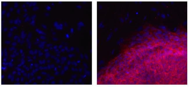
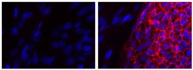
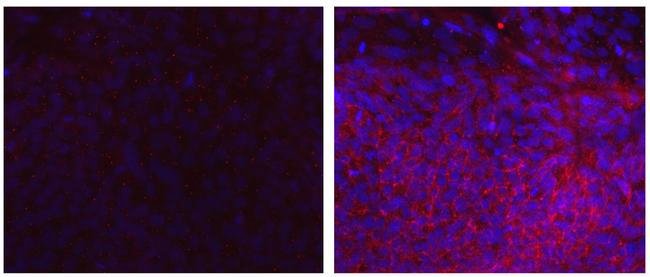
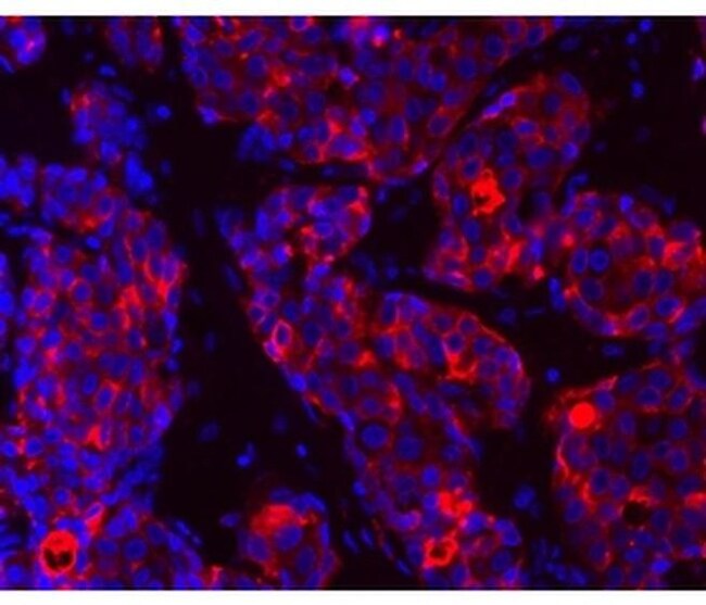
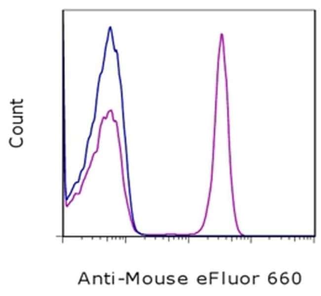
Description: The goat anti-mouse IgG polyclonal antibody reacts with the heavy and light chains of mouse IgG, with minimal cross reactivity to human, rat, bovine, goat, hamster, and rabbit serum proteins. Reactivity has been shown to IgG1, IgG2a, IgG2b, and IgG3, as well as some reactivity to IgM. The antibody is a F(ab')2 fragment which contains the 2 Fab domains still linked through the hinge region of the immunoglobulin. Removal of the Fc region helps to reduce issues typically found with Fc-mediated binding. This polyclonal has been validated as a secondary reagent (its ability to recognize cells stained with a mouse IgG followed by this polyclonal antibody). Applications Reported: This polyclonal antibody has been reported for use in flow cytometric analysis, immunohistochemistry and immunocytochemistry. Applications Tested: This polyclonal antibody has been tested by flow cytometric analysis of normal human peripheral blood cells stained first with a mouse anti-human antibody. This can be used at less than or equal to 0.5 μg per test. A test is defined as the amount (μg) of antibody that will stain a cell sample in a final volume of 100 μL. Cell number should be determined empirically but can range from 10^5 to 10^8 cells/test. This polyclonal antibody has been tested by immunohistochemistry on formalin-fixed paraffin embedded sections that have been stained first with a mouse anti-human antibody. This can be used at less than or equal to 10 ...
Specifications
Specifications
| Antigen | Mouse IgG (H+L) |
| Applications | Flow Cytometry, Immunohistochemistry (Paraffin), Immunocytochemistry |
| Classification | Polyclonal |
| Concentration | 0.2 mg/mL |
| Conjugate | eFluor 660 |
| Formulation | PBS with 0.09% sodium azide; pH 7.2 |
| Host Species | Goat |
| Purification Method | Affinity chromatography |
| Quantity | 100 μg |
| Regulatory Status | RUO |
| Show More |
Frequently Asked Questions (FAQs)
Our options will depend on the samples you are analyzing.
If cell viability is not critical, you can store your stained samples at 4 degrees C or on ice overnight in the dark and analyze the following day.
For samples stained with eFluor organic fluorochromes, we recommend that cells be suspended in 100 uL of Flow Cytometry Staining Buffer (Cat. No. 00-4222) and 100 uL of eBioscience IC Fixation Buffer (Cat. No. 00-8222); samples can be incubated for up to 3 days at 4 degrees C in the dark. Alternatively, the 1-step Fix/Lyse Solution (Cat. No. 00-5333) can be used. This is a great option when working with whole blood but also works for other cell types.
Yes, the eFluor Organic fluorochromes can be used for intracellular staining. The eFluor organic fluorochromes maintain bright signal and require minimal changes in compensation when fixed with eBioscience IC Fixation Buffer (Cat. No. 00-8222-49) and Permeabilization Buffer (Cat. No. 00-8333-56) or 1-step Fix/Lyse Solution (Cat. No. 00-5333-54, 00-5333-57) (as compared to live cells).
Yes, in-house studies have demonstrated that the eFluor 660 fluorochrome is recognized by Anti-Cy5/Alexa Fluor 647 beads. Side by side studies with Alexa Fluor 647 versus eFluor 660 conjugated antibodies have demonstrated comparable results.
The eFluor Organic Dyes (eFluor 450, APC-eFluor 780, PerCP-eFluor 710, eFluor 710) are conventional fluorochromes. In contrast, the eVolve line of products are Quantum dots.
As with other fluorochromes, we recommend minimal exposure to light to maintain optimal signal.
By clicking Submit, you acknowledge that you may be contacted by Fisher Scientific in regards to the feedback you have provided in this form. We will not share your information for any other purposes. All contact information provided shall also be maintained in accordance with our Privacy Policy.