Promotional price valid on web orders only. Your contract pricing may differ. Interested in signing up for a dedicated account number?
Learn More
Learn More
Description
The probe is intended to be used in combination with the CISH Implementation Kit 2, which provides necessary reagents for specimen pretreatment and post-hybridization processing.
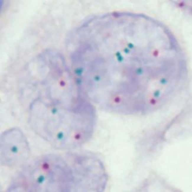
Hybridization signals of digoxigenin-labeled polynucleotides appear as dark green colored distinct dots (FGFR2 gene region), and dinitrophenyl-labeled polynucleotides appear as bright red colored distinct dots (CEN 10).
Normal situation: In interphases of normal cells or cells without an amplification involving the FGFR2 gene locus, two green signals and two red signals appear.
Aberrant situation: In a cell with amplification of the FGFR2 gene locus, multiple copies of the green signal or green signal clusters will be observed. Other signal distribution may be observed in some abnormal samples which might result in a different signal pattern than described above, indicating variant rearrangements.
Unexpected signal patterns should be further investigated.
Specifications
Specifications
| Content And Storage | The product is ready-to-use. No reconstitution, mixing, or dilution is required. Bring probe to room temperature (18 to 25°C) and mix briefly before use. |
| For Use With (Application) | CISH-P |
Product Title
By clicking Submit, you acknowledge that you may be contacted by Fisher Scientific in regards to the feedback you have provided in this form. We will not share your information for any other purposes. All contact information provided shall also be maintained in accordance with our Privacy Policy.
Spot an opportunity for improvement?