Learn More
Description
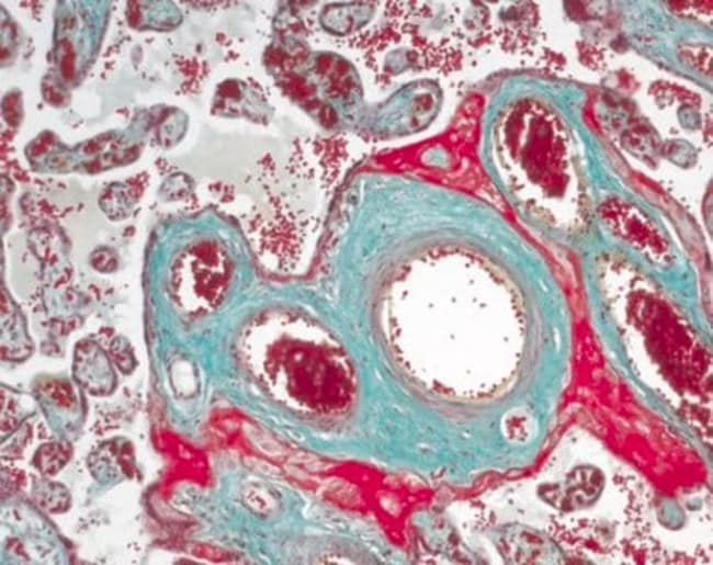
The Masson-Goldner staining kit for the visualization of connective tissue with trichromic staining, is used for human-medical cell diagnosis and serves the purpose of the histological investigation of sample material of human origin. Using a combination of three different staining solutions, muscle fibers, collagenous fibers, fibrin and erythrocytes can be selectively visualized.
The original methods were primarily used to differentiate collagenous and muscle fibers. The stains used have different molecular sizes and enable the individual tissues to be stained differentially.
The Masson-Goldner staining technique can be carried out using formalin fixed material. Subsequent to staining the nucleus with Weigert's iron hematoxylin, components such as muscle, cytoplasm and erythrocytes are stained with azophloxin and orange G solution. Connective tissue is then counter stained using light green SF solution.
The package is sufficient for 400 to 500 applications. The product is registered as IVD and CE product and can be used in diagnostics and laboratory accreditation.
Specifications
Specifications
| No. of Reactions | 400 to 500 Stainings |
| Product Type | Masson-Goldner Stain Kit |
By clicking Submit, you acknowledge that you may be contacted by Fisher Scientific in regards to the feedback you have provided in this form. We will not share your information for any other purposes. All contact information provided shall also be maintained in accordance with our Privacy Policy.