Promotional price valid on web orders only. Your contract pricing may differ. Interested in signing up for a dedicated account number?
Learn More

Invitrogen™ Rat anti-Mouse IgG1 Secondary Antibody, eFluor™ 660, eBioscience™

Catalog No. 501124348
Encompass_Preferred
Change view
Click to view available options
Quantity:
100 μg
25 μg
2 product options available for selection
Product selection table with 2 available options. Use arrow keys to navigate and Enter or Space to select.
Catalog No. Quantity
50-112-4348 25 μg
50-401-582 100 μg
Use arrow keys to navigate between rows. Press Enter or Space to select a product option. 2 options available.
2 options
Catalog No. 50-112-4348 Supplier Invitrogen™ Supplier No. 50401580
Only null left
Add to Cart
Add to Cart

Rat Monoclonal Secondary Antibody

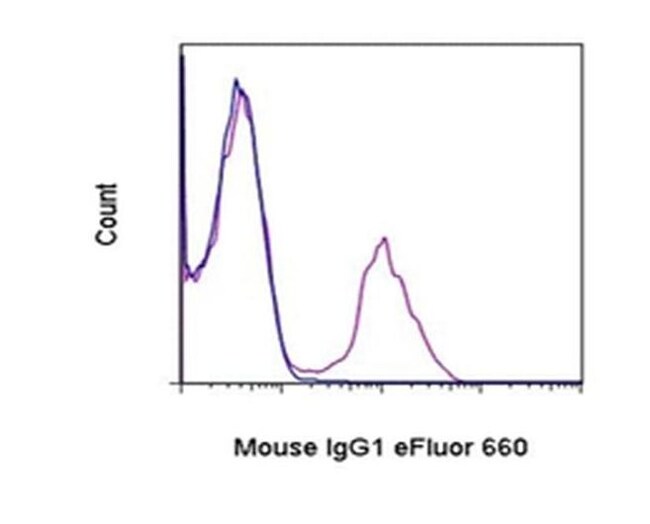
Description: The monoclonal antibody M1-14D12 recognizes Mouse IgG1 antibodies and can be used as a second step reagent in flow cytometry and microscopy. The monoclonal does not recognize other mouse isotypes nor does it crossreact to rat IgG1 or any rat isotype antibodies. Applications Reported: This M1-14D12 antibody has been reported for use in flow cytometric analysis, microscopy, immunohistochemical staining, and immunocytochemistry. Applications Tested: This M1-14D12 antibody has been tested by flow cytometric analysis of normal human peripheral blood cells stained first with a primary antibody of the mouse IgG1 isotype. This can be used at less than or equal to 0.25 μg per test. A test is defined as the amount (μg) of antibody that will stain a cell sample in a final volume of 100 μg. Cell number should be determined empirically but can range from 10^5 to 10^8 cells/test. This M1-14D12 antibody has also been tested by immunocytochemistry on fixed and permeabilized cells that have been stained first with a primary antibody of the mouse IgG1 isotype. For immunocytochemistry and immunohistochemistry, this can be used at less than or equal to 10 μg/mL. It is recommended that the antibody be carefully titrated for optimal performance in the assay of interest. Excitation: 633-647 nm; Emission: 668 nm; Laser: Red Laser. Filtration: 0.2 μm post-manufacturing filtered.

Anti-Mouse secondary antibodies are affinity-purified antibodies with well-characterized specificity for mouse immunoglobulins and are useful in the detection, sorting or purification of its specified target. Secondary antibodies offer increased versatility enabling users to use many detection systems (e.g. HRP, AP, fluorescence). They can also provide greater sensitivity through signal amplification as multiple secondary antibodies can bind to a single primary antibody. Most commonly, secondary antibodies are generated by immunizing the host animal with a pooled population of immunoglobulins from the target species and can be further purified and modified (i.e. immunoaffinity chromatography, antibody fragmentation, label conjugation, etc.) to generate highly specific reagents.
TRUSTED_SUSTAINABILITY

Specifications

Antigen Mouse IgG1
Applications Flow Cytometry, Immunohistochemistry, Immunocytochemistry
Classification Monoclonal
Clone M1-14D12
Concentration 0.2 mg/mL
Conjugate eFluor 660
Formulation PBS with 0.09% sodium azide; pH 7.2
Host Species Rat
Purification Method Affinity chromatography
Quantity 25 μg
Regulatory Status RUO
Primary or Secondary Secondary
Target Species Mouse
Content And Storage 4°C, store in dark, DO NOT FREEZE!
Product Type Secondary Antibody
Form Liquid
Isotype IgG
Show More Show Less
I am unable to analyze my cells stained with eFluor Organic Dyes today. What options do I have?

Our options will depend on the samples you are analyzing.

If cell viability is not critical, you can store your stained samples at 4 degrees C or on ice overnight in the dark and analyze the following day.

For samples stained with eFluor organic fluorochromes, we recommend that cells be suspended in 100 uL of Flow Cytometry Staining Buffer (Cat. No. 00-4222) and 100 uL of eBioscience IC Fixation Buffer (Cat. No. 00-8222); samples can be incubated for up to 3 days at 4 degrees C in the dark. Alternatively, the 1-step Fix/Lyse Solution (Cat. No. 00-5333) can be used. This is a great option when working with whole blood but also works for other cell types.

Can the eFluor Organic fluorochromes be used for intracellular staining?

Yes, the eFluor Organic fluorochromes can be used for intracellular staining. The eFluor organic fluorochromes maintain bright signal and require minimal changes in compensation when fixed with eBioscience IC Fixation Buffer (Cat. No. 00-8222-49) and Permeabilization Buffer (Cat. No. 00-8333-56) or 1-step Fix/Lyse Solution (Cat. No. 00-5333-54, 00-5333-57) (as compared to live cells).

Is the eFluor 660 fluorochrome compatible with Anti-Cy5/Alexa Fluor 647 beads?

Yes, in-house studies have demonstrated that the eFluor 660 fluorochrome is recognized by Anti-Cy5/Alexa Fluor 647 beads. Side by side studies with Alexa Fluor 647 versus eFluor 660 conjugated antibodies have demonstrated comparable results.

What buffers are compatible with the eFluor Organic fluorochromes and eVolve QDots?

The eFluor Organic fluorochromes and eVolve QDots can be used with flow staining buffers containing PBS and protein.

What is the difference between eFluor Organic Dyes and eVolve Qdots?

The eFluor Organic Dyes (eFluor 450, APC-eFluor 780, PerCP-eFluor 710, eFluor 710) are conventional fluorochromes. In contrast, the eVolve line of products are Quantum dots.

Are the eFluor Organic Dyes photo-labile?

As with other fluorochromes, we recommend minimal exposure to light to maintain optimal signal.


WARNING: Reproductive Harm - www.P65Warnings.ca.gov
Product Title
Select an issue

By clicking Submit, you acknowledge that you may be contacted by Fisher Scientific in regards to the feedback you have provided in this form. We will not share your information for any other purposes. All contact information provided shall also be maintained in accordance with our Privacy Policy.