
Incubator Accessories
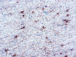
Diagnostic Biosystems HRP LABEL Glial Fibrillary Acidic Protein (GFAP)
This antibody recognizes a protein of ~50kDa which is identified as Glial Fibrillary Acidic Protein (GFAP). It shows no cross-reaction with other intermediate filament proteins. GFAP is specifically found in astroglia. GFAP is a very popular marker for localizing benign astrocyte and neoplastic cells of glial origin in the central nervous system. Antibody to GFAP is useful in differentiating primary gliomas from metastatic lesions in the brain and for documenting astrocytic differentiation in tumors outside the CNS.

Diagnostic Biosystems CA 19-9 CA 19-9
This antibody reacts specifically with sialyl Lewisa and recognizes an epitope being designated CA 19-9. It does not cross-reacts with Lewisa and Lewisb. CA 19-9 has been shown to be useful marker in the diagnosis and management of gastrointestinal cancers.
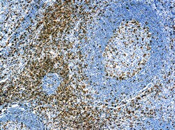
Diagnostic Biosystems CD5 CD5 (CD5/54/F6)
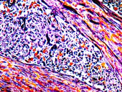
This antibody is Recognizes a 67kDa transmembrane protein, which is identified as CD5. The CD5 antigen is found on 95% of thymocytes and 72% of peripheral blood lymphocytes. In lymph nodes, Important in identification of T cells and most T cell lymphomas, some low-grade B cell lymphomas, thymic carcinoma.
Diagnostic Biosystems SATB2 SATB2 (EP 281)
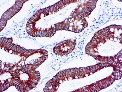
Special AT-rich sequence-binding protein 2 (SATB2) is a recently described marker that functions as a nuclear matrix-associated transcription factor. It has been reported that SATB2, in combination with CK20, could identify almost all colorectal carcinomas, including poorly differentiated colorectal carcinomas. Upper gastrointestinal (GI) carcinomas and pancreatic ductal carcinomas are usually negative for SATB2, and ovarian carcinomas, lung adenocarcinomas, and adenocarcinomas from other origin are rarely positive for SATB2. Therefore, SATB2 is a good marker for identifying a carcinoma of colorectal origin when working on a tumor of unknown primary. It can help differentiate colorectal metastasis (SATB2+) from primary pulmonary adenocarcinoma of mucinous or enteric type (SATB2- but often CDX2+)\n

Diagnostic Biosystems NUCLEAR FAST RED FOR IHC Nuclear Fast Red
Nuclear Fast Red is a histological staining reagent suitable for visualization of nuclei in tissue sections. Nuclear Fast Red provides a red counterstain designed for optimal performance when used in immunohistochemistry.\n\nDBS’s Nclear Fast Red is intended for use in the histologic demonstration of nuclear staining. This staining technique is used to make the critical distinction between a normal nucleus and an abnormal one. When used as a counterstain in immunohstochemical procedures, Nuclear Fast Red is compatible with most commonly used chromogens including DBS HRP-Yellow, HRP-Green, HRP-Black, and DAB. Nuclear Fast Red is particularly useful with blue chromogens, such as DBS HRP-Blue.
Diagnostic Biosystems PTAH PTAH
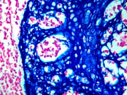
The PTAH Stain Kit is intended for use in the histological visualization of collagen, striated muscle, glial fibers and collagen without using Zenker’s Fixative containing Mercuric Chloride. This kit may be used on formalin-fixed, paraffin-embedded or frozen sections.
Diagnostic Biosystems PERMABLUE/HRP PermaBlue/HRP
PermaBlue/HRP is a substrate-chromogen system designed to be used for either IHC or ISH when utilizing horseradish peroxidase. PermaBlue/HRP can be permanently mounted to produce a strong azure blue color that can be easily distinguished from other stains.


Diagnostic Biosystems PMS2 PMS2
Postmeiotic segregation increased 2 or PMS2 was originally discovered in S. cerevisiae and is part of the mismatch repair system. It resides on 7p22.2 and its gene product partners with MLH1 to help detect mismatches in DNA. Mutations in PMS2 have been reported in about two percent of families with Lynch syndrome (hereditary nonpolyposis colorectal cancer).
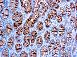
Diagnostic Biosystems CYTOKERATIN 18 Cytokeratin 18 (DC10)
This antibody is specific to human cytokeratin 18 of 45 kDa. Cytokeratin 18 belongs to a family of acidic type A keratins and exists along with cytokeratin 8 in most simple ductal and glandular epithelia. This antibody does not react with squamous epithelium. It reacts with benign and malignant epithelial lesions as well as a majority of adenocarcinomas and basal cell carcinomas.
Diagnostic Biosystems 10X EDTA BUFFER PH 80 10X EDTA Buffer For Heat Induced Epitope Recovery, pH 8.0
10X EDTA Buffer For Heat Induced Epitope Recovery, pH 8.0 is designed for use during the heat induced epitope retrieval (HIER) step prior to immunohistochemistry on formalin-fixed paraffin embedded tissue sections. The use of this buffer in combination with heat (often by microwave, water bath, or pressure cooker) has been shown to restore the antigenicity of proteins modified during the formalin fixation of tissue. This buffer is supplied as a 10X stock solution.
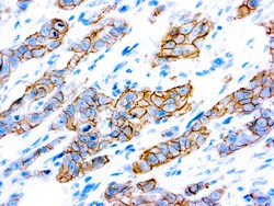
Diagnostic Biosystems HER2/NEU C-ERBB-2 c-erbB-2 Oncoprotein (SP3)
This antibody recognizes a c-erbB-2 protein, which is a receptor tyrosine kinase of the c-erbB family. It is closely related in structure to the epidermal growth factor receptor. c-erbB-2 oncoprotein is detectable in a subset of breast and other adenocarcinomas, as well astransitional cell carcinomas. In the case of breast cancer, expression determined byimmunohistochemistry has been shown to be associated with poor prognosis.
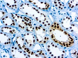
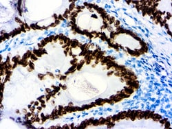
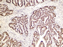
Diagnostic Biosystems PAX-8 PAX-8
PAX 8 is expressed in a high percentage of renal cell carcinomas and ovarian cancers. This mouse monoclonal PAX 8 antibody [4H7B3] has been designed to target restricted epitopes, and exhibits higher specificity and provides sharper staining than the PAX 8 rabbit polyclonal antibody. Unlike the polyclonal PAX 8, this mouse monoclonal antibody does not stain B-cells, and does not recognize epitopes of pancreatic origin and neuroendocrine cells in stomach and colon; thus providing superior specificity. The expression of the mouse monoclonal PAX 8 targets antigens found in normal kidney, thyroid and cervix, but not normal ovary. PAX 8 stains nuclei exclusively and performs well in formalin-fixed paraffin-embedded tissues.