
Rabbit Secondary Antibodies


Biotium CD28(C28/77), CF594 conjugate, 0.1mg/mL
Recognizes a glycoprotein of 44-88 kDa, which is identified as CD28. It is the critical T-cell co-stimulatory receptor which provides to the cell the important second activation signal by binding CD80 and CD86 that are expressed by antigen presenting cells. Besides its co-stimulation role, CD28 functions in preventing T-cells from anergic hyporesponsive state or from undergoing premature apoptotic cell death. CD28 is also expressed on human fetal NK cells and some NK cell lines, whereas on murine NK cells the CD28 expression is much broader.Primary antibodies are available purified, or with a selection of fluorescent CF Dyes and other labels. CF Dyes offer exceptional brightness and photostability. Note: Conjugates of blue fluorescent dyes like CF405S and CF405M are not recommended for detecting low abundance targets, because blue dyes have lower fluorescence and can give higher non-specific background than other dye colors.

Biotium CD27(203.6), CF594 conjugate, 0.1mg/mL
Recognizes a protein of a disulfide-linked 120 kDa dimer, identified as CD27 (Workshop VI; Code 6T-028). CD27 is expressed on the majority of peripheral T cells, medullary thymocytes, memory-type B cells, and natural killer cells. It is a transmembrane phosphoglycoprotein that belongs to the tumor necrosis factor receptor (TNFR) superfamily. CD27 binds to its ligand CD70, a member of the TNF family, and induces T-cell co-stimulation and B-cell activation. It also interacts with TRAFs and is involved in activation of NFB and SAPK/JNK and induces apoptosis.Primary antibodies are available purified, or with a selection of fluorescent CF Dyes and other labels. CF Dyes offer exceptional brightness and photostability. Note: Conjugates of blue fluorescent dyes like CF405S and CF405M are not recommended for detecting low abundance targets, because blue dyes have lower fluorescence and can give higher non-specific background than other dye colors.
Biotium CD48(156-4H9), CF594 conjugate, 0.1mg/mL
Reacts with human CD48, a 45 kDa glycosyl phophatidyl-inositol (GPI)-anchored cell surface protein. CD48 is strongly expressed on lymphocytes and monocytes and weakly on granulocytes but is absent on platelets, fibroblasts, epithelium and endothelium. CD48 is one of marker for detecting the defects of GPI anchoring structure on the patients with paroxysmal nocturnal hemoglobulinuria (PNH) and serves as a low affinity ligand for CD2.Primary antibodies are available purified, or with a selection of fluorescent CF Dyes and other labels. CF Dyes offer exceptional brightness and photostability. Note: Conjugates of blue fluorescent dyes like CF405S and CF405M are not recommended for detecting low abundance targets, because blue dyes have lower fluorescence and can give higher non-specific background than other dye colors.
Biotium CD33(C33/68), CF594 conjugate, 0.1mg/mL
Recognizes a 67 kDa glycoprotein, which is identified as CD33. It is a transmembrane protein of the sialic acid-binding immunoglobulin-like lectin (Siglec) family. It belongs to the immunoreceptor tyrosine-based inhibitory motif (ITIM)-containing molecules able of recruiting protein tyrosine phosphatases SHP-1 and SHP-2 to signal assemblies; these ITIMs are also used for ubiquitin-mediated removal of the receptor from the cell surface. CD33 is expressed on cells of myelomonocytic lineage, binds sialic acid residues in N- and O-glycans on cell surfaces, and is a therapeutic target for acute myeloid leukemia. CD33 is expressed on myeloid progenitors, monocytes, granulocytes, dendritic cells and mast cells. It is absent on platelets, lymphocytes, erythrocytes and hematopoietic stem cells.Primary antibodies are available purified, or with a selection of fluorescent CF Dyes and other labels. CF Dyes offer exceptional brightness and photostability. Note: Conjugates of blue fluor
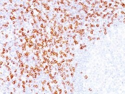
Biotium CD8(C8/144B), CF594 conjugate, 0.1mg/mL
CD8 is a cell surface receptor expressed either as a heterodimer with the CD8 β chain (CD8 α/β) or as a homodimer (CD8 α/α). A majority of thymocytes and a subpopulation of mature T cells and NK cells express CD8a. CD8 binds to MHC class 1 and through its association with protein tyrosine kinase p56lck plays a role in T cell development and activation of mature T cells. For mature T-cells, CD4 and CD8 are mutually exclusive, so anti-CD8, generally used in conjunction with anti-CD4. It is a useful marker for distinguishing helper/inducer T-lymphocytes, and most peripheral T-cell lymphomas are CD4 /CD8-. Anaplastic large cell lymphoma is usually CD4 and CD8-, and in T-lymphoblastic lymphoma/leukemia, CD4 and CD8 are often co-expressed. CD8 is also found in littoral cell angioma of the spleen.Primary antibodies are available purified, or with a selection of fluorescent CF Dyes and other labels. CF Dyes offer exceptional brightness and photostabilit
Biotium Alkaline Phosphatase(V17.1), CF594 conjugate, 0.1mg/mL
This antibody reacts with tissue non-specific alkaline phosphatase. There are at least four distinct but related alkaline phosphatases: intestinal, placental, placental-like, and liver/bone/kidney (tissue non-specific). The first three are located together on chromosome 2, while the tissue non-specific form is located on chromosome 1. The product of this gene is a membrane bound glycosylated enzyme that is not expressed in any particular tissue and is, therefore, referred to as the tissue-nonspecific form of the enzyme. The exact physiological function of the alkaline phosphatases is not known. A proposed function of this form of the enzyme is matrix mineralization; however, mice that lack a functional form of this enzyme show normal skeletal development. This enzyme has been linked directly to hypo-phosphatasia, a disorder that is characterized by hypercalcemia and includes skeletal defects. The character of this disorder can vary, however, depending on the specific mutation
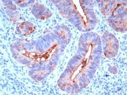
Biotium CD66 (CEA)(C66/195), CF594 conjugate, 0.1mg/mL
This antibody recognizes proteins of 80-200 kDa, identified as different members of CEA family. CEA is synthesized during development in the fetal gut and is re-expressed in increased amounts in intestinal carcinomas and several other tumors. This MAb reacts with nonspecific cross-reacting antigen (NCA) and shows a cross-reaction with human polymorphonuclear leucocytes. It shows no reaction with a variety of normal tissues and is suitable for staining of formalin/paraffin tissues. CEA is not found in benign glands, stroma, or malignant prostatic cells. Antibody to CEA is useful in detecting early foci of gastric carcinoma and in distinguishing pulmonary adenocarcinomas (60-70% are CEA ) from pleural mesotheliomas (rarely or weakly CEA ). Anti-CEA positivity is seen in adenocarcinomas from the lung, colon, stomach, esophagus, pancreas, gallbadder, urachus, salivary gland, ovary, and endocervix.Primary antibodies are available purified, or with a selection of fluorescent CF Dy
Immunoreagents Inc RABBIT A-GOAT IGG HRP 1 MG
Secondary Antibody; Rabbit anti-goat IgG Fc, Affinity Pure, min x w/Human Serum Proteins, HRP Conjugate, Host: Rabbit, Format: Lyo, Label: HRP, Applications: WB, ELISA, IHC/ICC
Immunoreagents Inc RABBIT A-MOUSE IGG FITC
Secondary Antibody; Rabbit anti-mouse IgG (H&L) - Affinity Pure, FITC Conjugate, Host: Rabbit, Format: Liq, Label: FITC, Applications: WB, ELISA, IHC/ICC, IF, IP, FC, FACS
Immunoreagents Inc RABBIT A-GOAT IGG HRP 0.5MG
Secondary Antibody; Rabbit anti-goat IgG (H&L), F(ab)'2 fragment, Affinity Pure, HRP Conjugate, Host: Rabbit, Format: Lyo, Label: HRP, Applications: WB, ELISA, IHC/ICC
Immunoreagents Inc RABBIT A-GOAT IGG BIO 1MG
Secondary Antibody; Rabbit anti-goat IgG Fc, Affinity Pure, min x w/Human Serum Proteins, Biotin Conjugate, Host: Rabbit, Format: Liq, Label: Biotin, Applications: WB, ELISA, IHC/ICC
Immunoreagents Inc RABBIT A-RAT IGG BIO
Secondary Antibody: Rabbit anti-rat IgG (H&L) -Affinity Pure, Biotin Conjugate, Host: Rabbit, Format: Liq, Label: Biotin, Applications: WB, ELISA, IHC/ICC
Immunoreagents Inc RABBIT A-GOAT IGG(H&L) ALP 1MG
Secondary Antibody; Rabbit anti-goat IgG (H&L), Affinity Pure, ALP Conjugate, Host: Rabbit, Format: Liq, Label: ALP, Applications: WB, ELISA
Immunoreagents Inc RABBIT A-GOAT IGG HRP 1 MG
Secondary Antibody; Rabbit anti-goat IgG (H&L), Affinity Pure, min x w/Hu, Mu or Rt lgG/SP, HRP Conjugate, Host: Rabbit, Format: Lyo, Label: HRP, Applications: WB, ELISA, IHC/ICC

Electron Microscopy Sciences Aurion ImmunoGold Rabbit-anti-Goat IgG (H&L), EM-grade 6nm 1 ML
Conventional ImmunoGold Reagents are monodisperse size population makes the conjugates suited for multiple labeling with no overlap. The Conventional Immunogold Reagents are the classical conjugates in immuno electron microscopy; they are a good choice when the antigen is abundant and the accessibility of the antigen is relatively good.
The conventional labeling approach in transmission and scanning electron microscopy utilizes secondary immunogold reagents based on particles that can be observed without enhancement. The particle population is monodisperse and thus shows minimal size variation and overlap.
The reagents are supplied in PBS with 1% Bovine Serum Albumin and 15 mM NaN3.
Particle size: EM-grade 6nm