Learn More
Invitrogen™ CD223 (LAG-3) Monoclonal Antibody (eBioC9B7W (C9B7W)), Super Bright™ 600, eBioscience™, Invitrogen™


Description
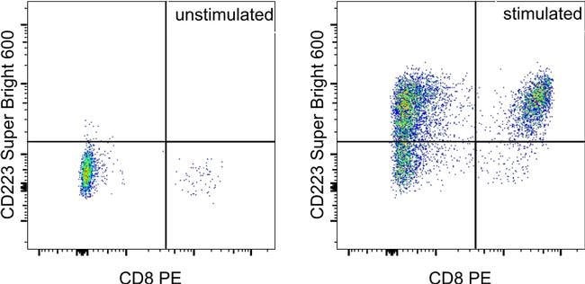
Description: The eBioC9B7W monoclonal antibody recognizes mouse CD223 (LAG-3, LAG3) protein expressed by activated alpha/beta-TCR bearing T cells, a subset of gamma/delta-TCR bearing T cells and a subset of NK cells. CD223 is a 70 kDa type I transmembrane protein with a structure that is similar to CD4. However, a soluble form of human CD223 has been detected by ELISA in human serum, and data suggest that mouse CD223 is proteolytically cleaved in the D4 domain. This results in a 54 kDa fragment containing all the extracellular domains, and a 16 kDa fragment containing the intracellular and transmembrane domains. The 54 kDa can remain membrane-associated or be released as soluble CD223. CD223 binds to MHC class II with higher affinity than CD4, and it is thought that this interaction is involved in the negative regulation of T-cell activation and homeostatic proliferation. Furthermore, CD223 is expressed by CD4+CD25+ regulatory T cells, and it has been suggested that CD223 may be involved in their regulatory function. Applications Reported: This eBioC9B7W antibody has been reported for use in flow cytometric analysis. Applications Tested: This eBioC9B7W antibody has been tested by flow cytometric analysis of stimulated mouse splenocytes. This may be used at less than or equal to 1.0 μg per test. A test is defined as the amount (μg) of antibody that will stain a cell sample in a final volume of 100 μL.
Specifications
Specifications
| Antigen | CD223 (LAG-3) |
| Applications | Flow Cytometry |
| Classification | Monoclonal |
| Clone | eBioC9B7W (C9B7W) |
| Concentration | 0.2 mg/mL |
| Conjugate | Super Bright 600 |
| Formulation | PBS with BSA and 0.09% sodium azide; pH 7.2 |
| Gene | LAG3 |
| Gene Accession No. | Q61790 |
| Gene Alias | Activation-induced cytidine deaminase-linked autoimmunity protein; Aida; CD223; FDC; LAG3; LAG-3; Ly66; lymphocyte activating 3; lymphocyte activation gene 3 protein; lymphocyte-activation gene 3; Secreted lymphocyte activation gene 3 protein; sLAG 3; sLAG3; sLAG-3; soluble LAG 3lymphocyte activating 3; soluble LAG3 |
| Show More |
Safety and Handling
By clicking Submit, you acknowledge that you may be contacted by Fisher Scientific in regards to the feedback you have provided in this form. We will not share your information for any other purposes. All contact information provided shall also be maintained in accordance with our Privacy Policy.