Learn More

Invitrogen™ CD107a (LAMP-1) Monoclonal Antibody (eBioH4A3), eFluor™ 660, eBioscience™


Description
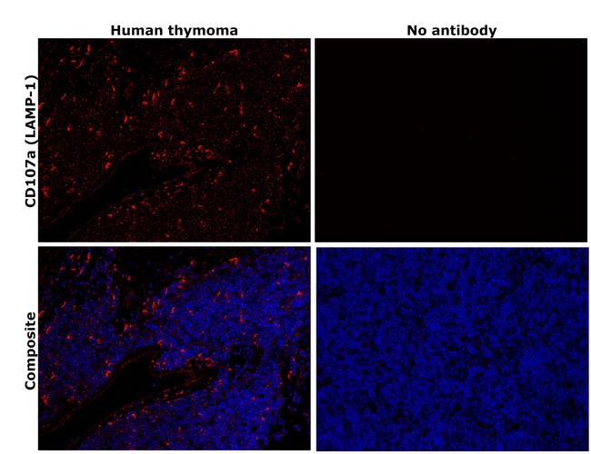
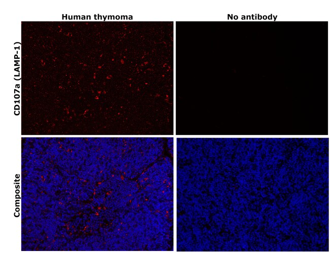
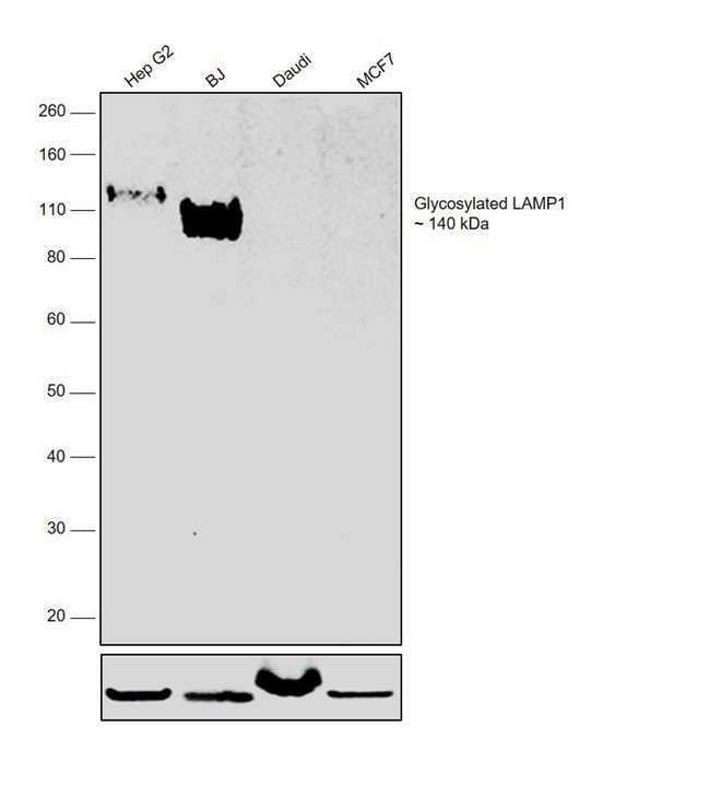
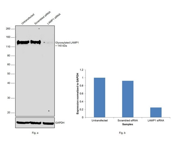
Description: The eBioH4A3 monoclonal antibody reacts with human CD107a, also known as lysosomal-associated membrane protein-1 (LAMP-1). CD107a is a highly glycosylated protein of approximately 110kDa. It is predominantly expressed intracellularly in the lysosomal/endosomal membrane in nearly all cells. CD107a is transiently expressed on the cell surface of degranulating cytolytic T cells, and is also upregulated on the surface of activated platelets and some cancer cells. Applications Reported: This H4A3 antibody has been reported for use in intracellular staining followed by flow cytometric analysis. Applications Tested: This eBioH4A3 antibody has been pre-titrated and tested by intracellular staining and flow cytometric analysis of the Jurkat cell line. This can be used at 5 μL (0.125 μg) per test. A test is defined as the amount (μg) of antibody that will stain a cell sample in a final volume of 100 μL. Cell number should be determined empirically but can range from 10^5 to 10^8 cells/test. eFluor™ 660 is a replacement for Alexa Fluor™ 647. eFluor™ 660 emits at 659 nm and is excited with the red laser (633 nm). Please make sure that your instrument is capable of detecting this fluorochome. Excitation: 633-647 nm; Emission: 668 nm; Laser: Red Laser. Filtration: 0.2 μm post-manufacturing filtered.
Specifications
Specifications
| Antigen | CD107a (LAMP-1) |
| Applications | Flow Cytometry, Immunohistochemistry (Paraffin), Western Blot |
| Classification | Monoclonal |
| Clone | eBioH4A3 |
| Concentration | 5 μL/Test |
| Conjugate | eFluor 660 |
| Formulation | PBS with BSA and 0.09% sodium azide; pH 7.2 |
| Gene | LAMP1 |
| Gene Accession No. | P11279 |
| Gene Alias | 120 kDa lysosomal membrane glycoprotein; AI196048; CD107 antigen-like family member A; CD107a; I79_011073; Lamp I; LAMP1; LAMP-1; LAMPA; LGP120; LGP-120; LGPA; LGP-A; lysosomal associated membrane protein 1; Lysosomal associated membrane protein 1 (120 kDa); lysosomal membrane glycoprotein 1; lysosomal membrane glycoprotein A; lysosomal-associated membrane protein 1; lysosome-associated membrane glycoprotein 1; LYSOSOME-ASSOCIATED MEMBRANE GLYCOPROTEIN 1 PRECURSOR (LAMP-1) (LGP-A) (LGP-120) (CD107A) (P2B); lysosome-associated membrane protein 1; P2B |
| Show More |
Frequently Asked Questions (FAQs)
Our options will depend on the samples you are analyzing.
If cell viability is not critical, you can store your stained samples at 4 degrees C or on ice overnight in the dark and analyze the following day.
For samples stained with eFluor organic fluorochromes, we recommend that cells be suspended in 100 uL of Flow Cytometry Staining Buffer (Cat. No. 00-4222) and 100 uL of eBioscience IC Fixation Buffer (Cat. No. 00-8222); samples can be incubated for up to 3 days at 4 degrees C in the dark. Alternatively, the 1-step Fix/Lyse Solution (Cat. No. 00-5333) can be used. This is a great option when working with whole blood but also works for other cell types.
Yes, the eFluor Organic fluorochromes can be used for intracellular staining. The eFluor organic fluorochromes maintain bright signal and require minimal changes in compensation when fixed with eBioscience IC Fixation Buffer (Cat. No. 00-8222-49) and Permeabilization Buffer (Cat. No. 00-8333-56) or 1-step Fix/Lyse Solution (Cat. No. 00-5333-54, 00-5333-57) (as compared to live cells).
Yes, in-house studies have demonstrated that the eFluor 660 fluorochrome is recognized by Anti-Cy5/Alexa Fluor 647 beads. Side by side studies with Alexa Fluor 647 versus eFluor 660 conjugated antibodies have demonstrated comparable results.
The eFluor Organic fluorochromes and eVolve QDots can be used with flow staining buffers containing PBS and protein.
The eFluor Organic Dyes (eFluor 450, APC-eFluor 780, PerCP-eFluor 710, eFluor 710) are conventional fluorochromes. In contrast, the eVolve line of products are Quantum dots.
By clicking Submit, you acknowledge that you may be contacted by Fisher Scientific in regards to the feedback you have provided in this form. We will not share your information for any other purposes. All contact information provided shall also be maintained in accordance with our Privacy Policy.