Learn More
Invitrogen™ CD4 Monoclonal Antibody (GK1.5), eFluor™ 660, eBioscience™, Invitrogen™


Description
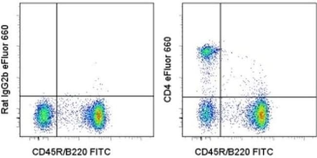
Description: The GK1.5 monoclonal antibody reacts with the mouse CD4 molecule, a 55 kDa cell surface receptor expressed by a majority of thymocytes, subpopulation of mature T cells and dendritic cells. CD4 binds to MHC class II on the surface of antigen presenting cells and plays an important role both in T cell development and in optimal functioning of mature T cells. In T cells, CD4 associates with protein tyrosine kinase p56lck through its cytoplasmic tail. Binding of GK1.5 is blocked by RM4-5. Applications Reported: This GK1.5 antibody has been reported for use in flow cytometric analysis. Applications Tested: This GK1.5 antibody has been tested by flow cytometric analysis of mouse splenocytes. This can be used at less than or equal to 0.125 μg per test. A test is defined as the amount (μg) of antibody that will stain a cell sample in a final volume of 100 μL. Cell number should be determined empirically but can range from 10^5 to 10^8 cells/test. It is recommended that the antibody be carefully titrated for optimal performance in the assay of interest. eFluor™ 660 is a replacement for Alexa Fluor™ 647. eFluor™ 660 emits at 659 nm and is excited with the red laser (633 nm). Please make sure that your instrument is capable of detecting this fluorochome. Excitation: 633-647 nm; Emission: 668 nm; Laser: Red Laser. Filtration: 0.2 μm post-manufacturing filtered.
Specifications
Specifications
| Antigen | CD4 |
| Applications | Flow Cytometry |
| Classification | Monoclonal |
| Clone | GK1.5 |
| Concentration | 0.2 mg/mL |
| Conjugate | eFluor 660 |
| Formulation | PBS with 0.09% sodium azide; pH 7.2 |
| Gene | CD4 |
| Gene Accession No. | P06332 |
| Gene Alias | Activation B7-1 antigen; B7; B7.1; B7-1; BB1; B-lymphocyte activation antigen B7; CD28LG; CD28LG1; CD4; CD4 antigen; CD4 antigen (p55); CD4 antigen p55; Cd4 molecule; CD4 precursor; CD4 receptor; CD4, allele 1; cd4a; CD4mut; CD80; CD80 antigen (CD28 antigen ligand 1, B7-1 antigen); CD80 molecule; cell surface glycoprotein CD4; costimulatory factor CD80; costimulatory molecule variant IgV-CD80; CTLA-4 counter-receptor B7.1; fCD4; L3T4; LAB7; Leu-3; Ly-4; lymphocyte antigen CD4; lymphocyte antigen CD4 precursor; membrane protein; p55; T-cell differentiation antigen L3T4; T-cell surface antigen T4/Leu-3; T-cell surface glycoprotein CD4; T-cell surface glycoprotein CD4 precursor (T-cell surface antigen T4/Leu-3) (T-cell differentiation antigen L3T4); T-lymphocyte activation antigen CD80; W3/25; W3/25 antigen |
| Show More |
For Research Use Only.
By clicking Submit, you acknowledge that you may be contacted by Fisher Scientific in regards to the feedback you have provided in this form. We will not share your information for any other purposes. All contact information provided shall also be maintained in accordance with our Privacy Policy.