Learn More
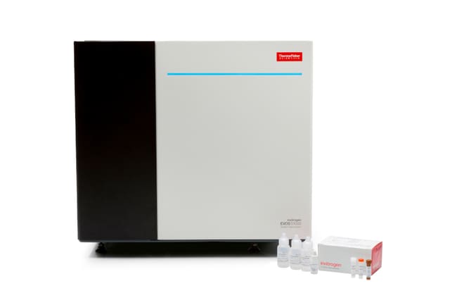
Invitrogen™ EVOS™ S1000 Spatial Imaging System Package Solution
Description
The Invitrogen EVOS S1000 Spatial Imaging System Package Solution includes the EVOS S1000 Spatial Imaging System plus additional service benefits and spatial imaging reagents to ensure a more comprehensive user experience.
The additions include installation and training, extended service coverage, and Aluora Spatial Rainbow Kit with ProLong Glass (Cat. No. A40002450). This kit contains all reagent components needed to perform 9-plex tyramide-based amplification detection for 100 slides.
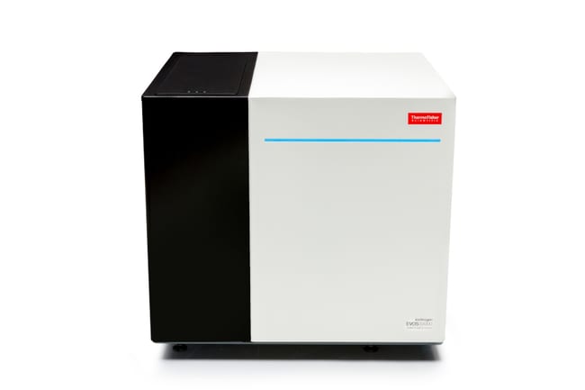
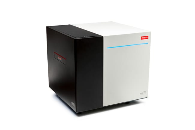
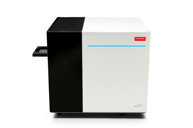
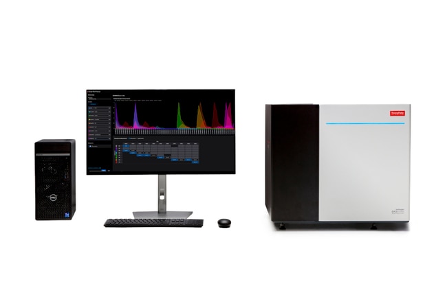
EVOS S1000 Spatial Imaging System
The EVOS S1000 Spatial Imaging System is a high performance, multi-modal instrument for imaging and exploration of tissue slides using multiplex spectral fluorescence, transmitted brightfield, phase contrast, and color brightfield modes. The spectral capabilities of the EVOS S1000 system allow simultaneous capturing and resolving of up to eight fluorescence targets plus nuclear stain (9-plex). This is performed in one single acquisition round, avoiding the need for repeated labeling and imaging cycles that may impact tissue integrity.
The EVOS S1000 system features a flexible hardware configuration that includes a custom LED-based light engine and multiple dichroic and emission filters, as well as a high sensitivity sCMOS 4.2-megapixel camera (6.5-μm pixel size) to detect even the most intricate tissue architecture. The system accommodates up to four slides and includes three pre-set objectives (2.5X ,10X, and 20X) with the option of adding other apochromatic-type 5X and 40X objectives.
The instrument is controlled by the EVOS S1000 spatial software, which provides a sophisticated yet simple interface for acquisition of whole tissue scans with integrated spectral unmixing. The software offers fast overview scans in both transmitted brightfield and fluorescence modes and 'periscope' mode for live tissue navigation. It also provides a sophisticated autofocus functionality to enable stitching and automated unmixing of whole slide tissue scans, all seamlessly integrated to avoid any unnecessary post-acquisition, offline processing steps.
The EVOS S1000 Spatial Imaging System offers these important advantages:
- User-friendly protocol builder that allows easy selection of fluorophores from a dropdown menu to automatically set the optical configuration needed by the instrument to capture the desired plexity
- Intuitive auto-expose and powerful auto-focus (laser-based) functions to promptly locate and identify the targets of interest
- Straightforward and consistent guided workflow for spectrally resolving (via linear unmixing) up to 9-plex samples, using unstained and single-color controls
- Off-the-shelf and ready-to-use protocols for 9-plex and 7-plex imaging, pre-created from reference tissues, that can be used to start imaging archived tissue samples with minimal effort
- Noticeably fast 9-plex acquisition of customizable area scans or whole slides with unmixing and stitching performed automatically, as part of the imaging acquisition workflow
- Output of high-quality, OME-TIFF images (with a resolution of 325 nm/pixel at 20X) that are already stitched, spectrally resolved, and ready to use with any third-party software for downstream analysis, eliminating the need for intermediate adjustments
- Full compatibility with fluorescent dyes from multiple vendors, including Alexa Fluor, Alexa Fluor Plus, Aluora Spatial Amplification, and Opalä dyes, allowing the use of either primary antibody fluorescence conjugates or spatial amplification labeling methods
- Full compatibility with standard tissue slides and coverslips and no proprietary consumables needed.
Spectral imaging for spatial biology, made simple
The EVOS S1000 Spatial Imaging System takes advantage of the power of spectral imaging to simultaneously resolve 9 fluorophores (8 protein targets and DAPI) in a single acquisition round. The system is designed to allow dye selection from a choice of more than 30 different fluorophores with emission spectra between 450 and 810 nm. It uses built-in spectral separation algorithms (based upon linear unmixing), allowing protein targets to be clearly and confidently resolved without the need for iterative labeling and imaging methods, helping preserve valuable tissue samples.
Future-proof and dependable hardware
The EVOS S1000 Spatial Imaging System has a 4-position precision slide holder on a motorized, software-controlled, automated stage. The instrument can spectrally detect more than 60 distinct excitation and emission wavelength combinations, ensuring that no additional filters are required for future spatial proteomics imaging projects. With a 5-position objective capacity, magnifications can be customized between 2.5X and 40X with apochromat-type objectives.
A high sensitivity sCMOS camera (2040 x 2040 pixel resolution) is used to capture 16-bit monochrome OME-TIFF images. Generated images can be saved on the internal hard drive (2X 8-TB SSDs for data), an external USB device, or customer IT-approved cloud solutions and local networking. An external Dell XE4 PC computer with 12th generation Intel Core™ i9-12900 processor, 128 GB DDR4 RAM, and NVIDIA Quadro RTX™ A4000 graphics card is used to operate the EVOS S1000 Spatial Imaging System.
Powerful software designed for sophisticated simplicity
The EVOS S1000 Spatial Software presents users with the opportunity to access multiplex imaging on tissue in the simplest possible way. The acquisition interface has been designed with a strong emphasis on usability, even for inexperienced users and those new to spectral unmixing. It starts with an extremely fast overview scan that can be performed either in transmitted or fluorescent mode. For example, four slides can be scanned in fluorescent mode, including the labels, in ~180 seconds.
Live ('periscope') mode is available for quickly navigating the tissue. Finding samples is fast and precise, using a powerful combination of custom IR laser focus and software autofocus. A guided workflow allows for the unmixing matrix creation process to be performed efficiently and confidently, including the creation of an unmixing quality metrics report with qualitative and quantitative parameters to evaluate the quality of the generated spectral unmixing matrix.
The EVOS S1000 Spatial Software can be used to run variable magnification scans of FOV and ROI with multiple options for automated routines. Whole slide stitching and unmixing is conducted automatically as part of the imaging acquisition.
Readiness for downstream analysis
Images acquired on the EVOS S1000 Spatial Imaging System can be further analyzed using any third-party software available that supports pyramidal OME-TIFF file output for handling of large multidimensional image data.
Additional components included in the package solution
The EVOS S1000 Spatial Imaging System Package Solution also features installation and training provided by our field and application service organization.
In addition to the Standard warranty (1 year), an extra year of coverage is included, with one planned maintenance being performed, among other benefits.
To get started with labeling, the package solution additionally includes an Aluora Spatial Rainbow Kit with ProLong Glass (Cat. No. A40002450), which comprises all reagents needed to perform 9-plex tyramide-based amplification detection, including all Aluora colors with 4X GAM and 4X GAR secondary detection core reagents, for 100 slides.
Specifications
Specifications
| Type | Configured Imaging System |
| Product Line | EVOS |
| Camera | 4.2 MP Highly Sensitive sCMOS Camera |
| For Use With (Application) | Fluorescence, Brightfield, Phase Contrast, and Color Imaging of Tissue Slides |
| For Use With (Equipment) | EVOS |
| High-throughput Compatibility | No |
| Light Source | Custom LED engine with the following excitation spectra: 375 nm, 405 nm, 440 nm, 500 nm, 530 nm, 570 nm, 630 nm and 730 nm |
| Objectives | 5-position automated (includes 2.5x, 10x and 20x) |
| Pixel Size | 6.5 μm |
| Resolution | 2040 x 2040, 4.2 MP |
| Show More |
For Research Use Only. Not for use in diagnostic procedures.
By clicking Submit, you acknowledge that you may be contacted by Fisher Scientific in regards to the feedback you have provided in this form. We will not share your information for any other purposes. All contact information provided shall also be maintained in accordance with our Privacy Policy.