Learn More
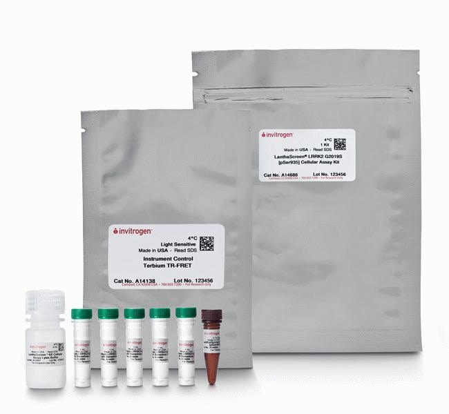
Thermo Scientific™ LanthaScreen™ LRRK2 G2019S [pSer935] Cellular Assay Kit

Description
The LanthaScreen™ LRRK2 G2019S [pSer935] Cellular Assay Kit combines the flexibility of the BacMam gene delivery system with the robustness and power of our LanthaScreen™ TR-FRET technology. It allows researchers to interrogate phosphorylation at serine 935 on a disease-relevant mutant (G2019S) Leucine-Rich Repeat Kinase-2 (LRRK2) protein in a variety of cell backgrounds to identify LRRK2 kinase inhibitors.
This assay format provides a convenient tool for the expression of G2019S LRRK2-GFP fusion protein in the cell background of your choice. The resulting cellular assay can be used to identify LRRK2 G2019S inhibitors, aiding in Parkinson's disease and other research focuses.
This kit provides enough material to evaluate the technology in a 384-well plate; larger sizes of all reagents are available separately.
The kit includes:
- LanthaScreen™ Tb-anti-LRRK2 [pSer935] Antibody, 5 μg
- BacMam LRRK2-GFP G2019S Reagent, 5 x 1 mL
- LanthaScreen™ 6X Lysis Buffer, 6 mL
- Instrument Control Terbium TR-FRET kit
A kit for interrogating wild-type LRRK2 protein (Cat. No. A14685) is also available.
Benefits of this assay include:
- More Physiologically Relevant: cell-based LRRK2 kinase activity and choice of cell background
- More Convenient: compatible with High Throughput Screening (HTS); LanthaScreen™ technology reduces interference and noise
- Disease Relevant: screen compound libraries for Parkinson's disease-relevant molecules
More Physiologically Relevant
The LanthaScreen™ Tb-Anti-LRRK2 [pSer935] Antibody allows the investigation of phosphorylation at serine 935 on LRRK2 protein kinase. When paired with LRRK2 BacMam reagents, you have the freedom to choose the cellular background for your assay, including primary cells. This enables screening for potential inhibitors of LRRK2 proteins in their natural complexes in a physiologically relevant cell type.
More Convenient
Assays are run in a fully homogenous, addition-only format without any of the washing, lysate transfer, or separation procedures required for traditional methods such as western blotting and ELISA, making this assay ideal for HTS applications. The LanthaScreen™ technology provides all of the advantages of TR-FRET ratiometric detection, including reduced data noise, less interference from fluorescent compounds, and high sensitivity, which results in the use of fewer cells than traditional Western or ELISA methods.
Disease Relevant
The LanthaScreen™ Tb-Anti-LRRK2 [pSer935] Antibody can be used by Parkinson's and other disease researchers to perform HTS, cell-based assays to generate inhibition curves (IC50) for their compound libraries. This will aid drug discovery efforts by reducing the time to results from 4 hrs to typically 1.5 hrs compared to alternative ELISAs.

Specifications
Specifications
| Assay | Cell-Based Assay |
| Assay Entry | Cell-based terbium modification activity (transient) |
| Cell State | Cells Not Included |
| Content And Storage | • BacMam LRRK2-GFP G2019S, 5 x 1 mL (Store at 4°C) • LanthaScreen™ 6X Lysis Buffer, 1 tube (Store at 4 °C) • LanthaScreen™ Tb-anti-LRRK2 [pSer935] Antibody, 1 tube (Store at -20°C) • Instrument Control Tb TR-FRET Kit (Store at 4°C) • LanthaScreen™ LRRK2 [pSer935] G2019S Cellular Asasy Kit Protocol (Store at room temperature) |
| Detection Method | Fluorescence |
| For Use With (Application) | TR-FRET |
| For Use With (Equipment) | Microplate Reader |
| Product Line | LanthaScreen |
| Quantity | 1 Kit |
| Readout | TR-FRET (Activity) |
| Show More |
Frequently Asked Questions (FAQs)
We performed a comparison between the LanthaScreen assay and other commercially available TR-FRET assays from 2 different suppliers for the PKC kinase target. Our data revealed that the assays performed comparably, but that the LanthaScreen assay was simpler to optimize and contained fewer components that required optimization. The LanthaScreen assay is a two component system, whereas the other assay formats utilize a trimolecular mechanism which is more time consuming to optimize and has added costs.
Yes, this is possible depending on the concentrations of reagents used and the time for which they are mixed. We recommend developing and optimizing the assay by using separate reagent additions, because this method will work under the widest range of conditions. Once the assay is optimized, the performance of the assay using pre-mixed antibody and EDTA can be evaluated. We have successfully developed robust assays in which the antibody and EDTA were pre-mixed and then stored overnight at 4 degrees C prior to use the following day. A loss of signal intensity was observed in this case, however, by using the ratiometric readout, this effect was minimal.
The chelate is completely stable to Mg2+. The amount of Mn2+ or EDTA that the chelate can tolerate depends largely on how long they are mixed together and the combination of additives used in the reaction. If a reaction requires either Mg2+ or Mn2+ for activation, it is best to stop the reaction by adding an equimolar amount (or slight excess) of EDTA to chelate the metal ions present. This will then essentially eliminate any interference on the terbium chelate by EDTA or Mn2+. Regardless, when LanthaScreen assays are performed using a ratiometric readout (division of the acceptor signal by the donor signal), any interference caused by Mn2+ or EDTA is largely cancelled out.
The Förster radius, the distance at which energy transfer efficiency is half-maximal, is around 50-angstroms for the terbiumÆ fluorescein pair. However, the Förster radius does not give a complete indication of energy transfer efficiency when using long lifetime fluorophores such as terbium chelates. When using terbium chelates, energy transfer efficiency is determined by the distance of closest approach between the donor and acceptor during the excited state lifetime of the donor. In many assay systems, such as those designed using antibodies or peptides, there is a large degree of conformational freedom that allows the donor and acceptor to approach one another, effectively enhancing the FRET signal. Additionally, it is important to note that as the donor/acceptor pair approach one another and the efficiency of energy transfer increases, the fluorescent lifetime decreases to a comparable extent. From a practical standpoint, this means that when energy transfer is extremely efficient, FRET cannot be measured in time-resolved mode (because the energy transfer is complete before the measurement is made). This is another reason why TR-FRET assays based around terbium-labeled antibodies or streptavidin perform so well, because there exist a range of donor/acceptor distances, several of which are optimal for measuring FRET.
It varies, depending on the concentration of substrate used in the assay. But in general, for the peptide substrates, 1 mg of peptide will run approximately 250,000 wells (10 µL reaction, 200 nM peptide). For Poly GT or GAT, the 1 mL of 30 µM size we sell is approximately 1 mg. With these substrates, 1 mL of 30 µM will run approximately 16,700
wells (10 µL reaction, 200 µM substrate).
20 nmol of our physiological protein substrates is sufficient for approximately 10,000 wells (10 µL reaction, 200 µM substrate).
For Research Use Only. Not for use in diagnostic procedures.
By clicking Submit, you acknowledge that you may be contacted by Fisher Scientific in regards to the feedback you have provided in this form. We will not share your information for any other purposes. All contact information provided shall also be maintained in accordance with our Privacy Policy.