Promotional price valid on web orders only. Your contract pricing may differ. Interested in signing up for a dedicated account number?
Learn More

Invitrogen™ Phospho-STAT3 (Tyr705) Monoclonal Antibody (LUVNKLA), PerCP-eFluor™ 710, eBioscience™
GREENER_CHOICE

Catalog No. 46903342
Encompass_Preferred
Change view
Click to view available options
Quantity:
100 Tests
1 product options available for selection
Product selection table with 1 available options. Use arrow keys to navigate and Enter or Space to select.
Catalog No. Quantity
46-903-342 100 Tests
Use arrow keys to navigate between rows. Press Enter or Space to select a product option. 1 options available.
1 options
Catalog No. 46-903-342 Supplier Invitrogen™ Supplier No. 46903342
Add to Cart
Add to Cart

Mouse Monoclonal Antibody

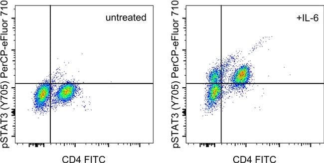
Description: This LUVNKLA monoclonal antibody recognizes human and mouse signal transducer and activator of transcription 3 (STAT3) when phosphorylated on tyrosine 705 (Y705). The STAT family represents seven transcription factors (STATs 1, 2, 3, 4, 5A, 5B, and 6) that are involved in many cellular processes including apoptosis, cell differentiation, and proliferation in a cell type- and cytokine-specific manner. STAT proteins are activated by ligand binding to cytokine receptors that associate with Janus kinase (JAK) family members. Following their phosphorylation by JAKs, STAT proteins translocate to the nucleus where they bind to DNA and regulate transcription of specific genes in a cell type- and cytokine-specific manner. STAT3 is activated downstream of numerous cytokines including interferons, IL-5, IL-6, IL-10, and LIF. STAT3 is important for the differentiation of Th17 cells and mediates a variety of cellular processes including cell growth and survival. The importance of STAT3 is highlighted by both loss-of-function and gain-of-function mutations. Deletion of STAT3 in T cells results in decreased IL-6- and IL-2-mediated proliferation, while deletion of STAT3 in neutrophils and macrophages results in increased susceptibility to LPS-induced endotoxic shock and increased production of the pro-inflammatory cytokines IL-6 and TNF alpha. Hyper STAT3 activity is associated with poor prognosis of many different cancers. Applications Reported: This LUVNKLA antibody has bee...

STAT3 belongs to the family of STAT (signal transducers and activators of transcription) proteins which are phosphorylated by receptor associated kinases, translocate to the nucleus, and act as transcription factors in response to cytokines and growth factors. Coactivators such as CREB-binding protein are required for the transcriptional activation by STAT3. STAT3 can also be activated by Interferon-alpha, Interferon-gamma, EGF, PDGF and IL6. Phosphorylation on tyrosine 705 by JAK1 and JAK2 is essential for STAT3 dimer formation, nuclear translocation, and DNA binding activity. In humans, the STAT3 gene is located on the q arm of chromosome 17. STAT3 has been shown to be activated by IFN-alpha but not IFN-beta. The transcription factors associated with STAT3 are c-Jun and cyclic AMP-responsive enhancer binding protein (CREB). STAT3 mediates the expression of a variety of genes in response to cell stimuli, and thus plays a key role in many cellular processes such as cell growth and apoptosis. The small GTPase Rac1 has been shown to bind and regulate the activity of STAT3 while the PIAS3 protein is a specific inhibitor of STAT3. Three alternatively spliced transcript variants encoding distinct isoforms of STAT3 have been described. Deletion of the STAT3 gene in knock-out mice was lethal at the early embryonic stage.
TRUSTED_SUSTAINABILITY

Specifications

Antigen Phospho-STAT3 (Tyr705)
Applications Flow Cytometry
Classification Monoclonal
Clone LUVNKLA
Concentration 5 μL/Test
Conjugate PerCP-eFluor 710
Formulation PBS with BSA and 0.09% sodium azide; pH 7.2
Gene STAT3
Gene Accession No. P40763, P42227
Gene Alias 1110034C02Rik; acute phase response factor; acute-phase response factor; ADMIO; ADMIO1; APRF; AW109958; DNA-binding protein APRF; FLJ20882; HIES; MGC16063; signal transducer and activator of transcription 3; signal transducer and activator of transcription 3 (acute-phase response factor); signal transducer; activator of transcription; acute-phase response factor; signal transduction and activation of transcription 3; STAT; STAT3; STAT-3; stat3 protein; STAT3b1; STAT3b2; transcription factor; Unknown (protein for MGC:128731); wu:fc15d02; wu:fl59g06; z-Stat3
Gene Symbols STAT3
Host Species Mouse
Purification Method Affinity chromatography
Quantity 100 Tests
Regulatory Status RUO
Primary or Secondary Primary
Gene ID (Entrez) 20848, 6774
Target Species Human, Mouse
Content And Storage 4°C, store in dark, DO NOT FREEZE!
Product Type Antibody
Form Liquid
Isotype IgG2b κ
Show More Show Less
Can the eFluor Organic fluorochromes be used for intracellular staining?

Yes, the eFluor Organic fluorochromes can be used for intracellular staining. The eFluor organic fluorochromes maintain bright signal and require minimal changes in compensation when fixed with eBioscience IC Fixation Buffer (Cat. No. 00-8222-49) and Permeabilization Buffer (Cat. No. 00-8333-56) or 1-step Fix/Lyse Solution (Cat. No. 00-5333-54, 00-5333-57) (as compared to live cells).

How does eBioscience determine the specificity of their phosphorylation-specific antibodies?

Antibodies are tested and validated in a variety of ways. First, we verify that in a panel of cells in which different pathways have been induced, we see phosphorylation specific staining only in the cells in which the specific pathway of interest has been activated. Second, we verify that phosphorylation specific staining is observed only in cell types in which the protein is expressed and not in cell types in which the protein is not expressed. Third, whenever possible, western immunoblotting is used to confirm the presence of a band(s) of the appropriate size(s) in stimulated/treated cells (and not in unstimulated/untreated cells, as appropriate).

What buffers are compatible with the eFluor Organic fluorochromes and eVolve QDots?

The eFluor Organic fluorochromes and eVolve QDots can be used with flow staining buffers containing PBS and protein.

What is the difference between eFluor Organic Dyes and eVolve Qdots?

The eFluor Organic Dyes (eFluor 450, APC-eFluor 780, PerCP-eFluor 710, eFluor 710) are conventional fluorochromes. In contrast, the eVolve line of products are Quantum dots.

How long can cells that have been fixed and placed in methanol be stored for use with eBioscience antibodies? What is the recommended storage temperature? How about storage of fixed cells in eBioscience fixation buffers?

We have data demonstrating that fixed cells stored in methanol are stable at -20 degrees C or at -80 degrees C for several weeks. We do not recommend storing fixed cells in eBioscience Foxp3/Transcription Factor Buffer. However, cells fixed with eBioscience Foxp3/Transcription Factor Buffer or eBioscience Intracellular (IC) fixation buffer can be stored in eBioscience IC fixation buffer for up to 3 days at 4 degrees C in the dark.
Note: Please be aware that higher compensation values may be seen with tandem dyes if any other fixative is used apart from eBioscience IC fixation buffer, for 3-day storage.

What is the optimal protocol for eBioscience phosphorylation-specific antibodies?

Each antibody has been tested in three different buffer systems: IC fixation and permeabilization, Foxp3/Transcription Factor Buffer, and IC fixation Buffer/Methanol. The recommended buffer system(s) will be noted on the Technical Data Sheet for the specific antibody.

What is the best way to maintain surface staining when using the methanol-based protocol?

Because methanol can destroy the epitope recognized by some antibodies, ideally surface staining would be performed at the beginning of the protocol with a methanol-resistant fluorochrome (like eFluor 450, FITC, and eFluor 660).
Please recognize that surface staining before stimulation can have undesired effects due to activation of the cell caused by antibody binding. We recommend staining after stimulation/treatment, fixation, and methanol treatment.

Can I use other buffer systems from other companies when using eBioscience phosphorylation-specific antibodies?

In general, you can use other companies' buffer systems provided that their buffers are of a similar composition as the buffers recommended. Be aware that results will vary depending upon the buffer system used. eBioscience antibodies have been optimized for use in eBioscience buffers and we have not tested all of our antibodies in other buffer systems. Thus, we highly recommend using our optimized protocol and buffer systems.

What are the appropriate controls to run when using eBioscience phosphorylation-specific antibodies?

It is important to understand whether the stimulation/treatment results in an upregulation or a down regulation of the phosphorylation event. Thus, it is recommended that changes in phosphorylation levels in stimulated/treated versus unstimulated/untreated cells be compared. A histogram or density plot overlay these two treatments will provide a better assessment of a change in phosphorylation event than using isotype controls.

Can I perform phosphorylation specific staining and stain for intracellular cytokines at the same time when using eBioscience phosphorylation-specific antibodies?

In theory, if the buffer systems are compatible for the antibodies of interest, this can be done. In practice, cytokine translation and phosphorylation events do not typically occur in the same timeframe, therefore, it may be difficult to detect both events in the same sample. The results will be depended on the kinetics and should be determined for each system and protein of interest.

When using eBioscience phosphorylation-specific antibodies, can I perform phosphorylation specific staining together with transcription factor staining at the same time? If so, what buffer and protocol do I use?

In theory, this can be done if the antibodies against both the transcription factor and the phosphorylated protein work in the same buffer system. For example, Anti-Human/Mouse phsopho-H2AX and all transcription factor eBioscience antibodies offered will work in the Foxp3/Transcription Factor Buffer System (Cat. No. 00-5523-00).

For eBioscience phosphorylation-specific antibodies, can total and phosphorylated protein be analyzed in parallel?

In theory, this can be done if the antibodies to both proteins work in the same buffer system and if the antibodies recognize different epitopes. Please refer to the individual Technical Data Sheets for information about buffer compatibility. Also, consider using eBioscience InstantOne ELISA Kits for immunoassay quantitation of both total and phosphorylated proteins such as AKT1/2/3, JNK1/2/3, NFκB, STAT3, and STAT5.

Are the eFluor Organic Dyes photo-labile?

As with other fluorochromes, we recommend minimal exposure to light to maintain optimal signal.


Product Title
Select an issue

By clicking Submit, you acknowledge that you may be contacted by Fisher Scientific in regards to the feedback you have provided in this form. We will not share your information for any other purposes. All contact information provided shall also be maintained in accordance with our Privacy Policy.