Learn More
Invitrogen™ Phospho-Tau (Thr205) Polyclonal Antibody


Description
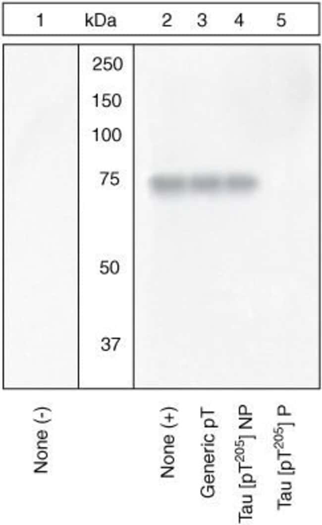
Purified from rabbit serum by sequential epitope-specific chromatography, this product contains enough material for 10 mini-blots. This antibody has been negatively preadsorbed using a non-phosphopeptide corresponding to the site of phosphorylation to remove antibody that is reactive with non-phosphorylated Tau. The final product is generated by affinity chromatography using a Tau-derived peptide that is phosphorylated at threonine 205. The antibody has been used in western blotting. Previous lots of this antibody have been used in immunohistochemistry. In western analysis the positive control used was recombinant human Tau treated with GSK3beta.
Specifications
Specifications
| Antigen | Phospho-Tau (Thr205) |
| Applications | Immunohistochemistry, Western Blot, Immunocytochemistry |
| Classification | Polyclonal |
| Conjugate | Unconjugated |
| Formulation | Dulbecco's PBS with 1mg/mL BSA and 0.05% sodium azide; pH 7.3 |
| Gene | MAPT |
| Gene Accession No. | P10636, P10637, P19332 |
| Gene Alias | AI413597; AW045860; DDPAC; FLJ31424; FTDP17; FTDP-17; G protein beta1/gamma2 subunit-interacting factor 1; map tau; Mapt; MAPTL; MGC138549; microtubule associated protein tau; microtubule-associated protein tau; microtubule-associated protein tau, isoform 4; microtubules; MSTD; Mtapt; MTBT1; MTBT2; Neurofibrillary tangle protein; neurofibrillary tangles; Neuronal Marker; paired helical filament-tau; PHFtau; PHF-tau; PPND; PPP1R103; protein phosphatase 1, regulatory subunit 103; pTau; RNPTAU; Tau; Tau microtubule-associated protein; tau protein; Tau-4; Tau5; Unknown (protein for MGC:134287) |
| Gene Symbols | MAPT |
| Host Species | Rabbit |
| Show More |
Frequently Asked Questions (FAQs)
Here is a procedure for the immunohistochemical staining of Beta-amyloid with paraffin embedded sections of transgenic mouse brain. The following protocol was developed for use with paraffin embedded sections stained with a variety of our antibodies against Beta-amyloid. Applicable rabbit and mouse anti-Beta-amyloid antibodies are: 51-2700, 13-0200, 71-5800, 13-0100Z, 37-4200, 43-7900, 44-136, 700254, 36-6900, and AHB0121.
Beta-amyloid Staining:
- Transgenic mice (expressing the transgenes PS1-A246E + APP swe, APP swe alone, or PS1-A246E alone) were perfused with 1 x Dulbecco's phosphate buffered saline (D-PBS) followed by 4% paraformldehyde buffered with D-PBS. Brain tissues were then embedded in paraffin prior to microtome sectioning.
- After mounting on slides, the paraffin-embedded tissue sections were then deparaffinized with heat. The sections were then incubated for 3 minutes in 70% formic acid. Next, they were deparaffinized further with xylene followed with 100% ethanol. The sections were then re-hydrated in a graded ethanol series (100% ethanol, 95% ethanol, 70% ethanol, and then water).
- Endogenous peroxidase activity was quenched with 3% hydrogen peroxide in methanol. The sections were then heated in the microwave for 5-7 minutes in water, cooled at room temperature for 5 minutes and then rinsed in water. The sections were then washed in TBS (0.05 M Tris-HCl, pH 7.6, with 0.25 M NaCl) prior to blocking. Non-specific binding was blocked with 3% normal goat serum and 0.1% Triton X-100 in TBS for 1 hour at room temperature.
- The sections were then stained with anti-Beta-amyloid antibodies. The staining solution typically consisted of the antibody at a concentration of 5 µg/mL in TBS containing 2% normal goat serum. After incubation at room temperature for at least 1 hr., the sections were washed in TBS 3 times for 5 minutes each. An anti-mouse or anti-rabbit secondary antibody labeled with HRP was then used with DAB or AEC as substrates to stain the Beta-amyloid.
[Adapted from Borchelt, D.R., et al. (1997) Accelerated amyloid deposition in the brains of transgenic mice coexpressing mutant presenilin 1 and amyloid precursor proteins. Neuron 19:939-945.]
Tau and Synuclein Staining:
Here is a general procedure for the immunohistochemical staining of tau, its phosphorylated forms, and synuclein in frozen sections of rat brain. This protocol is adapted from one kindly contributed by Dr. Emil Adamec, M.D., Ph.D., McLean Hospital, Belmont, MA. The procedure was developed for use with cryostat sections of rat brain.
- Sections were fixed with 4% paraformaldehyde for 25 minutes. Sections were then extracted with 0.01% (v/v) Triton X-100. Endogenous peroxidase was blocked with 3% hydrogen peroxide. Sections were then treated with 80% formic acid for 5 to 10 minutes at room temperature to enhance staining. Note that formic acid treatment is also very useful prior to staining with anti-synuclein antibodies.
- The various antibodies were used at dilutions of 1:100 to 1:250. The sections were incubated with the diluted primary antibody overnight at 4°C. A species-specific secondary antibody labeled with HRP was then used with either DAB or AEC to stain the antigens.
- Applicable rabbit and mouse anti-tau and phospho-tau antibodies are: AHB0042, AHB0061, 44-738G, 39-1800, 13-6400, 13-1400, and 18-7461. Anti-synuclein antibodies useful for IHC are: 18-0215, 18-7461, 32-8100, 32-8200, 32-8500, 35-8300, 35-8400, 39-1800, and AHB0261.
By clicking Submit, you acknowledge that you may be contacted by Fisher Scientific in regards to the feedback you have provided in this form. We will not share your information for any other purposes. All contact information provided shall also be maintained in accordance with our Privacy Policy.