
Mouse Secondary Antibodies

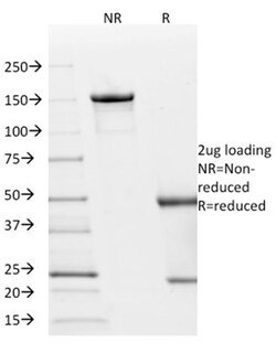
Biotium p21WAF1 (Tumor Suppressor Protein)(AC8), Biotin conjugate, 0.1mg/mL
This MAb recognizes a 21 kDa protein, identified as the p21WAF1 tumor suppressor protein. This MAb is highly specific to p21 and shows no cross-reaction with other closely related mitotic inhibitors. p21WAF1 is a specific inhibitor of cdk's and a tumor suppressor involved in the pathogenesis of a variety of malignancies. The expression of this gene acts as an inhibitor of the cell cycle during G1 phase and is tightly controlled by the tumor suppressor protein p53. Its expression is induced by the wild type, but not mutant, p53 suppressor protein. Normal cells generally display a rather intense nuclear p21 expression. Loss of p21 expression has been reported in many carcinomas (gastric carcinoma, non-small cell lung carcinoma, thyroid carcinoma). Primary antibodies are available purified, or with a selection of fluorescent CF Dyes and other labels. CF Dyes offer exceptional brightness and photostability. Note: Conjugates of blue fluorescent dyes like CF405S and CF405M are not

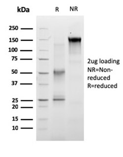
Biotium RET Proto-oncogene(RET/2663), CF568 conjugate, 0.1mg/mL
The Ret proto-oncogene is structurally related to the growing family of tyrosine kinase transmembrane receptors and is involved in GDNF signaling. RET expression is reported in several regions of the central nervous system; in the developing cranial nerve ganglia and a subset of cells within dorsal root ganglia, in motor neurons in the spinal cord and hindbrain, in neuro-retina and the growing tips of the renal collecting ducts in developing kidney. Alterations in RETgene are associated with diseases including papillary thyroid carcinoma, multiple endocrine neoplasia (type 2A and 2B), familial medullary thyroid carcinoma, and a congenital developmental disorder known as Hirschsprung's disease. Primary antibodies are available purified, or with a selection of fluorescent CF Dyes and other labels. CF Dyes offer exceptional brightness and photostability. Note: Conjugates of blue fluorescent dyes like CF405S and CF405M are not recommended for detecting low abundance targets,
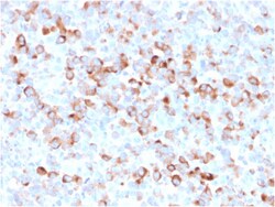
Biotium Tyrosinase (Melanoma Marker)(rOCA1/812), CF488A conjugate, 0.1mg/mL
Recognizes a cluster of proteins between 70-80 kDa, identified as tyrosinase. Occasionally a minor band at 55 kDa is also detected. This MAb shows no cross-reaction with MAGE-1 and tyrosinase-related protein 1, TRP-1/gp75. Tyrosinase is a copper-containing metalloglycoprotein that catalyzes several steps in the melanin pigment biosynthetic pathway; the hydroxylation of tyrosine to L-3, 4-dihydroxy-phenylalanine (dopa), and the subsequent oxidation of dopa to dopaquinone. Mutations of the tyrosinase gene occur in various forms of albinism. Tyrosinase is one of the targets for cytotoxic T-cell recognition in melanoma patients. Staining of melanomas with this MAb shows tyrosinase in melanotic as well as amelanotic variants. This MAb is a useful marker for melanocytes and melanomas. Primary antibodies are available purified, or with a selection of fluorescent CF Dyes and other labels. CF Dyes offer exceptional brightness and photostability. Note: Conjugates of blue fluorescent dyes
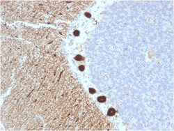
Biotium Carbonic Anhydrase VIII(CPTC-CA8-2), Biotin conjugate, 0.1mg/mL
Carbonic anhydrase VIII (carbonic anhydrase 8) was named because of sequence similarity to other known carbonic anhydrase genes. However, the gene product lacks carbonic anhydrase activity (i. e. , the reversible hydration of carbon dioxide). The gene product continues to carry a carbonic anhydrase designation based on clear sequence identity to other members of the carbonic anhydrase gene family. Mutations in the gene cause neurological disorders in mice. Primary antibodies are available purified, or with a selection of fluorescent CF Dyes and other labels. CF Dyes offer exceptional brightness and photostability. Note: Conjugates of blue fluorescent dyes like CF405S and CF405M are not recommended for detecting low abundance targets, because blue dyes have lower fluorescence and can give higher non-specific background than other dye colors.
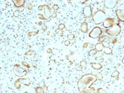
Biotium STAT6 (Solitary Fibrous Tumor Marker)(STAT6/2410), CF488A conjugate, 0.1mg/mL
STAT6 is a transcription factor in the Jak/STAT signal transduction pathway responsible for mediating IL-4 immune signaling. STAT6 was recently suggested to be a reliable marker to distinguish solitary fibrous tumors from other soft tissue neoplasms. Gene fusions are common in solitary fibrous tumors. Recent next generation sequencing studies demonstrated the presence of a NAB2-STAT6 fusion, formed by an intrachromosomal inversion fusing two neighboring genes on chromosome 12q13, in 55-100% of solitary fibrous tumors, regardless of tumor morphology or anatomical site. By immunohistochemistry, nuclear STAT6 expression can discriminate solitary fibrous tumors from its morphological mimics in the meninges, including meningioma, glioblastoma, gliosarcoma, haemangioblastoma, schwannoma and haemangioma. A recent study by Cheah, et al. using the rabbit monoclonal STAT6 antibody (Clone YE361) observed expression in all solitary fibrous tumors (54/54) tested, regardless of histology, anato
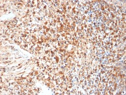
Biotium HSV1 (Herpes Simplex Virus Type I)(HSVI/2095), Biotin conjugate, 0.1mg/mL
The antibody reacts with HSV type 1 specific antigen. It is suitable for detection of HSV in human cellular material obtained from superficial lesions or biopsies and for the early identification of HSV in infected tissue cultures. The herpes simplex virus (HSV) (also known as cold sore, night fever or fever blister) is a virus that causes a contagious disease. There are two main types of Herpes Simplex Virus (HSV), 1 and 2. The HSV-1 strain generally appears in the orafacial organs. HSV2 usually resides in the sacral ganglion at the base of the spine. All herpes viruses are morphologically identical: they have a large double-stranded DNA genome and the virion consists of an icosahedral nucleo-capsid, which is surrounded by a lipid bilayer envelope. UL42, the processivity subunit of the HSV-1DNA polymerase binds DNA as a monomer and is essential for the replication of the virus. UL42 reduces the rate of dissociation from primer-template DNA, but it does not reduce the rate of elon
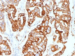
Biotium Cytokeratin 7 (Glandular and Transitional Epithelial Marker)(KRT7/2200), Biotin conjugate, 0.1mg/mL
Recognizes an intermediate filament protein (IFP) of 55 kDa, which is identified as cytokeratin 7. This MAb is highly specific to cytokeratin 7 and shows no cross-reaction with other IFPs. Cytokeratin 7 is a basic cytokeratin, which is found in most glandular and transitional epithelia but not in the stratified squamous epithelia. Keratin 7 is expressed in the epithelial cells of ovary, lung, and breast but not of colon, prostate, or gastrointestinal tract. MAb to cytokeratin 7 is highly useful in distinguishing ovarian carcinomas (keratin 7 ) from colon carcinomas (keratin 7-). Primary antibodies are available purified, or with a selection of fluorescent CF Dyes and other labels. CF Dyes offer exceptional brightness and photostability. Note: Conjugates of blue fluorescent dyes like CF405S and CF405M are not recommended for detecting low abundance targets, because blue dyes have lower fluorescence and can give higher non-specific background than other dye colors.
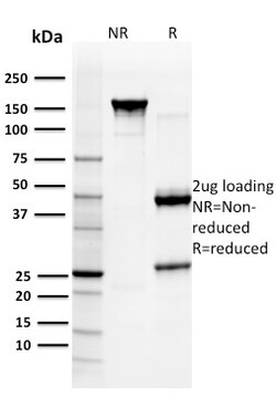
Biotium Glutathione S-Transferase Mu3 (GSTM3)(CPTC-GSTMu3-1), Biotin conjugate, 0.1mg/mL
Recognizes Glutathione S-Transferase of the mu class, which is involved in the detoxification of toxins, drugs, and products of oxidative stress by conjugation with glutathione. Primary antibodies are available purified, or with a selection of fluorescent CF Dyes and other labels. CF Dyes offer exceptional brightness and photostability. Note: Conjugates of blue fluorescent dyes like CF405S and CF405M are not recommended for detecting low abundance targets, because blue dyes have lower fluorescence and can give higher non-specific background than other dye colors.
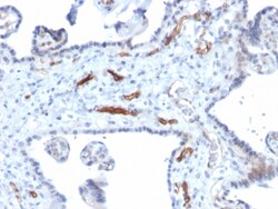
Biotium Podocalyxin (PODXL) (Hematopoietic Stem Cell Marker)(PODXL/2184), CF488A conjugate, 0.1mg/mL
Podocalyxin is a member of the CD34 transmembrane sialomucin family. It is over-expressed on the podocyte foot projections and plays essential roles in kidney development and homeostasis, blood filtration and urine formation. It is also expressed on vascular endothelia, hematopoietic progenitors and a subset of neurons. Overexpression of podocalyxin may be linked to more aggressive tumor behavior. Podocalyxin antibody can identify podocytes in the urine (podocyturia) that may indicate glomerular disease, pre-eclampsia, and other kidney pathology. Primary antibodies are available purified, or with a selection of fluorescent CF Dyes and other labels. CF Dyes offer exceptional brightness and photostability. Note: Conjugates of blue fluorescent dyes like CF405S and CF405M are not recommended for detecting low abundance targets, because blue dyes have lower fluorescence and can give higher non-specific background than other dye colors.
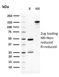
Biotium Squamous Cell Carcinoma Antigen 1(CPTC-SERPINB3-2), Biotin conjugate, 0.1mg/mL
Metastasis of a primary tumor to a distant site is determined through signaling cascades that break down interactions between the cell and extracellular matrix proteins. Among the proteins mediating metastasis are serine proteases, such as neutrophil elastase. Serpins are family of serine protease inhibitors, contain a stretch of peptide that mimics a true substrate for a corresponding serine protease. Serine proteases bind to this substrate mimic in a 1:1 stoichiometric fashion and become catalytically inactive. Aberrant expression of serpin family members can contribute to a number of conditions, including emphysema (-1 antitrypsin deficiency), fatal bleeding (elastase to Thrombin specificity) and thrombosis (antithrombin deficiency), and are indicators of cancer stage phenotypes (circulating levels of squamous cell carcinoma antigen, known as SCCA1, increase in advancing stages of some cervical, lung, esophageal and head and neck cancers). SCCA1 expression has been demonstrated
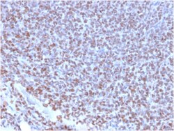
Biotium NKX2.2 (Neuroendocrine & Ewing's Sarcoma Marker)(NX2/2198R), CF488A conjugate, 0.1mg/mL
Expression of NKX2. 2 has been found in neuroendocrine tumors of the gut, making it a potential marker for the study of gastrointestinal neuroendocrine tumors. More recently, NKX2. 2 protein was identified as a target of EWS-FLI-1, the fusion protein specific to Ewing sarcoma, and was shown to be differentially upregulated in Ewing sarcoma on the basis of array-based gene expression analysis. It acts as a valuable marker for Ewing sarcoma, with a sensitivity of 93% and a specificity of 89%, and aids in the differential diagnosis of small round cell tumors. Primary antibodies are available purified, or with a selection of fluorescent CF Dyes and other labels. CF Dyes offer exceptional brightness and photostability. Note: Conjugates of blue fluorescent dyes like CF405S and CF405M are not recommended for detecting low abundance targets, because blue dyes have lower fluorescence and can give higher non-specific background than other dye colors.
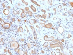
Biotium ARF1 (Golgi Apparatus Marker)(ARF1/2117), Biotin conjugate, 0.1mg/mL
The ADP-ribosylation factor (ARF) family comprises a group of structurally and functionally conserved proteins, which are members of the Ras superfamily of regulatory GTP-binding proteins. The ARF family is divided functionally into the ARF and the ARF-like proteins. ARF's share more than 60% sequence identity, appear to be ubiquitous in eukaryotes, and are highly conserved evolutionarily. ARF is involved in intracellular protein traffic to and within the Golgi complex. ARF has a number of disparate activities including maintenance of organelle integrity, assembly of coat proteins, as a co-factor for cholera toxin and as an activator of phospholipase D. Primary antibodies are available purified, or with a selection of fluorescent CF Dyes and other labels. CF Dyes offer exceptional brightness and photostability. Note: Conjugates of blue fluorescent dyes like CF405S and CF405M are not recommended for detecting low abundance targets, because blue dyes have lower fluorescence and
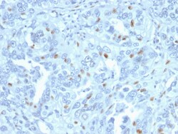
Biotium Cyclin A2 (S- & G2-phase Cyclin)(CCNA2/2333), CF488A conjugate, 0.1mg/mL
Recognizes a protein of 54 kDa, which is identified as cyclin A. Its epitope is located amino acids 144-148 of human Cyclin A2. Cyclins are regulatory subunits of the cyclin-dependent kinases (cdk's) and they control transition at different specific phases of the cell cycle. The temporal expression of cyclins is tightly regulated and subsequently plays a critical role in controlling the enzymatic activity of cdk's. These cyclin/cdk complexes are essential for passage through specific stages in the cell cycle. In mammalian somatic cells, cyclin A is required for S-phase and passage through G2-phase. The D and E type cyclins regulate the passage of G1, while cyclin B is a critical regulator of mitosis. Mutation or disruption of normal cyclin A expression causes cells to arrest in G2-phase. Primary antibodies are available purified, or with a selection of fluorescent CF Dyes and other labels. CF Dyes offer exceptional brightness and photostability. Note: Conjugates of blue fluores
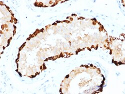
Biotium Melanoma Antigen Family A, 4 / MAGEA4(CPTC-MAGEA4-1), CF568 conjugate, 0.1mg/mL
The melanoma-associated antigen (MAGE) family consists of a number of antigens recognized by cytotoxic T lymphocytes. The MAGE genes were initially isolated from different kinds of tumors, and based on their virtually exclusive tumor-specific expression in adult tissues, they have been used as targets for cancer immunotherapy. MAGE genes encode for tumor-rejection antigens and are expressed in tumors of different histologic types, but not in normal tissues, with the exception of testis and placenta. Primary antibodies are available purified, or with a selection of fluorescent CF Dyes and other labels. CF Dyes offer exceptional brightness and photostability. Note: Conjugates of blue fluorescent dyes like CF405S and CF405M are not recommended for detecting low abundance targets, because blue dyes have lower fluorescence and can give higher non-specific background than other dye colors.
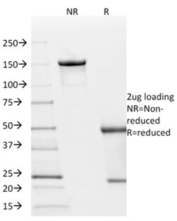
Biotium p21WAF1 (Tumor Suppressor Protein)(AC8), CF568 conjugate, 0.1mg/mL
This MAb recognizes a 21 kDa protein, identified as the p21WAF1 tumor suppressor protein. This MAb is highly specific to p21 and shows no cross-reaction with other closely related mitotic inhibitors. p21WAF1 is a specific inhibitor of cdk's and a tumor suppressor involved in the pathogenesis of a variety of malignancies. The expression of this gene acts as an inhibitor of the cell cycle during G1 phase and is tightly controlled by the tumor suppressor protein p53. Its expression is induced by the wild type, but not mutant, p53 suppressor protein. Normal cells generally display a rather intense nuclear p21 expression. Loss of p21 expression has been reported in many carcinomas (gastric carcinoma, non-small cell lung carcinoma, thyroid carcinoma). Primary antibodies are available purified, or with a selection of fluorescent CF Dyes and other labels. CF Dyes offer exceptional brightness and photostability. Note: Conjugates of blue fluorescent dyes like CF405S and CF405M are not