Protein Labeling Reagents
- (108)
- (3)
- (2)
- (17)
- (2)
- (2)
- (1)
- (7)
- (1)
- (38)
- (2)
- (1)
- (17)
- (7)
- (4)
- (15)
- (16)
- (8)
- (2)
- (1)
- (10)
- (5)
- (17)
- (1)
- (2)
- (16)
- (6)
- (6)
- (5)
- (15)
- (88)
- (17)
- (8)
- (66)
- (2)
- (2)
- (38)
- (1)
- (4)
- (18)
- (3)
- (17)
- (5)
- (6)
- (1)
- (22)
- (2)
- (2)
- (2)
- (6)
- (6)
- (11)
- (27)
- (2)
- (2)
- (1)
- (11)
- (1)
- (4)
- (30)
- (1)
- (1)
- (2)
- (8)
- (6)
- (5)
- (5)
- (5)
- (5)
- (1)
- (6)
- (47)
- (2)
- (3)
- (3)
- (5)
- (5)
- (41)
- (44)
- (35)
Filtered Search Results
Biotium CD34(QBEnd/10 + HPCA1/763), CF568 conjugate, 0.1mg/mL
This MAb recognizes a single chain, transmembrane, heavily glycosylated protein of 90-120 kDa, which is identified as CD34. Its expression is a hallmark for identifying pluripotent hematopoietic stem or progenitor cells. Its expression is gradually lost as lineage committed progenitors differentiate. CD34 is a marker of choice for staining blasts in acute myeloid leukemia. In addition, CD34 is expressed by soft tissue tumors, such as solitary fibrous tumor and gastrointestinal stromal tumor. Its expression is also found in vascular endothelium. Additionally, it appears that proliferating endothelial cells express this molecule more than the non-proliferating endothelial cells. Anti-CD34 labels > 85% of angiosarcoma and Kaposi's sarcoma, but with a lower specificity.Primary antibodies are available purified, or with a selection of fluorescent CF Dyes and other labels. CF Dyes offer exceptional brightness and photostability. Note: Conjugates of blue fluorescent dyes like CF
Non-distribution item offered as a customer accommodation; additional freight charges may apply.
Learn More
Biotium CD1a(CB-T6), CF488A conjugate, 0.1mg/mL
At least five CD1 genes (CD1a, b, c, d, and e) are identified. CD1 proteins have been demonstrated to restrict T cell response to non-peptide lipid and glycolipid antigens and play a role in non-classical antigen presentation. CD1a is a non-polymorphic MHC Class 1 related cell surface glycoprotein, expressed in association with Beta-2 microglobulin. Anti-CD1a labels Langerhans cell histiocytosis (Histiocytosis X), extranodal histiocytic sarcoma, a subset of T-lymphoblastic lymphoma/leukemia, and interdigitating dendritic cell sarcoma of the lymph node. When combined with antibodies against TTF-1 and CD5, anti-CD1a is useful in distinguishing between pulmonary and thymic neoplasms since CD1a is consistently expressed in thymic lymphocytes in both typical and atypical thymomas, but only focally in 1/6 of thymic carcinomas and not in lymphocytes in pulmonary neoplasms. Anti-CD1a is reported to be a new marker for perivascular epithelial cell tumor (PEComa).Primary antibodies
Non-distribution item offered as a customer accommodation; additional freight charges may apply.
Learn More
Biotium CD1a(CB-T6), CF488A conjugate, 0.1mg/mL
At least five CD1 genes (CD1a, b, c, d, and e) are identified. CD1 proteins have been demonstrated to restrict T cell response to non-peptide lipid and glycolipid antigens and play a role in non-classical antigen presentation. CD1a is a non-polymorphic MHC Class 1 related cell surface glycoprotein, expressed in association with Beta-2 microglobulin. Anti-CD1a labels Langerhans cell histiocytosis (Histiocytosis X), extranodal histiocytic sarcoma, a subset of T-lymphoblastic lymphoma/leukemia, and interdigitating dendritic cell sarcoma of the lymph node. When combined with antibodies against TTF-1 and CD5, anti-CD1a is useful in distinguishing between pulmonary and thymic neoplasms since CD1a is consistently expressed in thymic lymphocytes in both typical and atypical thymomas, but only focally in 1/6 of thymic carcinomas and not in lymphocytes in pulmonary neoplasms. Anti-CD1a is reported to be a new marker for perivascular epithelial cell tumor (PEComa).Primary antibodies
Non-distribution item offered as a customer accommodation; additional freight charges may apply.
Learn More
Biotium Fascin-1(FSCN1/416), CF488A conjugate, 0.1mg/mL
Recognizes a protein of 55 kDa, which is identified as fascin-1. Its actin binding ability is regulated by phosphorylation. Antibody to fascin-1 is a very sensitive marker for Reed-Sternberg cells and variants in nodular sclerosis, mixed cellularity, and lymphocyte depletion Hodgkin's disease. It is uniformly negative in lymphoid cells, plasma cells, and myeloid cells. Fascin-1 is also expressed in dendritic cells. This marker may be helpful to distinguish between Hodgkin lymphoma and non-Hodgkin lymphoma in difficult cases. Also, the lack of expression of fascin-1 in the neoplastic follicles in follicular lymphoma may be helpful in distinguishing these lymphomas from reactive follicular hyperplasia in which the number of follicular dendritic cells is normal or increased. Antibody to fascin-1 has been suggested as a prognostic marker in neuroendocrine neoplasms of the lung as well as in ovarian cancer. Fascin-1 expression may be induced by Epstein-Barr virus (EBV) infection o
Non-distribution item offered as a customer accommodation; additional freight charges may apply.
Learn More
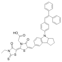
eMolecules 786643-20-7 | Medchem Express | D149 Dye | 50mg | 446271272 | HY-50938 | MFCD28016704 | 741.94 | C42H35N3O4S3
Medchem Express | D149 Dye | 50mg | 446271272 | HY-50938 | 786643-20-7 | MFCD28016704 | 741.940 | C42H35N3O4S3
Non-distribution item offered as a customer accommodation; additional freight charges may apply.
Learn More
Biotium Insulin(2D11-H5), CF568 conjugate, 0.1mg/mL
Recognizes a polypeptide which is identified as insulin, a 51-amino acid polypeptide composed of A and B chains connected through the C-peptide. Proinsulin, which has very little biological activity, is cleaved by proteases within its cell of origin into the insulin molecule and the C-terminal basic residue. Insulin enhances membrane transport of glucose, amino acids, and certain ions. It also promotes glycogen storage, formation of triglycerides, and synthesis of proteins and nucleic acids. Deficiency of insulin results in diabetes mellitus. The main storage site for insulin is the pancreatic islets. Antibodies to insulin are important as beta-cell and insulinoma marker.Primary antibodies are available purified, or with a selection of fluorescent CF Dyes and other labels. CF Dyes offer exceptional brightness and photostability. Note: Conjugates of blue fluorescent dyes like CF405S and CF405M are not recommended for detecting low abundance targets, because blue dyes have
Non-distribution item offered as a customer accommodation; additional freight charges may apply.
Learn More
Medchemexpress LLC MEDCHEMEXPRESS LLC
Small and Specialty Supplier Partner
Small and/or specialty supplier based on Federal laws and SBA requirements.
Learn More
Small and/or specialty supplier based on Federal laws and SBA requirements.
Learn More
5000699738 IL-17A HUMAN BI 100UG
Non-distribution item offered as a customer accommodation; additional freight charges may apply.
Learn More
Biotium Bax(2D2), CF488A conjugate, 0.1mg/mL
This antibody recognizes a protein of 21 kDa, identified as the Bax protein. This MAb is highly specific to Bax and shows no cross-reaction with Bcl-2 or Bcl-X protein. Bcl-2 blocks cell death following a variety of stimuli. Bax has extensive amino acid homology with Bcl-2 and it homodimerizes and forms heterodimers with Bcl-2. Overexpression of Bax accelerates apoptotic death induced by cytokine deprivation in an IL-3 dependent cell line, and Bax also counters the death repressor activity of Bcl-2.Primary antibodies are available purified, or with a selection of fluorescent CF Dyes and other labels. CF Dyes offer exceptional brightness and photostability. Note: Conjugates of blue fluorescent dyes like CF405S and CF405M are not recommended for detecting low abundance targets, because blue dyes have lower fluorescence and can give higher non-specific background than other dye colors.
Non-distribution item offered as a customer accommodation; additional freight charges may apply.
Learn More
eMolecules 871514-32-8 | Cy3 hydrazide | Broadpharm543.580 | C30H40Cl2N4O | 96.000 | [Cl-].[Cl-].CN1\C(=C\C=C\C2=[N+](CCCCCC(=O)N[NH3+])c3ccccc3C2(C)C)C(C)(C)c2ccccc12 | 1mg | 296213684
Cy3 hydrazide | Broadpharm | 871514-32-8543.580 | C30H40Cl2N4O | 96.000 | [Cl-].[Cl-].CN1\C(=C\C=C\C2=[N+](CCCCCC(=O)N[NH3+])c3ccccc3C2(C)C)C(C)(C)c2ccccc12 | 1mg | 296213684
Non-distribution item offered as a customer accommodation; additional freight charges may apply.
Learn More
eMolecules 3326-32-7 | Fluorescein isothiocyanate isomer I, tech grade | Combi-Blocks | MFCD00005063 | 389.380 | C21H11NO5S | 98.000 | Oc1ccc2c(Oc3cc(O)ccc3C22OC(=O)c3cc(ccc23)N=C=S)c1 | 1g | 232307473
Fluorescein isothiocyanate isomer I, tech grade | Combi-Blocks | 3326-32-7 | MFCD00005063 | 389.380 | C21H11NO5S | 98.000 | Oc1ccc2c(Oc3cc(O)ccc3C22OC(=O)c3cc(ccc23)N=C=S)c1 | 1g | 232307473
Non-distribution item offered as a customer accommodation; additional freight charges may apply.
Learn More
Biotium HER-2 / CD340(HRB2/282), CF568 conjugate, 0.1mg/mL
Recognizes a protein of 185 kDa, which is identified as c-erbB-2/HER-2/neu. Its epitope is localized in the extracellular domain. C-erbB-2/HER-2 is a member of the EGFR family. This MAb is specific and shows minimal cross-reaction with other members of the EGFR-family. Receptors of this family are located on the plasma membrane and consist of an extracellular ligand-binding domain that is connected to a large intracellular domain by a single transmembrane sequence. c-erbB-2/HER-2 protein is over-expressed in a variety of carcinomas especially those of breast and ovary.Primary antibodies are available purified, or with a selection of fluorescent CF Dyes and other labels. CF Dyes offer exceptional brightness and photostability. Note: Conjugates of blue fluorescent dyes like CF405S and CF405M are not recommended for detecting low abundance targets, because blue dyes have lower fluorescence and can give higher non-specific background than other dye colors.
Non-distribution item offered as a customer accommodation; additional freight charges may apply.
Learn More
Biotium MyoD1(5.8A), CF568 conjugate, 0.1mg/mL
Recognizes a phosphor-protein of 45 kDa, identified as MyoD1. The epitope of this MAb maps between amino acid 180-189 in the C-terminal of mouse MyoD1 protein. It does not cross react with myogenin, Myf5, or Myf6. Antibody to MyoD1 labels the nuclei of myoblasts in developing muscle tissues. MyoD1 is not detected in normal adult tissue, but is highly expressed in the tumor cell nuclei of rhabdomyosarcomas. Occasionally nuclear expression of MyoD1 is seen in ectomesenchymoma and a subset of Wilm s tumors. Weak cytoplasmic staining is observed in several non-muscle tissues, including glandular epithelium and also in rhabdomyosarcomas, neuroblastomas, Ewing s sarcomas and alveolar soft part sarcomas.Primary antibodies are available purified, or with a selection of fluorescent CF Dyes and other labels. CF Dyes offer exceptional brightness and photostability. Note: Conjugates of blue fluorescent dyes like CF405S and CF405M are not recommended for detecting low abundance tar
Non-distribution item offered as a customer accommodation; additional freight charges may apply.
Learn More
Biotium CD45RA(PTPRC/1131), CF568 conjugate, 0.1mg/mL
Recognizes a protein of 205 kDa-220 kDa, identified as CD45RA (Workshop III). CD45RA is isoforms of the human leukocyte common antigen (CD45). Human CD45 contains three exons which encode peptide segments designated A, B and C, respectively. The differential splicing of the exons generates at least five isoforms, ABC, AB, BC, B and O. This antibody reacts with ABC and BC isoforms. CD45RA is expressed on 40-50% of peripheral CD4 T-cells, 50% of peripheral CD8 T-cells, B-cells, and leukemic B-cell lines. T-cells expressing CD45RA are naive or virgin T-cells. T-cells expressing CD45RO are memory T-cells. CD45RA and CD45RO define complementary, predominantly non-overlapping populations of resting peripheral T-cells. This MAb is useful in study on the subpopulation of CD4 or CD8 T-cells. It can especially be used to differentiate T-cell lymphomas (CD45RO positive) from B cell lymphomas (CD45RA positive).Primary antibodies are available purified, or with a selection of fluoresc
Non-distribution item offered as a customer accommodation; additional freight charges may apply.
Learn More
Biotium CD63(NKI-C3), CF488A conjugate, 0.1mg/mL
This MAb recognizes protein of 26 kDa-60 kDa, which is identified as CD63. Its epitope is different from that of MAb LAMP3/529. The tetraspanins are integral membrane proteins expressed on cell surface and granular membranes of hematopoietic cells and are components of multi-molecular complexes with specific integrins. The tetraspanin CD63 is a lysosomal membrane glycoprotein that translocates to the plasma membrane after platelet activation. CD63 is expressed on activated platelets, monocytes and macrophages, and is weakly expressed on granulocytes, T cell and B cells. It is located on the basophilic granule membranes and on the plasma membranes of lymphocytes and granulocytes. CD63 is a member of the TM4 superfamily of leukocyte glycoproteins that includes CD9, CD37 and CD53, which contain four transmembrane regions. CD63 may play a role in phagocytic and intracellular lysosome-phagosome fusion events. CD63 deficiency is associated with Hermansky-Pudlak syndrome and is stro
Non-distribution item offered as a customer accommodation; additional freight charges may apply.
Learn More
Biotium CD66 (CEA)(C66/261), CF568 conjugate, 0.1mg/mL
This antibody recognizes proteins of 80-200 kDa, identified as different members of CEA family. CEA is synthesized during development in the fetal gut and is re-expressed in increased amounts in intestinal carcinomas and several other tumors. This MAb does not react with nonspecific cross-reacting antigen (NCA) and with human polymorphonuclear leucocytes. It shows no reaction with a variety of normal tissues and is suitable for staining of formalin/paraffin tissues. CEA is not found in benign glands, stroma, or malignant prostatic cells. Antibody to CEA is useful in detecting early foci of gastric carcinoma and in distinguishing pulmonary adenocarcinomas (60-70% are CEA positive) from pleural mesotheliomas (rarely or weakly CEA positive). Anti-CEA positivity is seen in adenocarcinomas from the lung, colon, stomach, esophagus, pancreas, gallbadder, urachus, salivary gland, ovary, and endocervix.Primary antibodies are available purified, or with a selection of fluorescent CF
Non-distribution item offered as a customer accommodation; additional freight charges may apply.
Learn More