Promotional price valid on web orders only. Your contract pricing may differ. Interested in signing up for a dedicated account number?
Learn More
Learn More
PLC-γ2 Mouse anti-Human, PE, Clone: K86-1161, BD
Mouse Monoclonal Antibody
Supplier: BD Biosciences 560134
Description
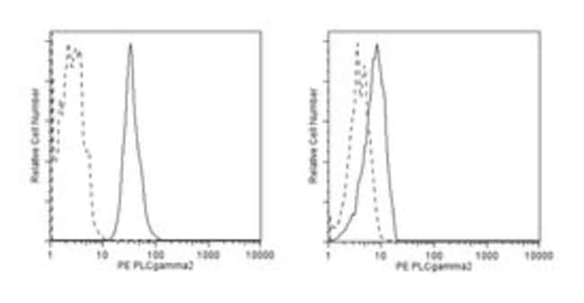
The Phospholipase C (PLC) isozymes hydrolyze phosphatidyl inositol biphosphate to inositol triphosphate and diacylglycerol. The former causes release of calcium from the endoplasmic reticulum, while the latter is an activator of Protein Kinase C. Within the PLC family, PLC-g is the only member that contains SH2 and SH3 domains. These domains enable it to interact with receptor tyrosine kinases and become enzymatically activated via phosphorylation. It exists as two isoforms: 1) PLC-g1, which is ubiquitously expressed, and 2) PLCg2, found primarily in the lymphoid system. PLC-g is essential for growth factor-induced cell motility and mitogenesis. Overexpression of PLC-g is evident in several forms of cancer, and it has been identified as a key mediator of PDGF-dependent cellular transformation. Thus regulation of PLC-g activity by growth factors is involved in cell growth and transformation. Although the immunogen for generation of the K86-1161 monoclonal antibody was a phosphorylated peptide, peptide blocking studies demonstrated that the mAb recognizes PLC-g2 regardless of phosphorylation status. This antibody was raised to a unique region of PLC-γ2 and is predicted not to crossreact with PLC-γ1.
Host Species: Mouse
Clone: K86-1161
Isotype: IgG1 κ
Species Reactivity: Human
Immunogen: Phosphorylated Human PLC-γ2 Peptide
Intracellular Staining
Specifications
| PLC-γ2 | |
| Monoclonal | |
| PE | |
| Mouse | |
| Affinity Purified | |
| RUO | |
| Primary | |
| Store undiluted at 4°C and protected from prolonged exposure to light. Do not freeze. |
| Flow Cytometry | |
| K86-1161 | |
| Aqueous buffered solution containing BSA and ≤0.09% sodium azide. | |
| Phosphorylated Human PLC-γ2 Peptide | |
| 50 Tests | |
| Cell Biology | |
| Human | |
| IgG1 κ |
Product Content Correction
Your input is important to us. Please complete this form to provide feedback related to the content on this product.
Product Title
Spot an opportunity for improvement?Share a Content Correction